Advertisement

Main menu

  • Home
  • Content
    • Current Issue
    • Accepted Manuscripts
    • Article Preview
    • Past Issue Archive
    • AJNR Case Collection
    • Case of the Week Archive
    • Classic Case Archive
    • Case of the Month Archive
  • Special Collections
    • Spinal CSF Leak Articles (Jan 2020-June 2024)
    • 2024 AJNR Journal Awards
    • Most Impactful AJNR Articles
  • Multimedia
    • AJNR Podcast
    • AJNR Scantastics
    • Video Articles
  • For Authors
    • Submit a Manuscript
    • Author Policies
    • Fast publishing of Accepted Manuscripts
    • Graphical Abstract Preparation
    • Manuscript Submission Guidelines
    • Imaging Protocol Submission
    • Submit a Case for the Case Collection
  • About Us
    • About AJNR
    • Editorial Board
  • More
    • Become a Reviewer/Academy of Reviewers
    • Subscribers
    • Permissions
    • Alerts
    • Feedback
    • Advertisers
    • ASNR Home
  • Other Publications
    • ajnr

User menu

  • Alerts
  • Log in

Search

  • Advanced search
American Journal of Neuroradiology

American Journal of Neuroradiology

ASHNR American Society of Functional Neuroradiology ASHNR American Society of Pediatric Neuroradiology ASSR
  • Alerts
  • Log in

Advanced Search

  • Home
  • Content
    • Current Issue
    • Accepted Manuscripts
    • Article Preview
    • Past Issue Archive
    • AJNR Case Collection
    • Case of the Week Archive
    • Classic Case Archive
    • Case of the Month Archive
  • Special Collections
    • Spinal CSF Leak Articles (Jan 2020-June 2024)
    • 2024 AJNR Journal Awards
    • Most Impactful AJNR Articles
  • Multimedia
    • AJNR Podcast
    • AJNR Scantastics
    • Video Articles
  • For Authors
    • Submit a Manuscript
    • Author Policies
    • Fast publishing of Accepted Manuscripts
    • Graphical Abstract Preparation
    • Manuscript Submission Guidelines
    • Imaging Protocol Submission
    • Submit a Case for the Case Collection
  • About Us
    • About AJNR
    • Editorial Board
  • More
    • Become a Reviewer/Academy of Reviewers
    • Subscribers
    • Permissions
    • Alerts
    • Feedback
    • Advertisers
    • ASNR Home
  • Follow AJNR on Twitter
  • Visit AJNR on Facebook
  • Follow AJNR on Instagram
  • Join AJNR on LinkedIn
  • RSS Feeds

Welcome to the new AJNR, Updated Hall of Fame, and more. Read the full announcements.


AJNR is seeking candidates for the position of Associate Section Editor, AJNR Case Collection. Read the full announcement.


Research ArticleSpine

Exophytic Lumbar Vertebral Body Mass in an Adult with Back Pain

J.C. Benson, M.A. Vizcaino, D.K. Kim, C. Carr, P. Rose, L. Eckel and F. Diehn
American Journal of Neuroradiology October 2020, 41 (10) 1786-1790; DOI: https://doi.org/10.3174/ajnr.A6749
J.C. Benson
aFrom the Departments of Radiology (J.C.B., D.K.K., C.C., L.E., F.D.)
  • Find this author on Google Scholar
  • Find this author on PubMed
  • Search for this author on this site
  • ORCID record for J.C. Benson
M.A. Vizcaino
bPathology (M.A.V.)
  • Find this author on Google Scholar
  • Find this author on PubMed
  • Search for this author on this site
  • ORCID record for M.A. Vizcaino
D.K. Kim
aFrom the Departments of Radiology (J.C.B., D.K.K., C.C., L.E., F.D.)
  • Find this author on Google Scholar
  • Find this author on PubMed
  • Search for this author on this site
  • ORCID record for D.K. Kim
C. Carr
aFrom the Departments of Radiology (J.C.B., D.K.K., C.C., L.E., F.D.)
  • Find this author on Google Scholar
  • Find this author on PubMed
  • Search for this author on this site
  • ORCID record for C. Carr
P. Rose
cOrthopedic Surgery (P.R.), Mayo Clinic, Rochester, Minnesota
  • Find this author on Google Scholar
  • Find this author on PubMed
  • Search for this author on this site
  • ORCID record for P. Rose
L. Eckel
aFrom the Departments of Radiology (J.C.B., D.K.K., C.C., L.E., F.D.)
  • Find this author on Google Scholar
  • Find this author on PubMed
  • Search for this author on this site
  • ORCID record for L. Eckel
F. Diehn
aFrom the Departments of Radiology (J.C.B., D.K.K., C.C., L.E., F.D.)
  • Find this author on Google Scholar
  • Find this author on PubMed
  • Search for this author on this site
  • ORCID record for F. Diehn
  • Article
  • Figures & Data
  • Info & Metrics
  • Responses
  • References
  • PDF
Loading

Article Figures & Data

Figures

  • Tables
  • FIG 1.
    • Download figure
    • Open in new tab
    • Download powerpoint
    FIG 1.

    Axial MR imaging of the mass. T2 fat-saturated (A), T1 precontrast (B), and postcontrast (C) sequences show a mass centered within the mid and left aspects of the L3 vertebral body (asterisk). Most of the tumor demonstrates substantial T2 prolongation, which is particularly evident with fat saturation. Internal T2-hypointense septations are noted, which enhance with moderate avidity (straight arrows), while the fluid-filled regions lack enhancement. The tumor extends out of the left vertebral body into the adjacent soft tissues, displacing but not invading the left psoas muscle laterally (curved arrows).
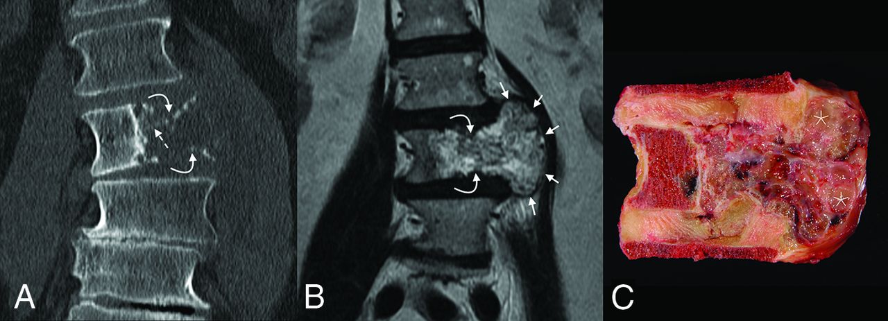
  • FIG 2.
    • Download figure
    • Open in new tab
    • Download powerpoint
    FIG 2.

    Coronal imaging and corresponding gross pathology of the tumor. On CT (A), the mass is destructive, with faint small foci of high attenuation, either representing amorphous calcifications or residual/partially destroyed vertebral body trabeculae (dashed arrow). Both the superior and inferior endplates are fractured (curved arrows). The degree of extraosseous extension is best seen on T2 MR imaging (B), where the soft-tissue components are seen to mushroom out along the adjacent intervertebral discs (between the short arrows), with pushing-type margins. The gross pathology specimen (C) confirms the presence of numerous high-water-content loculations (asterisk) separated by small septations, corresponding with areas of T2 hyperintensity.
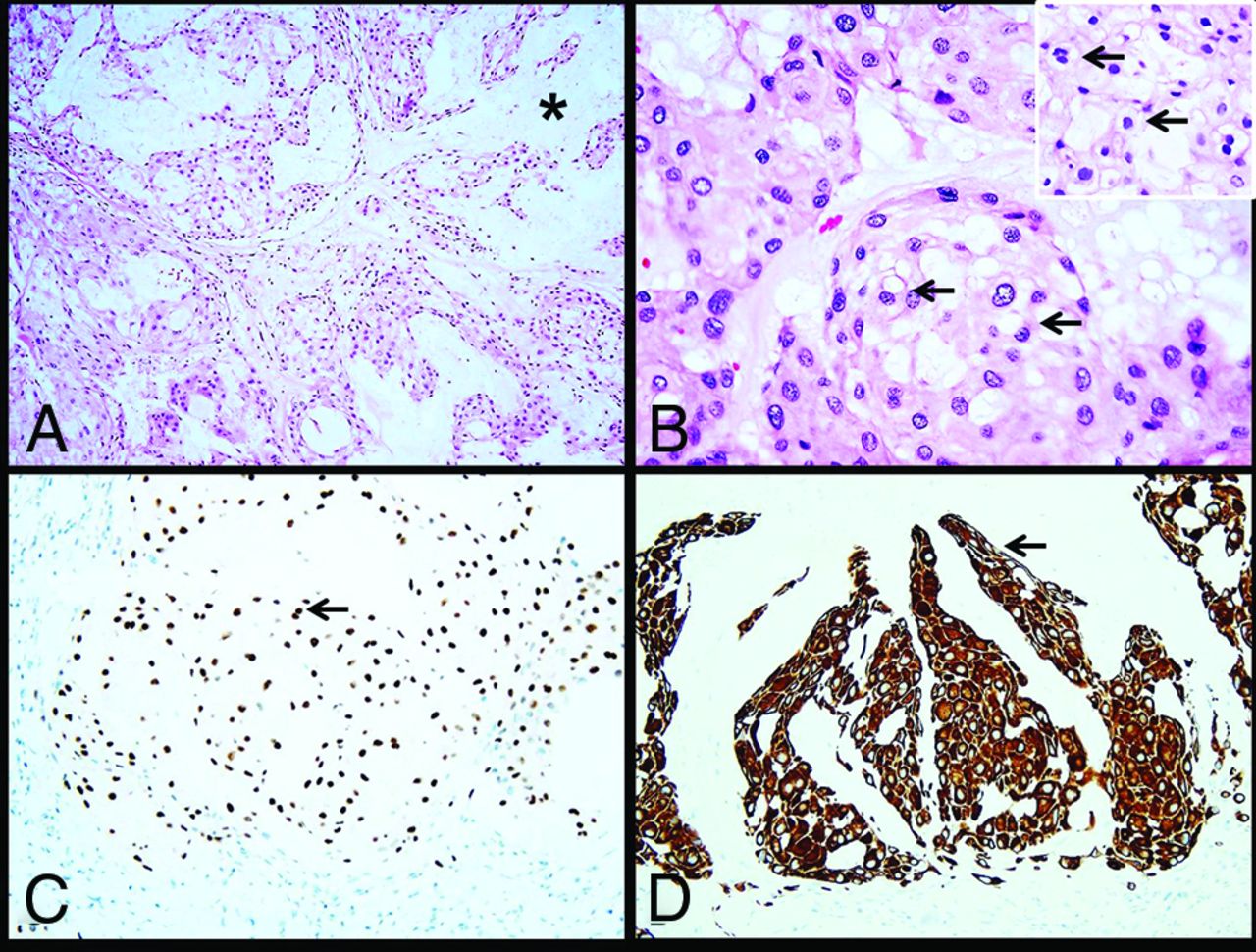
  • FIG 3.
    • Download figure
    • Open in new tab
    • Download powerpoint
    FIG 3.

    Histology images. The tumor consists of cells arranged in nests and cords (A) within an abundant mucoid matrix (A, asterisk). Most of the tumor cells are large with central nuclei and mildly vacuolated eosinophilic cytoplasm (B, arrows). Scattered physaliphorous cells are also present (B inset, arrows). Immunohistochemistry demonstrates nuclear brachyury (C, arrow) and cytoplasmic keratin CAM 5.2 (D, arrow) expression in tumor cells.
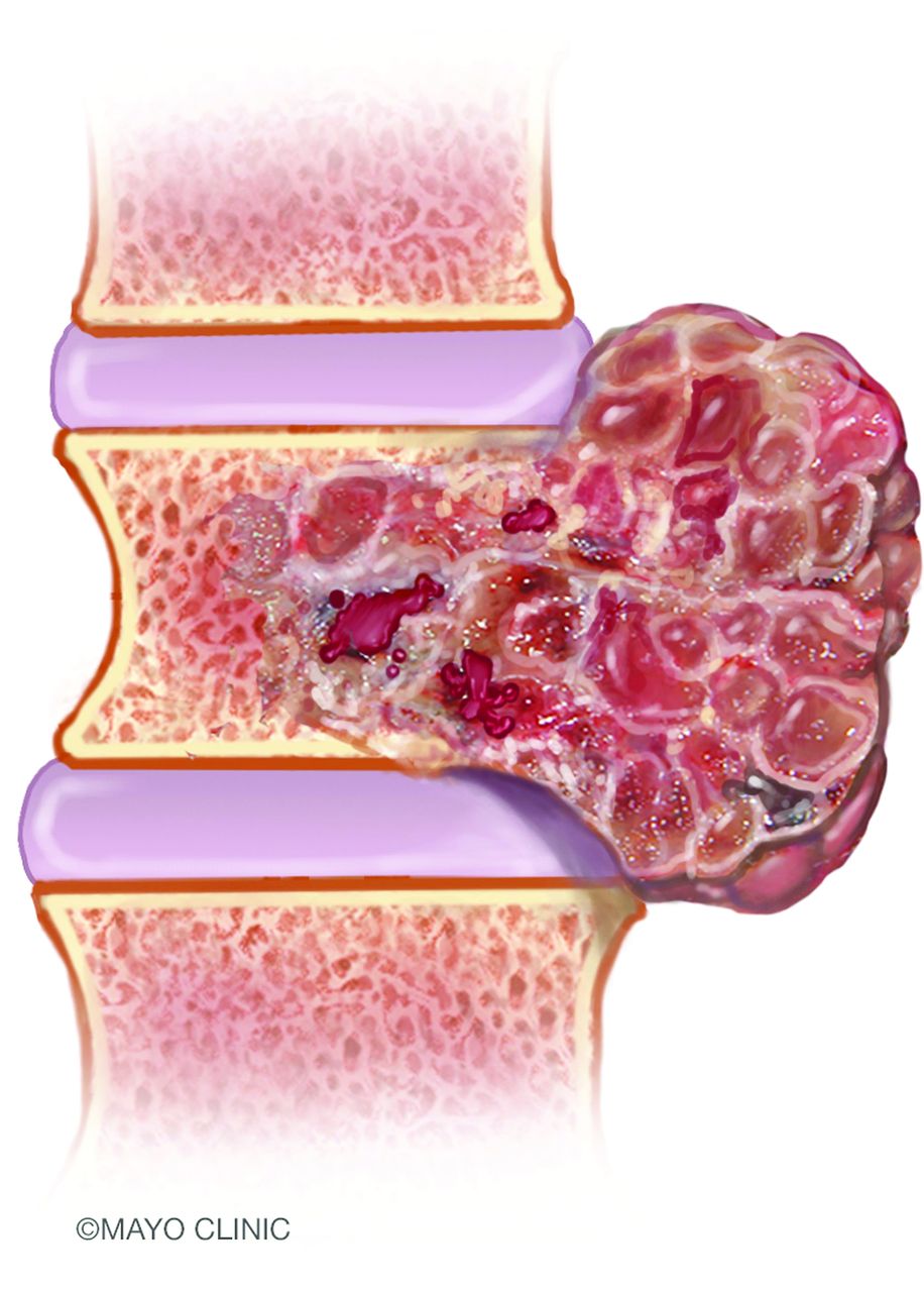
  • FIG 4.
    • Download figure
    • Open in new tab
    • Download powerpoint
    FIG 4.

    An artist’s illustration of a spinal chordoma. The tumors are generally sizable at the time of diagnosis. Extraosseous components are often larger than those within the vertebral bodies and extend along the adjacent spinal segments, compatible with the classically described dumbbell appearance. Image used with permission of Mayo Foundation for Medical Education and Research. All rights reserved.

Tables

  • Figures
  • Comparison of various demographic, location, and imaging features of chordomas with those of the most common imaging differential diagnoses

    ChordomaGCTChondrosarcomaPlasmacytoma
    Age at diagnosis (range) (peak yr)40–6020–3030–7030–60
    Most common location in mobile spineCervicalLumbar (sacrum much more common)LumbarThoracic
    Commonly involves posterior elements–+++
    Intratumoral calcificationAmorphous–Rings and arcs–
    T2WI↑↑↓→↑↑
    Extraosseous extension+++–
    Characteristic featureDumbbell or mushroom shapeLytic lesion without sclerotic rim; fluid-fluid levelsIntratumoral chondroid matrixMinibrain, soap bubble
    • Note:—GCT indicates Giant cell tumor; –, features absent; +, features present; ↓→, hypo- to iso-intense intralesional signal; ↑, hyperintense intralesional signal.

PreviousNext
Back to top

In this issue

American Journal of Neuroradiology: 41 (10)
American Journal of Neuroradiology
Vol. 41, Issue 10
1 Oct 2020
  • Table of Contents
  • Index by author
  • Complete Issue (PDF)
Advertisement
Print
Download PDF
Email Article

Thank you for your interest in spreading the word on American Journal of Neuroradiology.

NOTE: We only request your email address so that the person you are recommending the page to knows that you wanted them to see it, and that it is not junk mail. We do not capture any email address.

Enter multiple addresses on separate lines or separate them with commas.
Exophytic Lumbar Vertebral Body Mass in an Adult with Back Pain
(Your Name) has sent you a message from American Journal of Neuroradiology
(Your Name) thought you would like to see the American Journal of Neuroradiology web site.
CAPTCHA
This question is for testing whether or not you are a human visitor and to prevent automated spam submissions.
Cite this article
J.C. Benson, M.A. Vizcaino, D.K. Kim, C. Carr, P. Rose, L. Eckel, F. Diehn
Exophytic Lumbar Vertebral Body Mass in an Adult with Back Pain
American Journal of Neuroradiology Oct 2020, 41 (10) 1786-1790; DOI: 10.3174/ajnr.A6749

Citation Manager Formats

  • BibTeX
  • Bookends
  • EasyBib
  • EndNote (tagged)
  • EndNote 8 (xml)
  • Medlars
  • Mendeley
  • Papers
  • RefWorks Tagged
  • Ref Manager
  • RIS
  • Zotero
0 Responses
Respond to this article
Share
Bookmark this article
Exophytic Lumbar Vertebral Body Mass in an Adult with Back Pain
J.C. Benson, M.A. Vizcaino, D.K. Kim, C. Carr, P. Rose, L. Eckel, F. Diehn
American Journal of Neuroradiology Oct 2020, 41 (10) 1786-1790; DOI: 10.3174/ajnr.A6749
del.icio.us logo Twitter logo Facebook logo Mendeley logo
  • Tweet Widget
  • Facebook Like
  • Google Plus One
Purchase

Jump to section

  • Article
    • Abstract
    • Footnotes
    • References
  • Figures & Data
  • Info & Metrics
  • Responses
  • References
  • PDF

Related Articles

  • No related articles found.
  • PubMed
  • Google Scholar

Cited By...

  • Diagnostic Value of "Mushroom" Morphology in Vertebral Chordoma
  • Crossref (3)
  • Google Scholar

This article has been cited by the following articles in journals that are participating in Crossref Cited-by Linking.

  • Diagnostic Value of “Mushroom” Morphology in Vertebral Chordoma
    T.S. Ghosh, S. Tiwari, P.K. Garg, P.S. Khera, P. Elhence
    American Journal of Neuroradiology 2021 42 3
  • Chondroid chordoma within dorsal spine
    El Houssni Jihane, Moatassim Billah Nabil, Nassar Ittimade
    International Journal of Case Reports and Images 2022 13
  • Imaging of Primary Tumors of the Osseous Spine
    Wiem Abid, Filip Vanhoenacker
    2023

More in this TOC Section

  • Bern Score Validity for SIH
  • MP2RAGE 7T in MS Lesions of the Cervical Spine
  • Resisted Inspiration for CSF-Venous Fistula
Show more Spine

Similar Articles

Advertisement

Indexed Content

  • Current Issue
  • Accepted Manuscripts
  • Article Preview
  • Past Issues
  • Editorials
  • Editors Choice
  • Fellow Journal Club
  • Letters to the Editor

Cases

  • Case Collection
  • Archive - Case of the Week
  • Archive - Case of the Month
  • Archive - Classic Case

Special Collections

  • Special Collections

Resources

  • News and Updates
  • Turn around Times
  • Submit a Manuscript
  • Author Policies
  • Manuscript Submission Guidelines
  • Evidence-Based Medicine Level Guide
  • Publishing Checklists
  • Graphical Abstract Preparation
  • Imaging Protocol Submission
  • Submit a Case
  • Become a Reviewer/Academy of Reviewers
  • Get Peer Review Credit from Publons

Multimedia

  • AJNR Podcast
  • AJNR SCANtastic
  • Video Articles

About Us

  • About AJNR
  • Editorial Board
  • Not an AJNR Subscriber? Join Now
  • Alerts
  • Feedback
  • Advertise with us
  • Librarian Resources
  • Permissions
  • Terms and Conditions

American Society of Neuroradiology

  • Not an ASNR Member? Join Now

© 2025 by the American Society of Neuroradiology All rights, including for text and data mining, AI training, and similar technologies, are reserved.
Print ISSN: 0195-6108 Online ISSN: 1936-959X

Powered by HighWire