Advertisement

Main menu

  • Home
  • Content
    • Current Issue
    • Accepted Manuscripts
    • Article Preview
    • Past Issue Archive
    • AJNR Case Collection
    • Case of the Week Archive
    • Classic Case Archive
    • Case of the Month Archive
  • Special Collections
    • Spinal CSF Leak Articles (Jan 2020-June 2024)
    • 2024 AJNR Journal Awards
    • Most Impactful AJNR Articles
  • Multimedia
    • AJNR Podcast
    • AJNR Scantastics
    • Video Articles
  • For Authors
    • Submit a Manuscript
    • Author Policies
    • Fast publishing of Accepted Manuscripts
    • Graphical Abstract Preparation
    • Manuscript Submission Guidelines
    • Imaging Protocol Submission
    • Submit a Case for the Case Collection
  • About Us
    • About AJNR
    • Editorial Board
  • More
    • Become a Reviewer/Academy of Reviewers
    • Subscribers
    • Permissions
    • Alerts
    • Feedback
    • Advertisers
    • ASNR Home
  • Other Publications
    • ajnr

User menu

  • Alerts
  • Log in

Search

  • Advanced search
American Journal of Neuroradiology

American Journal of Neuroradiology

ASHNR American Society of Functional Neuroradiology ASHNR American Society of Pediatric Neuroradiology ASSR
  • Alerts
  • Log in

Advanced Search

  • Home
  • Content
    • Current Issue
    • Accepted Manuscripts
    • Article Preview
    • Past Issue Archive
    • AJNR Case Collection
    • Case of the Week Archive
    • Classic Case Archive
    • Case of the Month Archive
  • Special Collections
    • Spinal CSF Leak Articles (Jan 2020-June 2024)
    • 2024 AJNR Journal Awards
    • Most Impactful AJNR Articles
  • Multimedia
    • AJNR Podcast
    • AJNR Scantastics
    • Video Articles
  • For Authors
    • Submit a Manuscript
    • Author Policies
    • Fast publishing of Accepted Manuscripts
    • Graphical Abstract Preparation
    • Manuscript Submission Guidelines
    • Imaging Protocol Submission
    • Submit a Case for the Case Collection
  • About Us
    • About AJNR
    • Editorial Board
  • More
    • Become a Reviewer/Academy of Reviewers
    • Subscribers
    • Permissions
    • Alerts
    • Feedback
    • Advertisers
    • ASNR Home
  • Follow AJNR on Twitter
  • Visit AJNR on Facebook
  • Follow AJNR on Instagram
  • Join AJNR on LinkedIn
  • RSS Feeds

Welcome to the new AJNR, Updated Hall of Fame, and more. Read the full announcements.


AJNR is seeking candidates for the position of Associate Section Editor, AJNR Case Collection. Read the full announcement.


Research ArticleInterventional

Intraprocedural Flat Panel Detector Rotational Angiography and an Image Fusion Technique for Delivery of a Microcatheter into the Targeted Shunt Pouch of a Dural Arteriovenous Fistula

J.H. Choi, D.Y. Cho, Y.S. Shin and B.-S. Kim
American Journal of Neuroradiology October 2020, 41 (10) 1876-1878; DOI: https://doi.org/10.3174/ajnr.A6724
J.H. Choi
aFrom the Departments of Neurosurgery (J.H.C., Y.S.S.)
  • Find this author on Google Scholar
  • Find this author on PubMed
  • Search for this author on this site
  • ORCID record for J.H. Choi
D.Y. Cho
cDepartment of Neurosurgery (D.Y.C.), Ewha Womans University Seoul Hospital, College of Medicine, Ewha Womans University, Seoul, Republic of Korea
  • Find this author on Google Scholar
  • Find this author on PubMed
  • Search for this author on this site
  • ORCID record for D.Y. Cho
Y.S. Shin
aFrom the Departments of Neurosurgery (J.H.C., Y.S.S.)
  • Find this author on Google Scholar
  • Find this author on PubMed
  • Search for this author on this site
  • ORCID record for Y.S. Shin
B.-S. Kim
bRadiology (B.-S.K.), Seoul St. Mary’s Hospital, The Catholic University of Korea College of Medicine, Seoul, Republic of Korea
  • Find this author on Google Scholar
  • Find this author on PubMed
  • Search for this author on this site
  • ORCID record for B.-S. Kim
  • Article
  • Figures & Data
  • Supplemental
  • Info & Metrics
  • Responses
  • References
  • PDF
Loading

Abstract

SUMMARY: The accurate and safe delivery of a microcatheter to a targeted shunt pouch is essential for successful transvenous embolization of intracranial dural arteriovenous fistulas. However, complex anatomy and variations in head and neck veins and occluded sinuses can hinder intraprocedural microcatheter delivery. In this study, we introduce an intraprocedural flat panel detector rotational angiography and image fusion technique to aid precise navigation inside the veins and proper placement of the microcatheter in the targeted shunt pouch.

ABBREVIATIONS:

ACC
anterior condylar confluence
DAVF
dural arteriovenous fistula
FPCTRA
flat panel CT rotational angiography
IF
image fusion
TVE
transvenous embolization

For the successful treatment of intracranial dural arteriovenous fistulas (DAVFs), precise evaluation of the angioarchitecture using pretherapeutic DSA is important. However, identification of detailed angioarchitectures of some DAVFs using conventional 2D-DSA or 3D rotational angiography images is difficult because of overlapping vessels.1,2 With the evolution of angiography equipment and 3D postprocessing technology, performing a flat panel CT rotational angiography and image fusion (FPCTRA and IF) technique to obtain a fusion image using 2 different rotational angiographic images is feasible. This technique enables physicians to gain better pretherapeutic knowledge for establishing an appropriate treatment plan.1⇓-3 However, to our knowledge, no research has focused mainly on the value of the intraprocedural FPCTRA and IF technique. Especially, for transvenous embolization (TVE), the most important factor for successful occlusion is the accurate and safe delivery of a microcatheter to the shunt pouch. Complex anatomy and variations in head and neck veins,4 especially those located in the skull base,5 and sinus occlusion6,7 can hinder intraprocedural microcatheter delivery. In this study, we introduce an intraprocedural FPCTRA and IF technique to aid the navigation of a microcatheter inside veins and confirm the placement of the microcatheter in the targeted pouch.

Technique

For treating all DAVFs and image processing, we used Artis Zee biplane angiography equipment (Siemens). All procedures were conducted with the patient under general anesthesia. Systemic heparin (2000- to 3000-IU bolus) was routinely administrated after placement of a femoral sheath. Antiplatelet agents were not routinely administrated. We placed a 5F angiocatheter in the main feeding artery and a 7F guiding catheter in the internal jugular vein or the retromandibular vein according to the access route. Thereafter, we performed 3D dual rotation angiography with contrast injection to obtain subtraction (vessel only), mask (bone only), and native fill (vessel with bone) images as a baseline. We named this series DSA24, which irradiated at 0.24 µGy/frame and took 30 frame/s for 5 seconds. The rotational angle was 200° with 1.5° increments, resulting in 133 projections and a rotation velocity of 40°/s. For contrast, we infused iodixanol (Visipaque 270; GE Healthcare) via the external carotid artery with an injector at a rate of 2–3 mL/s for 5 seconds with a 1- to 2-second acquisition delay according to vessel diameter and shunt flow. Acquired data were sent to the workstation (syngo 3D workplace; Siemens), and 3D reconstruction was performed with the following parameters: kernel type, Hounsfield unit; image characteristics, normal; reconstruction mode, dual volume; section matrix, 512 × 512; and voxel size, 0.47 mm. A size 18 microcatheter (Excelsior 1018, Stryker) and a 0.014-inch microwire (Transend; Stryker) were usually used for navigation. During navigation, if needed, we performed 3D single-rotation angiography with a reduced FOV without contrast injection while keeping the microwire and microcatheter inside the vein and fused with the initial subtraction image to identify the microcatheter going in the right direction. This series was named DRcare, which was performed with a single rotation for 5 seconds and irradiated 0.10 μGy/frame. In the workstation, we performed 3D reconstruction: kernel type, EE; image characteristics, normal; reconstruction mode, native. Two different image datasets (DSA24 and DRcare) were fused by a mapping application syngo iDentify (Syngo 3D workplace, Siemens AG, Erlangern, Germany), which superimposes the spatial coordinate system of the initial dataset onto that of the second dataset. We used landmark-based automatic registration to avoid misalignment.8 When the microcatheter accessed the shunt pouch, we also checked DRcare and fused images to confirm the exact placement of the microcatheter in the target. Thereafter, we inserted the coil until the shunt disappeared.

Clinical Results

Details of the study population and outcome assessment are presented in the On-line Appendix. We used the FPCTRA and IF technique in 18 of 58 DAVFs treated with TVE between January 2009 and March 2020. Among them, 11 had ipsilateral sinus occlusion (8 in the cavernous sinus, 2 in the anterior condylar confluence [ACC], and 1 in the transverse-sigmoid sinus). We approached the shunt pouch through the occluded sinus by blinded navigation (n = 6) or through alternative venous routes (the facial or superficial temporal vein, n = 5). Seven DAVFs were located in the ACCs. The complete or near-complete occlusion rate and symptom-free or improved rate were 94.4% and 100%, respectively. There was no catheter-related vessel injury with or without using this technique. We could access the shunt via the transfemoral route in all cases treated using this technique (On-line Table). In contrast, we failed to reach the shunt pouch via the transfemoral approach in 5 cases without this technique. Therefore, surgical cannulation of the target sinus or the direct ophthalmic vein approach was additionally performed to reach the shunt. Illustrative cases are presented in the Figure and On-line Figs 1 and 2.

FIGURE.
  • Download figure
  • Open in new tab
  • Download powerpoint
FIGURE.

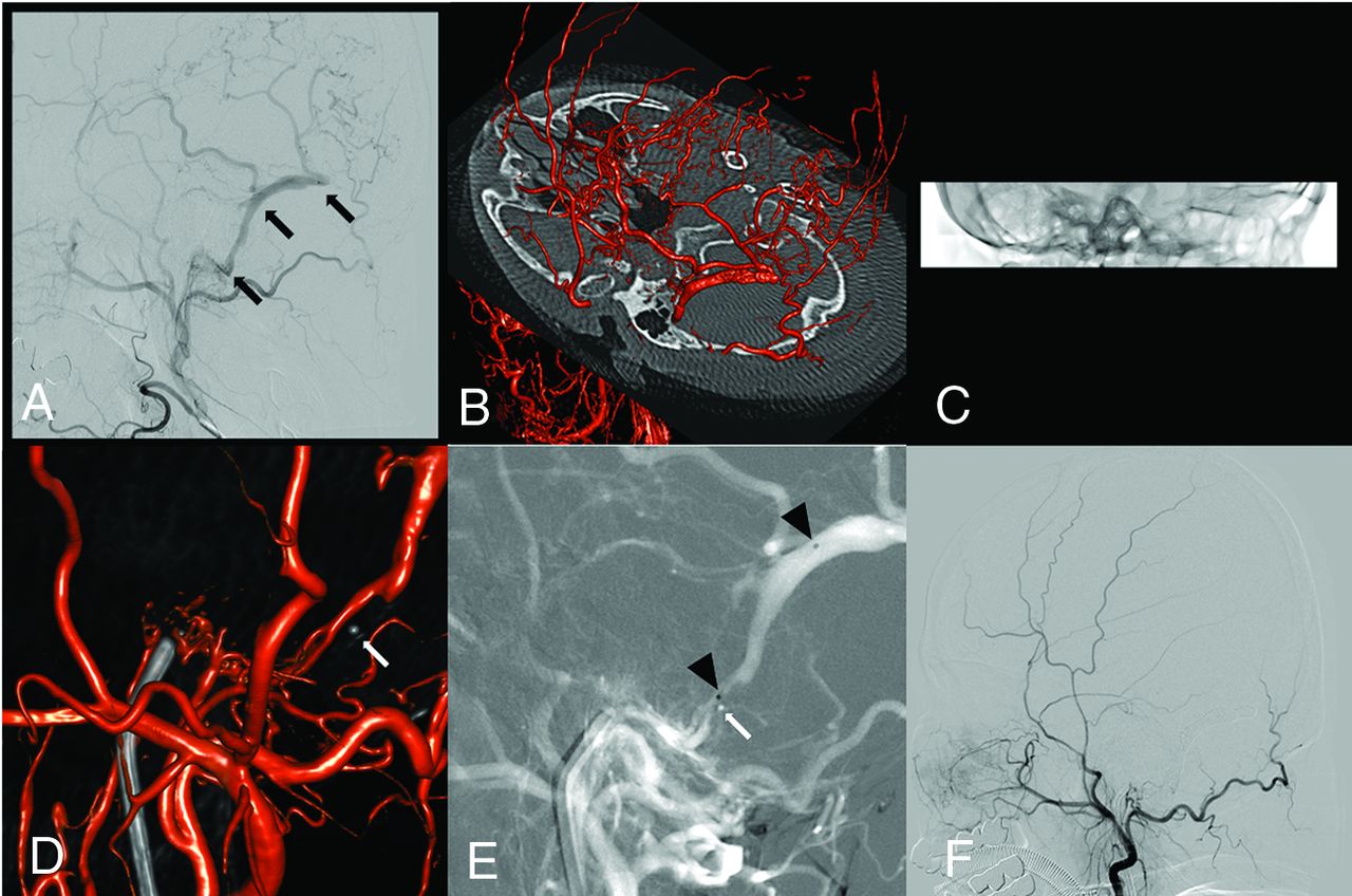
Case 1. A 61-year-old female patient presenting with intracranial hemorrhage. A, Initial right external carotid angiography shows a transverse sigmoid dural AVF with an isolated sinus (black arrow) and pial reflux. B, Intraprocedural 0.24-µG/frame FPCTRA (DSA24) is performed to obtain subtraction and mask images as a control. C, During the microcatheter navigation, we perform low-dose FPCTRA with a reduced FOV (DRcare). D, We found that the microcatheter was placed in the wrong direction (white arrow) by fusion of the initial DSA24 and follow-up DRcare images. E, After we pulled back the microcatheter (white arrow), we directed it the right way and placed the microcatheter in the target shunt (black arrowhead). F, Successful coil embolization is performed.

DISCUSSION

In this study, we described an intraprocedural FPCTRA and IF technique and demonstrated its usefulness during the TVE. The transvenous approach may have technical difficulties for various reasons. In cases with an occluded accessing route due to thrombosis or hypoplasia, TVE can be more challenging. Breaching the occluded sinus by blinded navigation can be attempted. However, breaching the occluded route is not always feasible, and serious complications, including vessel perforation, can occur.7 In addition, delivery of the microcatheter in the wrong direction can occur because breaching the occluded route is always performed by blinded navigation. Moreover, the complexity of head and neck veins that have numerous tributaries and confluences may prevent the microguidewire and microcatheter from advancing in the right direction to the shunt pouch.4 Especially, a DAVF involving the ACC can be challenging for navigation of the microcatheter because of complex venous anatomy around the hypoglossal canal.5 Additionally, roadmap images during TVE may have low image quality and efficacy due to an overlap of vessels on 2D-DSA. Therefore, blinded microcatheter navigation may be needed, and the microcatheter position for coil insertion can be inappropriately placed. These might lead to unexpected complications or incomplete occlusion. In this study, we used the FPCTRA and IF technique to overcome these obstacles. MPR or a partial MIP image was mainly used for identification of the microcatheter tip with bony landmarks during the blinded navigation. To confirm appropriate microcatheter placement, we used mainly 3D volume-rendering or MPR fusion images.

This study has some limitations. The retrospective nature and small sample size may not be enough to draw definite conclusions. Moreover, our technique was mainly used in DAVFs involving an ACC or with sinus occlusion needing blinded navigation. Other typical lesions could be successfully treated using conventional techniques. Therefore, we do not advocate our technique as a first-line use. Finally, our technique may require more radiation exposure. However, minimizing additional radiation exposure can be accomplished using low-dose protocols. Further large-volume data should be evaluated to confirm the efficacy and safety of our technique.

Intraprocedural use of FPCTRA and IF for TVE might be an effective and safe method to navigate the microcatheter into the targeted shunt pouch, especially in cases that require blinded microcatheter navigation.

References

  1. 1.↵
    1. Tanoue S,
    2. Kiyosue H,
    3. Mori H, et al
    . Fusion imaging using subtracted and unsubtracted rotational angiography for pretherapeutic evaluation of dural arteriovenous fistulas. Jpn J Radiol 2014;32:600–07 doi:10.1007/s11604-014-0351-8 pmid:25149420
    CrossRefPubMed
  2. 2.↵
    1. Li H,
    2. Wan F,
    3. Li J, et al
    . Flat detector computed tomography-based “dual vessel fusion” technique for diagnosis and surgical planning in the management of dural arteriovenous fistula. World Neurosurg 2015;84:520–27 doi:10.1016/j.wneu.2 015.04.020 pmid:25900791
    CrossRefPubMed
  3. 3.↵
    1. Fukuda K,
    2. Higashi T,
    3. Okawa M, et al
    . Fusion technique using three-dimensional digital subtraction angiography in the evaluation of complex cerebral and spinal vascular malformations. World Neurosurg 2016;85:353–58 doi:10.1016/j.wneu.2015.08.075 pmid:26348565
    CrossRefPubMed
  4. 4.↵
    1. Choi JH,
    2. Shin YS,
    3. Kim BS
    . Making microguidewire loop facilitates navigation through tortuous or abruptly angulated head and neck veins to access cavernous sinus dural arteriovenous fistulas. World Neurosurg 2019;129:e561–65 doi:10.1016/j.wneu.2019.05.216 pmid:31158531
    CrossRefPubMed
  5. 5.↵
    1. Spittau B,
    2. Millán DS,
    3. El-Sherifi S, et al
    . Dural arteriovenous fistulas of the hypoglossal canal: systematic review on imaging anatomy, clinical findings, and endovascular management. J Neurosurg 2015;122:883–903 doi:10.3171/2014.10.JNS14377 pmid:25415064
    CrossRefPubMed
  6. 6.↵
    1. Biondi A,
    2. Milea D,
    3. Cognard C, et al
    . Cavernous sinus dural fistulae treated by transvenous approach through the facial vein: report of seven cases and review of the literature. AJNR Am J Neuroradiol 2003;24:1240–46 pmid:12812963
    Abstract/FREE Full Text
  7. 7.↵
    1. Cho YD,
    2. Rhim JK,
    3. Yoo DH, et al
    . Transvenous microguidewire looping technique for breach of ipsilateral inferior petrosal sinus occlusions en route to cavernous sinus dural arteriovenous fistulas. Interv Neuroradiol 2016;22:590–95 doi:10.1177/1591019916653251 pmid:27298011
    CrossRefPubMed
  8. 8.↵
    1. Yuki I,
    2. Ishibashi T,
    3. Dahmani C, et al
    . Combination of high-resolution cone beam computed tomography and metal artefact reduction software: a new image fusion technique for evaluating intracranial stent apposition after aneurysm treatment. BMJ Case Rep 2019;12:e230687 doi:10.1136/bcr-2019-230687 pmid:31533950
    CrossRefPubMed
  • Received April 3, 2020.
  • Accepted after revision June 3, 2020.
  • © 2020 by American Journal of Neuroradiology
View Abstract
PreviousNext
Back to top

In this issue

American Journal of Neuroradiology: 41 (10)
American Journal of Neuroradiology
Vol. 41, Issue 10
1 Oct 2020
  • Table of Contents
  • Index by author
  • Complete Issue (PDF)
Advertisement
Print
Download PDF
Email Article

Thank you for your interest in spreading the word on American Journal of Neuroradiology.

NOTE: We only request your email address so that the person you are recommending the page to knows that you wanted them to see it, and that it is not junk mail. We do not capture any email address.

Enter multiple addresses on separate lines or separate them with commas.
Intraprocedural Flat Panel Detector Rotational Angiography and an Image Fusion Technique for Delivery of a Microcatheter into the Targeted Shunt Pouch of a Dural Arteriovenous Fistula
(Your Name) has sent you a message from American Journal of Neuroradiology
(Your Name) thought you would like to see the American Journal of Neuroradiology web site.
CAPTCHA
This question is for testing whether or not you are a human visitor and to prevent automated spam submissions.
Cite this article
J.H. Choi, D.Y. Cho, Y.S. Shin, B.-S. Kim
Intraprocedural Flat Panel Detector Rotational Angiography and an Image Fusion Technique for Delivery of a Microcatheter into the Targeted Shunt Pouch of a Dural Arteriovenous Fistula
American Journal of Neuroradiology Oct 2020, 41 (10) 1876-1878; DOI: 10.3174/ajnr.A6724

Citation Manager Formats

  • BibTeX
  • Bookends
  • EasyBib
  • EndNote (tagged)
  • EndNote 8 (xml)
  • Medlars
  • Mendeley
  • Papers
  • RefWorks Tagged
  • Ref Manager
  • RIS
  • Zotero
0 Responses
Respond to this article
Share
Bookmark this article
Intraprocedural Flat Panel Detector Rotational Angiography and an Image Fusion Technique for Delivery of a Microcatheter into the Targeted Shunt Pouch of a Dural Arteriovenous Fistula
J.H. Choi, D.Y. Cho, Y.S. Shin, B.-S. Kim
American Journal of Neuroradiology Oct 2020, 41 (10) 1876-1878; DOI: 10.3174/ajnr.A6724
del.icio.us logo Twitter logo Facebook logo Mendeley logo
  • Tweet Widget
  • Facebook Like
  • Google Plus One
Purchase

Jump to section

  • Article
    • Abstract
    • ABBREVIATIONS:
    • DISCUSSION
    • References
  • Figures & Data
  • Supplemental
  • Info & Metrics
  • Responses
  • References
  • PDF

Related Articles

  • PubMed
  • Google Scholar

Cited By...

  • No citing articles found.
  • Crossref (4)
  • Google Scholar

This article has been cited by the following articles in journals that are participating in Crossref Cited-by Linking.

  • Angioarchitectural Analysis of Arteriovenous Shunts in Dural Arteriovenous Fistulas and Its Clinical Implications
    Junhyung Kim, Byung Moon Kim, Keun Young Park, Jae Whan Lee, Yong Bae Kim, Joonho Chung, Dong Joon Kim
    Neurosurgery 2022 91 5
  • Image fusion technique using flat panel detector rotational angiography for transvenous embolization of intracranial dural arteriovenous fistula
    Jai Ho Choi, Yong Sam Shin, Bum-soo Kim
    Journal of Cerebrovascular and Endovascular Neurosurgery 2023 25 3
  • Dual Volume Image Fusion Technique for Anatomical Evaluation of Cavernous Sinus Dural Arteriovenous Fistula: Usefulness for Transvenous Target Segment Embolization
    Hyeong Jin Lee, Jai Ho Choi, Bum Soo Kim, Yong Sam Shin
    World Neurosurgery 2024 190
  • Transvenous embolization along with intraprocedural image fusion technique for complex intracranial dural arteriovenous fistula
    Sol Hooy Oh, Jai Ho Choi, Bum Soo Kim, Yong Sam Shin
    Acta Neurochirurgica 2023 165 12

More in this TOC Section

  • SAVE vs. Solumbra Techniques for Thrombectomy
  • CT Perfusion&Reperfusion in Acute Ischemic Stroke
  • Delayed Reperfusion Post-Thrombectomy&Thrombolysis
Show more Interventional

Similar Articles

Advertisement

Indexed Content

  • Current Issue
  • Accepted Manuscripts
  • Article Preview
  • Past Issues
  • Editorials
  • Editors Choice
  • Fellow Journal Club
  • Letters to the Editor

Cases

  • Case Collection
  • Archive - Case of the Week
  • Archive - Case of the Month
  • Archive - Classic Case

Special Collections

  • Special Collections

Resources

  • News and Updates
  • Turn around Times
  • Submit a Manuscript
  • Author Policies
  • Manuscript Submission Guidelines
  • Evidence-Based Medicine Level Guide
  • Publishing Checklists
  • Graphical Abstract Preparation
  • Imaging Protocol Submission
  • Submit a Case
  • Become a Reviewer/Academy of Reviewers
  • Get Peer Review Credit from Publons

Multimedia

  • AJNR Podcast
  • AJNR SCANtastic
  • Video Articles

About Us

  • About AJNR
  • Editorial Board
  • Not an AJNR Subscriber? Join Now
  • Alerts
  • Feedback
  • Advertise with us
  • Librarian Resources
  • Permissions
  • Terms and Conditions

American Society of Neuroradiology

  • Not an ASNR Member? Join Now

© 2025 by the American Society of Neuroradiology All rights, including for text and data mining, AI training, and similar technologies, are reserved.
Print ISSN: 0195-6108 Online ISSN: 1936-959X

Powered by HighWire