Advertisement

Main menu

  • Home
  • Content
    • Current Issue
    • Accepted Manuscripts
    • Article Preview
    • Past Issue Archive
    • AJNR Case Collection
    • Case of the Week Archive
    • Classic Case Archive
    • Case of the Month Archive
  • Special Collections
    • Spinal CSF Leak Articles (Jan 2020-June 2024)
    • 2024 AJNR Journal Awards
    • Most Impactful AJNR Articles
  • Multimedia
    • AJNR Podcast
    • AJNR Scantastics
    • Video Articles
  • For Authors
    • Submit a Manuscript
    • Author Policies
    • Fast publishing of Accepted Manuscripts
    • Graphical Abstract Preparation
    • Manuscript Submission Guidelines
    • Imaging Protocol Submission
    • Submit a Case for the Case Collection
  • About Us
    • About AJNR
    • Editorial Board
  • More
    • Become a Reviewer/Academy of Reviewers
    • Subscribers
    • Permissions
    • Alerts
    • Feedback
    • Advertisers
    • ASNR Home
  • Other Publications
    • ajnr

User menu

  • Alerts
  • Log in

Search

  • Advanced search
American Journal of Neuroradiology

American Journal of Neuroradiology

ASHNR American Society of Functional Neuroradiology ASHNR American Society of Pediatric Neuroradiology ASSR
  • Alerts
  • Log in

Advanced Search

  • Home
  • Content
    • Current Issue
    • Accepted Manuscripts
    • Article Preview
    • Past Issue Archive
    • AJNR Case Collection
    • Case of the Week Archive
    • Classic Case Archive
    • Case of the Month Archive
  • Special Collections
    • Spinal CSF Leak Articles (Jan 2020-June 2024)
    • 2024 AJNR Journal Awards
    • Most Impactful AJNR Articles
  • Multimedia
    • AJNR Podcast
    • AJNR Scantastics
    • Video Articles
  • For Authors
    • Submit a Manuscript
    • Author Policies
    • Fast publishing of Accepted Manuscripts
    • Graphical Abstract Preparation
    • Manuscript Submission Guidelines
    • Imaging Protocol Submission
    • Submit a Case for the Case Collection
  • About Us
    • About AJNR
    • Editorial Board
  • More
    • Become a Reviewer/Academy of Reviewers
    • Subscribers
    • Permissions
    • Alerts
    • Feedback
    • Advertisers
    • ASNR Home
  • Follow AJNR on Twitter
  • Visit AJNR on Facebook
  • Follow AJNR on Instagram
  • Join AJNR on LinkedIn
  • RSS Feeds

Welcome to the new AJNR, Updated Hall of Fame, and more. Read the full announcements.


AJNR is seeking candidates for the position of Associate Section Editor, AJNR Case Collection. Read the full announcement.


Getting new auth cookie, if you see this message a lot, tell someone!
Research ArticlePediatrics
Open Access

COVID-19-Associated Cytotoxic Lesions of the Corpus Callosum

P. Gaur, L. Dixon, B. Jones, H. Lyall and W. Jan
American Journal of Neuroradiology October 2020, 41 (10) 1905-1907; DOI: https://doi.org/10.3174/ajnr.A6713
P. Gaur
aFrom the Departments of Imaging (P.G., L.D., B.J., W.J.)
  • Find this author on Google Scholar
  • Find this author on PubMed
  • Search for this author on this site
  • ORCID record for P. Gaur
L. Dixon
aFrom the Departments of Imaging (P.G., L.D., B.J., W.J.)
  • Find this author on Google Scholar
  • Find this author on PubMed
  • Search for this author on this site
  • ORCID record for L. Dixon
B. Jones
aFrom the Departments of Imaging (P.G., L.D., B.J., W.J.)
  • Find this author on Google Scholar
  • Find this author on PubMed
  • Search for this author on this site
  • ORCID record for B. Jones
H. Lyall
bPaediatric Infectious Diseases (H.L.), Imperial Healthcare NHS Trust, Charing Cross Hospital, London, United Kingdom
  • Find this author on Google Scholar
  • Find this author on PubMed
  • Search for this author on this site
  • ORCID record for H. Lyall
W. Jan
aFrom the Departments of Imaging (P.G., L.D., B.J., W.J.)
  • Find this author on Google Scholar
  • Find this author on PubMed
  • Search for this author on this site
  • ORCID record for W. Jan
  • Article
  • Figures & Data
  • Info & Metrics
  • Responses
  • References
  • PDF
Loading

Abstract

SUMMARY: There are very limited published data on the neurologic complications associated with coronavirus disease 2019 (COVID-19) in the pediatric population. Here we present the first 2 pediatric cases of presumed COVID-19 related cytotoxic lesions of the corpus callosum. Similar to reports in adults, these cases suggest that the COVID-19 infection in children may rarely mediate a hyperinflammatory response that can cause CNS pathology. As the pandemic continues further, the presentation of cytotoxic lesions of the corpus callosum should prompt radiologists to consider COVID-19, among other known causes.

ABBREVIATIONS:

CLOCC
cytotoxic lesions of the corpus callosum
COVID-19
coronavirus disease 2019
CRP
C-reactive protein
RT-PCR
real-time reverse transcriptase polymerase chain reaction
SARS-CoV-2
Severe Acute Respiratory Syndrome coronavirus 2

As of June 2, 2020, over 6.1 million cases of coronavirus disease 2019 (COVID-19) have been recorded and over 370,000 people have died.1 Most confirmed cases have been in the adult population with only a small percentage (1%–2%) of reported cases being in children.2 In the pediatric population, COVID-19 infection seems to be often asymptomatic or associated with mild respiratory symptoms.2 As yet, there have been very limited reports of neurologic complications in children. Here we report the first series of pediatric neurologic presentations with COVID-19-associated cytotoxic lesions of the corpus callosum (CLOCC). This presentation has been related to other viral infections, but not yet demonstrated in association with COVID-19.

CASE DETAILS

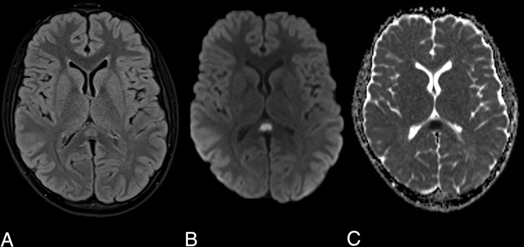
A 12-year-old boy presented with a 5-day history of fever, lethargy, severe headache, vomiting, lower abdominal pain, and diarrhea. On examination, he was febrile and tender in the left iliac fossa and there was bilateral conjunctival infection, periorbital swelling, and scrotal edema. Neurologic examination was normal. Blood tests demonstrated lymphopenia and thrombocytopenia, with evidence of a systemic inflammatory response (raised D-dimer, fibrinogen, C-reactive protein [CRP], ferritin, lactate dehydrogenase, and procalcitonin). Urine and blood cultures were negative. Chest radiograph was indeterminate for COVID-19. Real-time reverse transcriptase polymerase chain reaction (RT-PCR) analysis of 2 nasopharyngeal swabs was negative. A subsequent COVID-19 serology test, an immunoassay for the in vitro detection of IgG antibody specific to Severe Acute Respiratory Syndrome coronavirus 2 (SARS-CoV-2) in human serum and plasma, was positive. No other causative agent was isolated and serologic testing showed no evidence of other infection. In the context of the serology and supportive blood test results, a diagnosis of COVID-19 was reached. MR imaging of the brain, performed early into admission, showed a small, oval-shaped, T2-weighted hyperintense lesion in the splenium of the corpus callosum that exhibited restricted diffusion (Fig 1). There was no associated intrinsic susceptibility signal drop-out; postcontrast imaging was not performed. The patient was not on any medications or therapies that could have clearly accounted for the callosal lesion at the time of imaging. Shortly after admission he became hemodynamically shocked with raised troponin levels suggesting myocardial involvement. His level of consciousness and oxygen saturation worsened, without appreciable seizure phenomenon, necessitating intubation and mechanical ventilation. Based on the evidence of both cardiac and central nervous system pathology and blood tests supporting a hyperinflammatory response, a diagnosis of COVID-19-related pediatric multisystem inflammatory syndrome was made.3

FIG 1.
  • Download figure
  • Open in new tab
  • Download powerpoint
FIG 1.

Axial MR images demonstrate an oval T2-weighted hyperintensity in the splenium of the corpus callosum (A) with associated restricted diffusion on the DWI (B) and ADC maps (C).

The patient was treated with intravenous immunoglobulin, Anakinra (Interleukin-1 antagonist), infliximab, and steroids. The patient’s condition improved rapidly soon after commencing therapy and there were no neurologic symptoms at discharge. Repeat MR imaging has not yet been performed and was felt unwarranted acutely due to the clinical resolution.

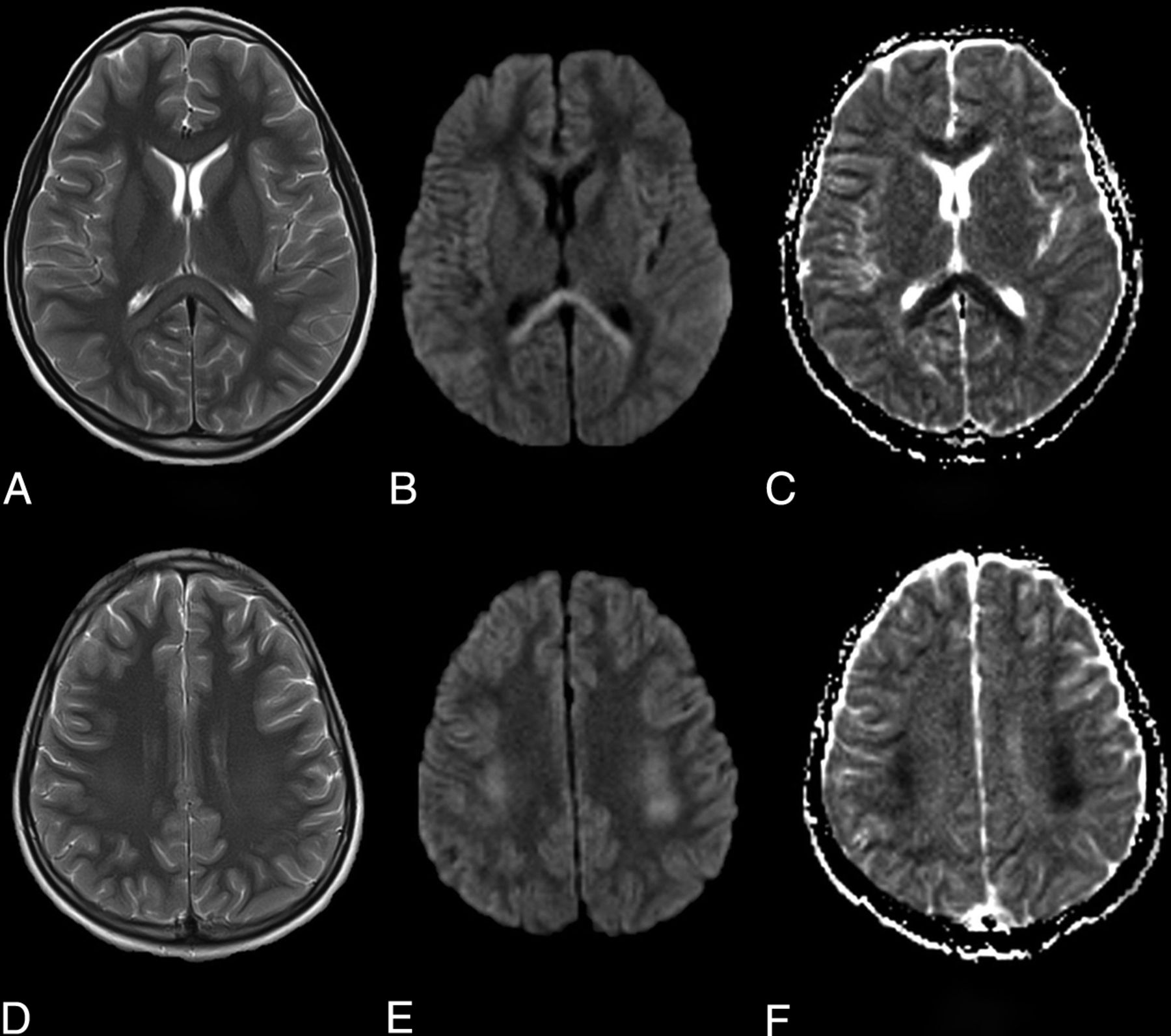
A second pediatric patient, a 9-year-old boy, presented with altered mental state, fever, and lethargy. On examination he was dysarthric and ataxic. Audible lung crepitations were noted on respiratory examination. Blood tests demonstrated a lymphopenia, raised C-reactive protein and serum ferritin. Urine and blood cultures were negative, and a chest radiograph was normal. An early head MR imaging showed similar but more extensive abnormal T2-weighted hyperintense signal and restricted diffusion in the entire corpus callosum and frontoparietal cerebral white matter. This had a dorsal predilection, with greatest in-volvement of the splenium, peri-atrial white matter, and posterior frontal and parietal centrum semiovale (Fig 2). There was no intrinsic susceptibility signal drop-out. Postcontrast imaging was not performed. SARS-CoV-2 on RT-PCR testing was subsequently confirmed on broncho-alveolar lavage, despite initial negative results from nasopharyngeal swabs. No other causative agent was isolated. This second patient’s symptoms improved rapidly and early follow-up MR imaging showed almost complete resolution of abnormal signal.1

FIG 2.
  • Download figure
  • Open in new tab
  • Download powerpoint
FIG 2.

Axial MR images demonstrate T2-weighted hyperintensity in the splenium and genu of the corpus callosum (A) and symmetrically in the deep cerebral white matter (B) with associated restricted diffusion on the DWI (B and E) and ADC maps (C and F).

DISCUSSION

CLOCC is a clinical-radiologic syndrome with a wide range of reported causes, including infection, trauma, drug-related, metabolic disorders, and malignancy.4 It typically manifests in children and can present with reduced consciousness, nuchal rigidity, and seizures.2,4

CLOCC has been described in association with many pathogens, including influenza, malaria, Ebstein-Barr virus, adenovirus, and E coli.5,6 In these cases where infection is thought to be the underlying cause, the diagnosis is also termed mild encephalitis/encephalopathy with a reversible splenial lesion. Our cases mirror clinical and radiologic features reported in a case series of 15 children with CLOCC of which 83% were presumed to be related to various different infections. Similar to our observations in presumptive COVID-19 related CLOCC, the authors of that series demonstrated evidence of systemic inflammation (raised CRP) and different degrees of nonenhancing, diffusion-restricting, white matter signal abnormality from isolated splenial lesions to more diffuse corpus callosal and extracallosal involvement.6 To our knowledge no cases of CLOCC related to the inflammatory response to SARS-CoV-2 or other coronaviruses have been reported. CLOCC is thought to be caused by a cytokine storm that triggers T cells to breach the blood-brain barrier causing inflammation and intramyelinic edema.5,7 This aligns with our cases of CLOCC that appeared to occur in the context of a multisystem inflammatory process with one of the cases showing more classic signs of pediatric inflammatory multisystem syndrome temporally associated with SARS-CoV-2, including myocardial and abdominal involvement.3 These cases reflect a new presentation of CNS inflammatory complication of SARS-CoV-2 exposure in children with pediatric inflammatory multisystem syndrome temporally associated with SARS-CoV-2. This inflammatory syndrome shares features with Kawasaki disease, toxic shock syndrome, and macrophage activation syndrome.8⇓-10 In our cases, the apparent rapid response to immunotherapy is encouraging and further supports the theory that this is an inflammatory and possibly cytokine-mediated process.

The diagnosis of CLOCC is based on the MR imaging demonstration of diffusion-restricting, nonenhancing lesions that usually resolve between a few days and weeks. Radiologically, as demonstrated in our cases, there is typically a lesion in the splenium of the corpus callosum with more variable involvement of the remaining corpus callosum and cerebral white matter.5 Often clinical recovery lags behind imaging, but is usually within 3 months.5

This is the first reported series of presumptive COVID-19-associated CLOCC that appeared to occur in the context of pediatric inflammatory multisystem syndrome temporally associated with SARS-CoV-2. Further investigation of COVID-19-related CNS pathology in children and the potential role of immunotherapy is required. Clinicians and radiologists should be aware of this potential presentation.

Indicates open access to non-subscribers at www.ajnr.org

References

  1. 1.↵
    1. Geneva S
    . World Health Organization. Coronavirus disease 2019 (COVID-19) situation report – 105. World Health Organization https://www.who.int/docs/default-source/coronaviruse/situation-reports/20200504-covid-19-sitrep-105.pdf?sfvrsn=4cdda8af_2. Published 2020
  2. 2.↵
    1. Parri N,
    2. Lenge M,
    3. Buonsenso D
    . Children with Covid-19 in pediatric emergency departments in Italy. N Engl J Med 2020;383:187–90 doi:10.1056/NEJMc2007617 pmid:32356945
    CrossRefPubMed
  3. 3.↵
    1. Whittaker E,
    2. Bamford A,
    3. Kenny J, et al
    . Clinical characteristics of 58 children with a pediatric inflammatory multisystem syndrome temporally associated with SARS-CoV-2. JAMA 2020;324:259–69 doi:10.1001/jama.2020.10369 pmid:32511692
    CrossRefPubMed
  4. 4.↵
    1. Takanashi JI
    . Two newly proposed infectious encephalitis/encephalopathy syndromes. Brain Dev 2009;31:521–28 doi:10.1016/j.braindev.2009.02.012 pmid:19339128
    CrossRefPubMed
  5. 5.↵
    1. Vanderschueren G,
    2. Schotsmans K,
    3. Maréchal E, et al
    . Mild encephalitis with reversible splenial (MERS) lesion syndrome due to influenza B virus. Pract Neurol 2018;18:391–92 doi:10.1136/practneurol-2018-001880 pmid:29599175
    Abstract/FREE Full Text
  6. 6.↵
    1. Chen WX,
    2. Liu HS,
    3. Yang SD, et al
    . Reversible splenial lesion syndrome in children: Retrospective study and summary of case series. Brain Dev 2016;38:915–27 doi:10.1016/j.braindev.2016.04.011 pmid:27591118
    CrossRefPubMed
  7. 7.↵
    1. Starkey J,
    2. Kobayashi N,
    3. Numaguchi Y, et al
    . Cytotoxic lesions of the corpus callosum that show restricted diffusion: mechanisms, causes, and manifestations. Radiographics 2017;37:562–76 doi:10.1148/rg.2017160085 pmid:28165876
    CrossRefPubMed
  8. 8.↵
    1. McGonagle D,
    2. Sharif K,
    3. O'Regan A, et al
    . The role of cytokines including interleukin-6 in COVID-19 induced pneumonia and macrophage activation syndrome-like disease. Autoimmun Rev 2020;19:102537 doi:10.1016/j.autrev.2020.102537 pmid:32251717
    CrossRefPubMed
  9. 9.↵
    1. Inciardi RM,
    2. Lupi L,
    3. Zaccone G, et al
    . Cardiac involvement in a patient with coronavirus disease 2019 (COVID-19). JAMA cardiology 2020 Mar 27. [Epub ahead of Print] doi:10.1001/jamacardio.2020.1096 pmid:32219357
    CrossRefPubMed
  10. 10.↵
    1. Jones VG,
    2. Mills M,
    3. Suarez D, et al
    . COVID-19 and Kawasaki disease: novel virus and novel case. Hosp Pediatr 2020;10:537–40 doi:10.1542/hpeds.2020-0123 pmid:32265235
    Abstract/FREE Full Text
  • Received June 3, 2020.
  • Accepted after revision June 11, 2020.
  • © 2020 by American Journal of Neuroradiology
View Abstract
PreviousNext
Back to top

In this issue

American Journal of Neuroradiology: 41 (10)
American Journal of Neuroradiology
Vol. 41, Issue 10
1 Oct 2020
  • Table of Contents
  • Index by author
  • Complete Issue (PDF)
Advertisement
Print
Download PDF
Email Article

Thank you for your interest in spreading the word on American Journal of Neuroradiology.

NOTE: We only request your email address so that the person you are recommending the page to knows that you wanted them to see it, and that it is not junk mail. We do not capture any email address.

Enter multiple addresses on separate lines or separate them with commas.
COVID-19-Associated Cytotoxic Lesions of the Corpus Callosum
(Your Name) has sent you a message from American Journal of Neuroradiology
(Your Name) thought you would like to see the American Journal of Neuroradiology web site.
CAPTCHA
This question is for testing whether or not you are a human visitor and to prevent automated spam submissions.
Cite this article
P. Gaur, L. Dixon, B. Jones, H. Lyall, W. Jan
COVID-19-Associated Cytotoxic Lesions of the Corpus Callosum
American Journal of Neuroradiology Oct 2020, 41 (10) 1905-1907; DOI: 10.3174/ajnr.A6713

Citation Manager Formats

  • BibTeX
  • Bookends
  • EasyBib
  • EndNote (tagged)
  • EndNote 8 (xml)
  • Medlars
  • Mendeley
  • Papers
  • RefWorks Tagged
  • Ref Manager
  • RIS
  • Zotero
0 Responses
Respond to this article
Share
Bookmark this article
COVID-19-Associated Cytotoxic Lesions of the Corpus Callosum
P. Gaur, L. Dixon, B. Jones, H. Lyall, W. Jan
American Journal of Neuroradiology Oct 2020, 41 (10) 1905-1907; DOI: 10.3174/ajnr.A6713
del.icio.us logo Twitter logo Facebook logo Mendeley logo
  • Tweet Widget
  • Facebook Like
  • Google Plus One
Purchase

Jump to section

  • Article
    • Abstract
    • ABBREVIATIONS:
    • CASE DETAILS
    • DISCUSSION
    • References
  • Figures & Data
  • Info & Metrics
  • Responses
  • References
  • PDF

Related Articles

  • No related articles found.
  • PubMed
  • Google Scholar

Cited By...

  • Clinical Features of Cytotoxic Lesions of the Corpus Callosum Associated with Aneurysmal Subarachnoid Hemorrhage
  • Neuroimaging Offers Low Yield in Children Positive for SARS-CoV-2
  • Crossref (48)
  • Google Scholar

This article has been cited by the following articles in journals that are participating in Crossref Cited-by Linking.

  • Neurological manifestations associated with SARS-CoV-2 and other coronaviruses: A narrative review for clinicians
    A. Maury, A. Lyoubi, N. Peiffer-Smadja, T. de Broucker, E. Meppiel
    Revue Neurologique 2021 177 1-2
  • Neurological complications in pediatric patients with SARS-CoV-2 infection: a systematic review of the literature
    L. Siracusa, A. Cascio, S. Giordano, A. A. Medaglia, G. A. Restivo, I. Pirrone, G. F. Saia, F. Collura, C. Colomba
    Italian Journal of Pediatrics 2021 47 1
  • The Spectrum of Neuroimaging Findings on CT and MRI in Adults With COVID-19
    Gul Moonis, Christopher G. Filippi, Claudia F. E. Kirsch, Suyash Mohan, Evan G. Stein, Joshua A. Hirsch, Amit Mahajan
    American Journal of Roentgenology 2021 217 4
  • Coronavirus Infections in the Nervous System of Children: A Scoping Review Making the Case for Long-Term Neurodevelopmental Surveillance
    Timothy G. Singer, Karen D. Evankovich, Kristen Fisher, Gail J. Demmler-Harrison, Sarah R. Risen
    Pediatric Neurology 2021 117
  • Thrombotic risk in children with COVID-19 infection: A systematic review of the literature
    Marco Zaffanello, Giorgio Piacentini, Luana Nosetti, Stefania Ganzarolli, Massimo Franchini
    Thrombosis Research 2021 205
  • Imaging of Multisystem Inflammatory Disease in Children (MIS-C) Associated With COVID-19
    Figen Palabiyik, Nihal Akcay, Esra Sevketoglu, Nevin Hatipoglu, Emine Ergul Sari, Ercan Inci
    Academic Radiology 2021 28 9
  • COVID-19 as a Cause of Acute Neonatal Encephalitis and Cerebral Cytotoxic Edema
    Diego Cardoso Fragoso, Catherine Marx, Bruna Garbugio Dutra, Carlos Jorge da Silva, Pollyanna Martins da Silva, Antônio Carlos Martins Maia Junior, Mariana Crema Tobara, Camila de Almeida Silva, Livio Dias, Angela Cristina Polycarpo, Rosana Richtmann
    Pediatric Infectious Disease Journal 2021 40 7
  • Subacute cerebellar ataxia following respiratory symptoms of COVID-19: a case report
    Jana Werner, Ina Reichen, Michael Huber, Irene A. Abela, Michael Weller, Ilijas Jelcic
    BMC Infectious Diseases 2021 21 1
  • Neurologic manifestations in children with COVID-19 from a tertiary center in Turkey and literature review
    Sibel Laçinel Gürlevik, Ceren Günbey, Yasemin Ozsurekci, Pembe Derin Oygar, Selman Kesici, Rahsan Gocmen, Orkun Aydin, Çağrı Temucin, Erennur Tufan, Kıvanc Terzi, Demet Baltu, Tugba Tastemel Ozturk, Ozlem Teksam, Seza Ozen, Kader Karlı Oguz, Ali Bulent Cengiz, Dilek Yalnızoglu
    European Journal of Paediatric Neurology 2022 37
  • International Prevalence and Mechanisms of SARS-CoV-2 in Childhood Arterial Ischemic Stroke During the COVID-19 Pandemic
    Lauren A. Beslow, Shannon C. Agner, Jonathan D. Santoro, Dipak Ram, Jenny L. Wilson, Dana Harrar, Brian Appavu, Stuart M. Fraser, Thomas Rossor, Marcela D. Torres, Manoëlle Kossorotoff, Yenny C. Zuñiga Zambrano, Marta Hernández-Chávez, Sahar M.A. Hassanein, Dimitrios Zafeiriou, Michael M. Dowling, Ilona Kopyta, Nicholas V. Stence, Timothy J. Bernard, Nomazulu Dlamini, Ahmed Abd El-Hamid Rihan, Maha Mohammed, Moustafa Farid, Wessam S.S. Guergues, Mohamed O.E. Babiker, Oded Hochberg, Paola Saracco, Thomas Main, Andrew Mallick, Selina Kala, Bryan L. Philbrook, Kartik Reddy, Rebecca N. Ichord, Evelyn K. Shih, Rachel P. Pearson, Mubeen F. Rafay, Mukta Sharma, Mary Allen Staat, Sudhakar Vadivelu, Marvid Duarte, Mary Suzanne Whitworth, Manish Parakh, Kevin Meesters, Charles-Joris Roud, Marianne Leruez- Ville, Vijeya Ganesan, Laura L. Lehman, Michael Rivkin, Zulma Hernandez, Fernanda Balut, Maria Celeste Buompadre, Heidy J. Gómez Naranjo, Veronica Gonzalez Alvarez, Nihal Bakeer, Stephanie Garrison, Christopher Belcher, Lorie Miller, Maria Whitmore, Giulia Amico, Mariasavina Severino, Marta Bertamino, Sara Signa, Lisa R. Sun, Ryan J. Felling, Pawan Kashyape, Lucia Gerstl, Gordana Kovacevic, Anna Basu, Yusri Taha, Warren D. Lo, Maggie L.Y. Yau, Deirdre Peake, Kim Stevenson, Samson Gwer, Andrea Andrade, Catherine Amlie- Lefond, Jacqueline Lee-Eng, Sarah Lee, Janette Mailo, Moran Hausman- Kedem, Kamna Jaiswal, Kellie Brown, Belinda Stojanovski, Mark T Mackay, Adriana Carolina Vargas Nino, Daune McGregor, Gabrielle deVeber, Ishvinder Bhathal, NP Liza Pulcine, Mahendra Moharir, Bruce Bjornson, Danny H.C. Kim, Adam Kirton, Amalia Floer, Christine K. Fox, Christiana Smith- Anderson, Kelly Wilt, Vivian Thompson, Michael L. Chang, Marilyn Tan, Lori C Jordan, Annette E. Grefe, Kristin Guilliams, Michael J. Noetzel
    Stroke 2022 53 8

More in this TOC Section

  • Comparison of Image Quality and Radiation Dose in Pediatric Temporal Bone CT Using Photon-Counting Detector CT and Energy-Integrating Detector CT
  • SyMRI & MR Fingerprinting in Brainstem Myelination
  • Venous Sinus Stenosis in Mucopolysaccharidosis I
Show more Pediatrics

Similar Articles

Advertisement

Indexed Content

  • Current Issue
  • Accepted Manuscripts
  • Article Preview
  • Past Issues
  • Editorials
  • Editors Choice
  • Fellow Journal Club
  • Letters to the Editor

Cases

  • Case Collection
  • Archive - Case of the Week
  • Archive - Case of the Month
  • Archive - Classic Case

Special Collections

  • Special Collections

Resources

  • News and Updates
  • Turn around Times
  • Submit a Manuscript
  • Author Policies
  • Manuscript Submission Guidelines
  • Evidence-Based Medicine Level Guide
  • Publishing Checklists
  • Graphical Abstract Preparation
  • Imaging Protocol Submission
  • Submit a Case
  • Become a Reviewer/Academy of Reviewers
  • Get Peer Review Credit from Publons

Multimedia

  • AJNR Podcast
  • AJNR SCANtastic
  • Video Articles

About Us

  • About AJNR
  • Editorial Board
  • Not an AJNR Subscriber? Join Now
  • Alerts
  • Feedback
  • Advertise with us
  • Librarian Resources
  • Permissions
  • Terms and Conditions

American Society of Neuroradiology

  • Not an ASNR Member? Join Now

© 2025 by the American Society of Neuroradiology All rights, including for text and data mining, AI training, and similar technologies, are reserved.
Print ISSN: 0195-6108 Online ISSN: 1936-959X

Powered by HighWire