Advertisement

Main menu

  • Home
  • Content
    • Current Issue
    • Accepted Manuscripts
    • Article Preview
    • Past Issue Archive
    • Video Articles
    • AJNR Case Collection
    • Case of the Week Archive
    • Case of the Month Archive
    • Classic Case Archive
  • Special Collections
    • AJNR Awards
    • Low-Field MRI
    • Alzheimer Disease
    • ASNR Foundation Special Collection
    • Photon-Counting CT
    • View All
  • Multimedia
    • AJNR Podcasts
    • AJNR SCANtastic
    • Trainee Corner
    • MRI Safety Corner
    • Imaging Protocols
  • For Authors
    • Submit a Manuscript
    • Submit a Video Article
    • Submit an eLetter to the Editor/Response
    • Manuscript Submission Guidelines
    • Statistical Tips
    • Fast Publishing of Accepted Manuscripts
    • Graphical Abstract Preparation
    • Imaging Protocol Submission
    • Author Policies
  • About Us
    • About AJNR
    • Editorial Board
    • Editorial Board Alumni
  • More
    • Become a Reviewer/Academy of Reviewers
    • Subscribers
    • Permissions
    • Alerts
    • Feedback
    • Advertisers
    • ASNR Home

User menu

  • Alerts
  • Log in

Search

  • Advanced search
American Journal of Neuroradiology

American Journal of Neuroradiology

ASHNR American Society of Functional Neuroradiology ASHNR American Society of Pediatric Neuroradiology ASSR
  • Alerts
  • Log in

Advanced Search

  • Home
  • Content
    • Current Issue
    • Accepted Manuscripts
    • Article Preview
    • Past Issue Archive
    • Video Articles
    • AJNR Case Collection
    • Case of the Week Archive
    • Case of the Month Archive
    • Classic Case Archive
  • Special Collections
    • AJNR Awards
    • Low-Field MRI
    • Alzheimer Disease
    • ASNR Foundation Special Collection
    • Photon-Counting CT
    • View All
  • Multimedia
    • AJNR Podcasts
    • AJNR SCANtastic
    • Trainee Corner
    • MRI Safety Corner
    • Imaging Protocols
  • For Authors
    • Submit a Manuscript
    • Submit a Video Article
    • Submit an eLetter to the Editor/Response
    • Manuscript Submission Guidelines
    • Statistical Tips
    • Fast Publishing of Accepted Manuscripts
    • Graphical Abstract Preparation
    • Imaging Protocol Submission
    • Author Policies
  • About Us
    • About AJNR
    • Editorial Board
    • Editorial Board Alumni
  • More
    • Become a Reviewer/Academy of Reviewers
    • Subscribers
    • Permissions
    • Alerts
    • Feedback
    • Advertisers
    • ASNR Home
  • Follow AJNR on Twitter
  • Visit AJNR on Facebook
  • Follow AJNR on Instagram
  • Join AJNR on LinkedIn
  • RSS Feeds

AJNR Awards, New Junior Editors, and more. Read the latest AJNR updates

Research ArticleAdult Brain
Open Access

Dynamic CTA-Derived Perfusion Maps Predict Final Infarct Volume: The Simple Perfusion Reconstruction Algorithm

C.C. McDougall, L. Chan, S. Sachan, J. Guo, R.G. Sah, B.K. Menon, A.M. Demchuk, M.D. Hill, N.D. Forkert, C.D. d’Esterre and P.A. Barber
American Journal of Neuroradiology November 2020, 41 (11) 2034-2040; DOI: https://doi.org/10.3174/ajnr.A6783
C.C. McDougall
aFrom the Department of Clinical Neurosciences (C.C.M., R.G.S., B.K.M., A.M.D., M.D.H., C.D.d., P.A.B.), Calgary Stroke Program
bDepartment of Radiology (C.C.M., B.K.M., N.D.F., C.D.d.E., P.A.B.)
cHotchkiss Brain Institute (C.C.M., B.K.M., A.M.D., M.D.H., N.D.F., C.C.d.E., P.A.B.)
dDepartment of Clinical Neurosciences (C.C.M., L.C., S.S., J.G., R.G.S., B.K.M., A.M.D., M.D.H., N.D.F., C.C.d.E.)
fSeaman Family Centre (C.C.M., R.G.S., B.K.M., A.M.D., M.D.H., C.D.d.E., P.A.B.), Foothills Medical Centre, Calgary, Alberta, Canada
  • Find this author on Google Scholar
  • Find this author on PubMed
  • Search for this author on this site
  • ORCID record for C.C. McDougall
L. Chan
dDepartment of Clinical Neurosciences (C.C.M., L.C., S.S., J.G., R.G.S., B.K.M., A.M.D., M.D.H., N.D.F., C.C.d.E.)
  • Find this author on Google Scholar
  • Find this author on PubMed
  • Search for this author on this site
  • ORCID record for L. Chan
S. Sachan
dDepartment of Clinical Neurosciences (C.C.M., L.C., S.S., J.G., R.G.S., B.K.M., A.M.D., M.D.H., N.D.F., C.C.d.E.)
  • Find this author on Google Scholar
  • Find this author on PubMed
  • Search for this author on this site
  • ORCID record for S. Sachan
J. Guo
dDepartment of Clinical Neurosciences (C.C.M., L.C., S.S., J.G., R.G.S., B.K.M., A.M.D., M.D.H., N.D.F., C.C.d.E.)
  • Find this author on Google Scholar
  • Find this author on PubMed
  • Search for this author on this site
  • ORCID record for J. Guo
R.G. Sah
aFrom the Department of Clinical Neurosciences (C.C.M., R.G.S., B.K.M., A.M.D., M.D.H., C.D.d., P.A.B.), Calgary Stroke Program
dDepartment of Clinical Neurosciences (C.C.M., L.C., S.S., J.G., R.G.S., B.K.M., A.M.D., M.D.H., N.D.F., C.C.d.E.)
fSeaman Family Centre (C.C.M., R.G.S., B.K.M., A.M.D., M.D.H., C.D.d.E., P.A.B.), Foothills Medical Centre, Calgary, Alberta, Canada
  • Find this author on Google Scholar
  • Find this author on PubMed
  • Search for this author on this site
  • ORCID record for R.G. Sah
B.K. Menon
aFrom the Department of Clinical Neurosciences (C.C.M., R.G.S., B.K.M., A.M.D., M.D.H., C.D.d., P.A.B.), Calgary Stroke Program
bDepartment of Radiology (C.C.M., B.K.M., N.D.F., C.D.d.E., P.A.B.)
cHotchkiss Brain Institute (C.C.M., B.K.M., A.M.D., M.D.H., N.D.F., C.C.d.E., P.A.B.)
dDepartment of Clinical Neurosciences (C.C.M., L.C., S.S., J.G., R.G.S., B.K.M., A.M.D., M.D.H., N.D.F., C.C.d.E.)
fSeaman Family Centre (C.C.M., R.G.S., B.K.M., A.M.D., M.D.H., C.D.d.E., P.A.B.), Foothills Medical Centre, Calgary, Alberta, Canada
  • Find this author on Google Scholar
  • Find this author on PubMed
  • Search for this author on this site
  • ORCID record for B.K. Menon
A.M. Demchuk
aFrom the Department of Clinical Neurosciences (C.C.M., R.G.S., B.K.M., A.M.D., M.D.H., C.D.d., P.A.B.), Calgary Stroke Program
cHotchkiss Brain Institute (C.C.M., B.K.M., A.M.D., M.D.H., N.D.F., C.C.d.E., P.A.B.)
dDepartment of Clinical Neurosciences (C.C.M., L.C., S.S., J.G., R.G.S., B.K.M., A.M.D., M.D.H., N.D.F., C.C.d.E.)
fSeaman Family Centre (C.C.M., R.G.S., B.K.M., A.M.D., M.D.H., C.D.d.E., P.A.B.), Foothills Medical Centre, Calgary, Alberta, Canada
  • Find this author on Google Scholar
  • Find this author on PubMed
  • Search for this author on this site
  • ORCID record for A.M. Demchuk
M.D. Hill
aFrom the Department of Clinical Neurosciences (C.C.M., R.G.S., B.K.M., A.M.D., M.D.H., C.D.d., P.A.B.), Calgary Stroke Program
cHotchkiss Brain Institute (C.C.M., B.K.M., A.M.D., M.D.H., N.D.F., C.C.d.E., P.A.B.)
dDepartment of Clinical Neurosciences (C.C.M., L.C., S.S., J.G., R.G.S., B.K.M., A.M.D., M.D.H., N.D.F., C.C.d.E.)
fSeaman Family Centre (C.C.M., R.G.S., B.K.M., A.M.D., M.D.H., C.D.d.E., P.A.B.), Foothills Medical Centre, Calgary, Alberta, Canada
  • Find this author on Google Scholar
  • Find this author on PubMed
  • Search for this author on this site
  • ORCID record for M.D. Hill
N.D. Forkert
bDepartment of Radiology (C.C.M., B.K.M., N.D.F., C.D.d.E., P.A.B.)
cHotchkiss Brain Institute (C.C.M., B.K.M., A.M.D., M.D.H., N.D.F., C.C.d.E., P.A.B.)
dDepartment of Clinical Neurosciences (C.C.M., L.C., S.S., J.G., R.G.S., B.K.M., A.M.D., M.D.H., N.D.F., C.C.d.E.)
eAlberta Children’s Hospital Research Institute (N.D.F.), University of Calgary, Calgary, Alberta, Canada
  • Find this author on Google Scholar
  • Find this author on PubMed
  • Search for this author on this site
  • ORCID record for N.D. Forkert
C.D. d’Esterre
aFrom the Department of Clinical Neurosciences (C.C.M., R.G.S., B.K.M., A.M.D., M.D.H., C.D.d., P.A.B.), Calgary Stroke Program
bDepartment of Radiology (C.C.M., B.K.M., N.D.F., C.D.d.E., P.A.B.)
cHotchkiss Brain Institute (C.C.M., B.K.M., A.M.D., M.D.H., N.D.F., C.C.d.E., P.A.B.)
dDepartment of Clinical Neurosciences (C.C.M., L.C., S.S., J.G., R.G.S., B.K.M., A.M.D., M.D.H., N.D.F., C.C.d.E.)
fSeaman Family Centre (C.C.M., R.G.S., B.K.M., A.M.D., M.D.H., C.D.d.E., P.A.B.), Foothills Medical Centre, Calgary, Alberta, Canada
  • Find this author on Google Scholar
  • Find this author on PubMed
  • Search for this author on this site
  • ORCID record for C.D. d’Esterre
P.A. Barber
aFrom the Department of Clinical Neurosciences (C.C.M., R.G.S., B.K.M., A.M.D., M.D.H., C.D.d., P.A.B.), Calgary Stroke Program
bDepartment of Radiology (C.C.M., B.K.M., N.D.F., C.D.d.E., P.A.B.)
cHotchkiss Brain Institute (C.C.M., B.K.M., A.M.D., M.D.H., N.D.F., C.C.d.E., P.A.B.)
fSeaman Family Centre (C.C.M., R.G.S., B.K.M., A.M.D., M.D.H., C.D.d.E., P.A.B.), Foothills Medical Centre, Calgary, Alberta, Canada
  • Find this author on Google Scholar
  • Find this author on PubMed
  • Search for this author on this site
  • ORCID record for P.A. Barber
  • Article
  • Figures & Data
  • Info & Metrics
  • Responses
  • References
  • PDF
Loading

Article Figures & Data

Figures

  • Tables
  • FIG 1.
    • Download figure
    • Open in new tab
    • Download powerpoint
    FIG 1.

    Bland-Altman plots for SPIRAL infarct volume (milliliters) (A) and Tmax infarct volume (milliliters) in cortical gray matter/white matter (i) and basal ganglia (ii). SPIRAL infarct volume (milliliters) (B) and follow-up DWI infarct volume (milliliters) in the cortical gray matter/white matter (i) and basal ganglia (ii). C, Tmax infarct volume and follow-up DWI infarct volume in cortical gray matter/white matter (i) and basal ganglia (ii).

  • FIG 2.
    • Download figure
    • Open in new tab
    • Download powerpoint
    FIG 2.

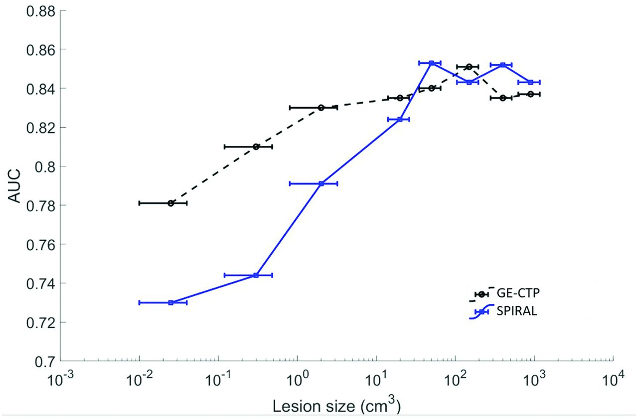
    Sensitivity for final infarction on 24-h our MR imaging versus lesion size for SPIRAL and the CTP Tmax map.

  • FIG 3.
    • Download figure
    • Open in new tab
    • Download powerpoint
    FIG 3.

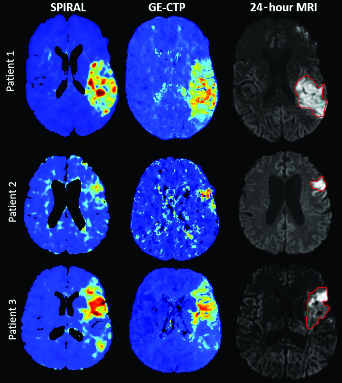
    Admission SPIRAL map and CTP Tmax map for 3 patients who underwent EVT for M1 occlusions who had quality/fast reperfusion. The final infarct volume is outlined on the 24-hour DWI.

Tables

  • Figures
    • View popup
    Table 1:

    Admission demographics, site of occlusion, and workflow metrics

    VariablesTotal (n = 72 Patients)
    Age (median) (minimum-maximum) (yr)68 (32–89)
    Men (No.) (%)37 (51.4)
    Stroke on awakening (No.) (%)27 (46.6)
    Site of occlusion (No.) (%) 
     MCA29 (40.3)
     ACA3 (4.0)
     ICA16 (22.2)
     Tandem5 (6.9)
    Affected hemisphere (No.) (%) 
     Right30 (41.7)
     Left39 (54.2)
    Coronary artery disease (No.) (%)12 (16.7)
    Congestive heart failure (No.) (%)6 (8.3)
    Valvular disease (No.) (%)2 (3.4)
    Hypertension (No.) (%)38 (52.8)
    Dyslipidemia (No.) (%)24 (33.3)
    Diabetes (No.) (%)1 (1.4)
    Smoking (No.) (%)20 (27.8)
    Statin (No.) (%)22 (37.9)
    EVT treatment (No.) (%)72 (100)
    tPA (alteplase) treatment (No.) (%)55 (76)
    Reperfusion (TICI 2b/3) (No.) (%)72 (100)
    Blood glucose (median) (minimum-maximum) (mmol)6 (4.4–20.0)
    NIHSS baseline (median) (minimum-maximum)17 (1–29)
    NIHSS 24 hours (median) (minimum-maximum)6 (0–24)
    MR spectroscopy baseline (median) (minimum-maximum)0 (0–3)
    MR spectroscopy (median) (minimum-maximum) (90 days)2 (0–6)
    CT to reperfusion time (median) (minimum-maximum) (hh:mm)1:28 (0:27–3:06)
    • Note:—hh:mm indicates hours: minutes; ACA, anterior cerebral artery.

    • View popup
    Table 2:

    ROC curve AUC for SPIRAL map, stratified by CT-to-reperfusion time for cortical gray and white matter tissue

    StatisticAUC, Patient LevelAUC, Cohort LevelCross-Validation SensitivityCross-Validation SpecificityCross-Validation Accuracy
    Early reperfusion, <90 minutes  (n = 48 patients)
     Mean0.830.820.820.720.77
     SD0.14NA0.060.030.06
    Late reperfusion, >90 minutes  (n = 24 patients)
     Mean0.840.810.790.700.74
     SD0.11NA0.080.060.07
    • Note:—NA indicates not applicable.

    • View popup
    Table 3:

    ROC curve AUC for SPIRAL map, stratified by CT-to-reperfusion time for basal ganglia regions

    StatisticAUC, Patient LevelAUC, Cohort LevelCross-Validation SensitivityCross-Validation SpecificityCross-Validation Accuracy
    Early reperfusion, >90 minutes  (21 patients)
     Mean0.820.810.820.810.82
     SD0.11NA0.050.060.06
    Late reperfusion, <90 minutes (7 patients)
     Mean0.840.800.860.710.78
     SD0.1NA0.080.090.09
    • Note:—NA indicates not applicable.

    • View popup
    Table 4:

    ROC curve AUC for SPIRAL map comparison with cine CTP maps for a 40-patient subcohort

    StatisticCortical GM and White Matter (WM)Basal Ganglia
    SPIRAL map
     AUC, patient level (mean) (SD)0.83 (0.14)0.79 (0.08)
     AUC, cohort level (mean)0.820.80
    CTP T-max map
     AUC, patient level0.82 (0.13)0.78 (0.11)
     AUC, cohort level0.810.74
    CTP blood flow map
     AUC, patient level0.74 (0.14)0.78 (0.09)
     AUC, cohort level0.720.77
PreviousNext
Back to top

In this issue

American Journal of Neuroradiology: 41 (11)
American Journal of Neuroradiology
Vol. 41, Issue 11
1 Nov 2020
  • Table of Contents
  • Index by author
  • Complete Issue (PDF)
Advertisement
Print
Download PDF
Email Article

Thank you for your interest in spreading the word on American Journal of Neuroradiology.

NOTE: We only request your email address so that the person you are recommending the page to knows that you wanted them to see it, and that it is not junk mail. We do not capture any email address.

Enter multiple addresses on separate lines or separate them with commas.
Dynamic CTA-Derived Perfusion Maps Predict Final Infarct Volume: The Simple Perfusion Reconstruction Algorithm
(Your Name) has sent you a message from American Journal of Neuroradiology
(Your Name) thought you would like to see the American Journal of Neuroradiology web site.
CAPTCHA
This question is for testing whether or not you are a human visitor and to prevent automated spam submissions.
Cite this article
C.C. McDougall, L. Chan, S. Sachan, J. Guo, R.G. Sah, B.K. Menon, A.M. Demchuk, M.D. Hill, N.D. Forkert, C.D. d’Esterre, P.A. Barber
Dynamic CTA-Derived Perfusion Maps Predict Final Infarct Volume: The Simple Perfusion Reconstruction Algorithm
American Journal of Neuroradiology Nov 2020, 41 (11) 2034-2040; DOI: 10.3174/ajnr.A6783

Citation Manager Formats

  • BibTeX
  • Bookends
  • EasyBib
  • EndNote (tagged)
  • EndNote 8 (xml)
  • Medlars
  • Mendeley
  • Papers
  • RefWorks Tagged
  • Ref Manager
  • RIS
  • Zotero
0 Responses
Respond to this article
Share
Bookmark this article
Dynamic CTA-Derived Perfusion Maps Predict Final Infarct Volume: The Simple Perfusion Reconstruction Algorithm
C.C. McDougall, L. Chan, S. Sachan, J. Guo, R.G. Sah, B.K. Menon, A.M. Demchuk, M.D. Hill, N.D. Forkert, C.D. d’Esterre, P.A. Barber
American Journal of Neuroradiology Nov 2020, 41 (11) 2034-2040; DOI: 10.3174/ajnr.A6783
del.icio.us logo Twitter logo Facebook logo Mendeley logo
  • Tweet Widget
  • Facebook Like
  • Google Plus One
Purchase

Jump to section

  • Article
    • Abstract
    • ABBREVIATIONS:
    • MATERIALS AND METHODS
    • RESULTS
    • DISCUSSION
    • CONCLUSIONS
    • Acknowledgments
    • Footnotes
    • References
  • Figures & Data
  • Info & Metrics
  • Responses
  • References
  • PDF

Related Articles

  • No related articles found.
  • PubMed
  • Google Scholar

Cited By...

  • Acute stroke imaging selection for mechanical thrombectomy in the extended time window: is it time to go back to basics? A review of current evidence
  • Crossref
  • Google Scholar

This article has not yet been cited by articles in journals that are participating in Crossref Cited-by Linking.

More in this TOC Section

Adult Brain

  • Diagnostic Neuroradiology of Monoclonal Antibodies
  • Clinical Outcomes After Chiari I Decompression
  • Segmentation of Brain Metastases with BLAST
Show more Adult Brain

Functional

  • Kurtosis and Epileptogenic Tubers: A Pilot Study
  • Glutaric Aciduria Type 1: DK vs. Conventional MRI
  • Multiparametric MRI in PEDS Pontine Glioma
Show more Functional

Similar Articles

Advertisement

Indexed Content

  • Current Issue
  • Accepted Manuscripts
  • Article Preview
  • Past Issues
  • Editorials
  • Editor's Choice
  • Fellows' Journal Club
  • Letters to the Editor
  • Video Articles

Cases

  • Case Collection
  • Archive - Case of the Week
  • Archive - Case of the Month
  • Archive - Classic Case

More from AJNR

  • Trainee Corner
  • Imaging Protocols
  • MRI Safety Corner
  • Book Reviews

Multimedia

  • AJNR Podcasts
  • AJNR Scantastics

Resources

  • Turnaround Time
  • Submit a Manuscript
  • Submit a Video Article
  • Submit an eLetter to the Editor/Response
  • Manuscript Submission Guidelines
  • Statistical Tips
  • Fast Publishing of Accepted Manuscripts
  • Graphical Abstract Preparation
  • Imaging Protocol Submission
  • Evidence-Based Medicine Level Guide
  • Publishing Checklists
  • Author Policies
  • Become a Reviewer/Academy of Reviewers
  • News and Updates

About Us

  • About AJNR
  • Editorial Board
  • Editorial Board Alumni
  • Alerts
  • Permissions
  • Not an AJNR Subscriber? Join Now
  • Advertise with Us
  • Librarian Resources
  • Feedback
  • Terms and Conditions
  • AJNR Editorial Board Alumni

American Society of Neuroradiology

  • Not an ASNR Member? Join Now

© 2025 by the American Society of Neuroradiology All rights, including for text and data mining, AI training, and similar technologies, are reserved.
Print ISSN: 0195-6108 Online ISSN: 1936-959X

Powered by HighWire