Advertisement

Main menu

  • Home
  • Content
    • Current Issue
    • Accepted Manuscripts
    • Article Preview
    • Past Issue Archive
    • Video Articles
    • AJNR Case Collection
    • Case of the Week Archive
    • Case of the Month Archive
    • Classic Case Archive
  • Special Collections
    • AJNR Awards
    • Low-Field MRI
    • Alzheimer Disease
    • ASNR Foundation Special Collection
    • Photon-Counting CT
    • View All
  • Multimedia
    • AJNR Podcasts
    • AJNR SCANtastic
    • Trainee Corner
    • MRI Safety Corner
    • Imaging Protocols
  • For Authors
    • Submit a Manuscript
    • Submit a Video Article
    • Submit an eLetter to the Editor/Response
    • Manuscript Submission Guidelines
    • Statistical Tips
    • Fast Publishing of Accepted Manuscripts
    • Graphical Abstract Preparation
    • Imaging Protocol Submission
    • Author Policies
  • About Us
    • About AJNR
    • Editorial Board
    • Editorial Board Alumni
  • More
    • Become a Reviewer/Academy of Reviewers
    • Subscribers
    • Permissions
    • Alerts
    • Feedback
    • Advertisers
    • ASNR Home

User menu

  • Alerts
  • Log in

Search

  • Advanced search
American Journal of Neuroradiology

American Journal of Neuroradiology

ASHNR American Society of Functional Neuroradiology ASHNR American Society of Pediatric Neuroradiology ASSR
  • Alerts
  • Log in

Advanced Search

  • Home
  • Content
    • Current Issue
    • Accepted Manuscripts
    • Article Preview
    • Past Issue Archive
    • Video Articles
    • AJNR Case Collection
    • Case of the Week Archive
    • Case of the Month Archive
    • Classic Case Archive
  • Special Collections
    • AJNR Awards
    • Low-Field MRI
    • Alzheimer Disease
    • ASNR Foundation Special Collection
    • Photon-Counting CT
    • View All
  • Multimedia
    • AJNR Podcasts
    • AJNR SCANtastic
    • Trainee Corner
    • MRI Safety Corner
    • Imaging Protocols
  • For Authors
    • Submit a Manuscript
    • Submit a Video Article
    • Submit an eLetter to the Editor/Response
    • Manuscript Submission Guidelines
    • Statistical Tips
    • Fast Publishing of Accepted Manuscripts
    • Graphical Abstract Preparation
    • Imaging Protocol Submission
    • Author Policies
  • About Us
    • About AJNR
    • Editorial Board
    • Editorial Board Alumni
  • More
    • Become a Reviewer/Academy of Reviewers
    • Subscribers
    • Permissions
    • Alerts
    • Feedback
    • Advertisers
    • ASNR Home
  • Follow AJNR on Twitter
  • Visit AJNR on Facebook
  • Follow AJNR on Instagram
  • Join AJNR on LinkedIn
  • RSS Feeds

AJNR Awards, New Junior Editors, and more. Read the latest AJNR updates

Research ArticleAdult Brain
Open Access

NAA is a Marker of Disability in Secondary-Progressive MS: A Proton MR Spectroscopic Imaging Study

B.S. Solanky, N.A. John, F. DeAngelis, J. Stutters, F. Prados, T. Schneider, R.A. Parker, C.J. Weir, A. Monteverdi, D. Plantone, A. Doshi, D. MacManus, I. Marshall, F. Barkhof, C.A.M. Gandini Wheeler-Kingshott, J. Chataway and for the MS-SMART Investigators
American Journal of Neuroradiology December 2020, 41 (12) 2209-2218; DOI: https://doi.org/10.3174/ajnr.A6809
B.S. Solanky
aFrom the Department of Neuroinflammation (B.S.S., N.A.J., F.D., J.S., F.P., D.P., A.D., D.M., C.A.M.G.W.-K., J.C.), Faculty of Brain Sciences, Queen Square Multiple Sclerosis Centre, UCL Queen Square Institute of Neurology
  • Find this author on Google Scholar
  • Find this author on PubMed
  • Search for this author on this site
  • ORCID record for B.S. Solanky
N.A. John
aFrom the Department of Neuroinflammation (B.S.S., N.A.J., F.D., J.S., F.P., D.P., A.D., D.M., C.A.M.G.W.-K., J.C.), Faculty of Brain Sciences, Queen Square Multiple Sclerosis Centre, UCL Queen Square Institute of Neurology
  • Find this author on Google Scholar
  • Find this author on PubMed
  • Search for this author on this site
  • ORCID record for N.A. John
F. DeAngelis
aFrom the Department of Neuroinflammation (B.S.S., N.A.J., F.D., J.S., F.P., D.P., A.D., D.M., C.A.M.G.W.-K., J.C.), Faculty of Brain Sciences, Queen Square Multiple Sclerosis Centre, UCL Queen Square Institute of Neurology
  • Find this author on Google Scholar
  • Find this author on PubMed
  • Search for this author on this site
  • ORCID record for F. DeAngelis
J. Stutters
aFrom the Department of Neuroinflammation (B.S.S., N.A.J., F.D., J.S., F.P., D.P., A.D., D.M., C.A.M.G.W.-K., J.C.), Faculty of Brain Sciences, Queen Square Multiple Sclerosis Centre, UCL Queen Square Institute of Neurology
  • Find this author on Google Scholar
  • Find this author on PubMed
  • Search for this author on this site
  • ORCID record for J. Stutters
F. Prados
aFrom the Department of Neuroinflammation (B.S.S., N.A.J., F.D., J.S., F.P., D.P., A.D., D.M., C.A.M.G.W.-K., J.C.), Faculty of Brain Sciences, Queen Square Multiple Sclerosis Centre, UCL Queen Square Institute of Neurology
bCentre for Medical Image Computing (F.P., F.B.), Department of Medical Physics and Biomedical Engineering, University College London, London, UK
jUniversitat Oberta de Catalunya (F.P.), Barcelona, Spain
  • Find this author on Google Scholar
  • Find this author on PubMed
  • Search for this author on this site
  • ORCID record for F. Prados
T. Schneider
iPhilips UK (T.S.), Guildford, Surrey, UK
  • Find this author on Google Scholar
  • Find this author on PubMed
  • Search for this author on this site
  • ORCID record for T. Schneider
R.A. Parker
cEdinburgh Clinical Trials Unit (R.A.P., C.J.W.), Usher Institute
  • Find this author on Google Scholar
  • Find this author on PubMed
  • Search for this author on this site
  • ORCID record for R.A. Parker
C.J. Weir
cEdinburgh Clinical Trials Unit (R.A.P., C.J.W.), Usher Institute
  • Find this author on Google Scholar
  • Find this author on PubMed
  • Search for this author on this site
  • ORCID record for C.J. Weir
A. Monteverdi
fDepartment of Brain and Behavioural Sciences (A.M., C.A.M.G.W.-K.), University of Pavia, Pavia, Italy
  • Find this author on Google Scholar
  • Find this author on PubMed
  • Search for this author on this site
  • ORCID record for A. Monteverdi
D. Plantone
aFrom the Department of Neuroinflammation (B.S.S., N.A.J., F.D., J.S., F.P., D.P., A.D., D.M., C.A.M.G.W.-K., J.C.), Faculty of Brain Sciences, Queen Square Multiple Sclerosis Centre, UCL Queen Square Institute of Neurology
  • Find this author on Google Scholar
  • Find this author on PubMed
  • Search for this author on this site
  • ORCID record for D. Plantone
A. Doshi
aFrom the Department of Neuroinflammation (B.S.S., N.A.J., F.D., J.S., F.P., D.P., A.D., D.M., C.A.M.G.W.-K., J.C.), Faculty of Brain Sciences, Queen Square Multiple Sclerosis Centre, UCL Queen Square Institute of Neurology
  • Find this author on Google Scholar
  • Find this author on PubMed
  • Search for this author on this site
  • ORCID record for A. Doshi
D. MacManus
aFrom the Department of Neuroinflammation (B.S.S., N.A.J., F.D., J.S., F.P., D.P., A.D., D.M., C.A.M.G.W.-K., J.C.), Faculty of Brain Sciences, Queen Square Multiple Sclerosis Centre, UCL Queen Square Institute of Neurology
  • Find this author on Google Scholar
  • Find this author on PubMed
  • Search for this author on this site
  • ORCID record for D. MacManus
I. Marshall
dCentre for Clinical Brain Sciences (I.M.), University of Edinburgh, Edinburgh, UK.
  • Find this author on Google Scholar
  • Find this author on PubMed
  • Search for this author on this site
  • ORCID record for I. Marshall
F. Barkhof
bCentre for Medical Image Computing (F.P., F.B.), Department of Medical Physics and Biomedical Engineering, University College London, London, UK
gNational Institute for Health Research (F.B.), University College London Hospitals Biomedical Research Centre, London, UK
hDepartment of Radiology and Nuclear Medicine (F.B., J.C.), MS Center Amsterdam, Amsterdam, the Netherlands
  • Find this author on Google Scholar
  • Find this author on PubMed
  • Search for this author on this site
  • ORCID record for F. Barkhof
C.A.M. Gandini Wheeler-Kingshott
aFrom the Department of Neuroinflammation (B.S.S., N.A.J., F.D., J.S., F.P., D.P., A.D., D.M., C.A.M.G.W.-K., J.C.), Faculty of Brain Sciences, Queen Square Multiple Sclerosis Centre, UCL Queen Square Institute of Neurology
eBrain MRI 3T Research Center (C.A.M.G.W.-K.), Scientific Institute for Research, Hospitalization and Healthcare Mondino National Neurological Institute Foundation, Pavia, Italy
fDepartment of Brain and Behavioural Sciences (A.M., C.A.M.G.W.-K.), University of Pavia, Pavia, Italy
  • Find this author on Google Scholar
  • Find this author on PubMed
  • Search for this author on this site
  • ORCID record for C.A.M. Gandini Wheeler-Kingshott
J. Chataway
aFrom the Department of Neuroinflammation (B.S.S., N.A.J., F.D., J.S., F.P., D.P., A.D., D.M., C.A.M.G.W.-K., J.C.), Faculty of Brain Sciences, Queen Square Multiple Sclerosis Centre, UCL Queen Square Institute of Neurology
hDepartment of Radiology and Nuclear Medicine (F.B., J.C.), MS Center Amsterdam, Amsterdam, the Netherlands
  • Find this author on Google Scholar
  • Find this author on PubMed
  • Search for this author on this site
  • ORCID record for J. Chataway
  • Article
  • Figures & Data
  • Supplemental
  • Info & Metrics
  • Responses
  • References
  • PDF
Loading

Article Figures & Data

Figures

  • Tables
  • FIG 1.
    • Download figure
    • Open in new tab
    • Download powerpoint
    FIG 1.

    Proton MR spectroscopy slice placement and mask. The upper panel demonstrates slice placement in the sagittal, coronal, and axial planes. The lower panel shows an example of the chemical shift mask for normal-appearing white matter and gray matter.

  • FIG 2.
    • Download figure
    • Open in new tab
    • Download powerpoint
    FIG 2.

    Sample spectra and the chemical shift grid show the position where representative spectra originate, including a lesion (A), accepted spectra in red and lesion in black (B), and spectra from a rejected voxel (C). The position is shown in green in A.

  • FIG 3.
    • Download figure
    • Open in new tab
    • Download powerpoint
    FIG 3.

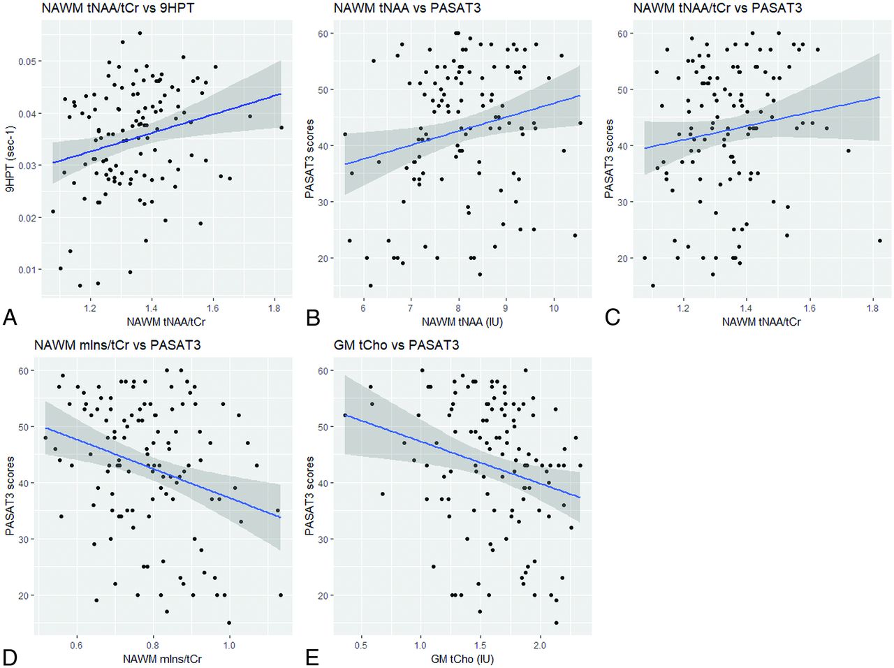
    Associations between neurometabolites and clinical disability measures. The scatterplots with the line of best fit and 95% confidence intervals are shown for the associations in normal-appearing white matter among the following: A, tNAA/tCr and 9HPT (ρ = 0.23; 95% CI, 0.06–0.40). B, tNAA and Paced Auditory Serial Addition Test (ρ  = 0.21; 95% CI, 0.03–0.38). C, tNAA/tCr and Paced Auditory Serial Addition Test (ρ  = 0.19; 95% CI, 0.01–0.36). D, mIns/tCr and Paced Auditory Serial Addition Test (ρ = –0.23; 95% CI,–0.39 to −0.05) and in gray matter between tCho and Paced Auditory Serial Addition Test (ρ = –0.24; 95% CI, –0.40 to −0.06) (E).

Tables

  • Figures
    • View popup
    Table 1:

    Baseline demographics and characteristics (n = 119)

    Clinical VariableMean (SD)
    Age (yr)54.2 (6.7)
    EDSSa6.0 (4.0–6.5)
    Normalized brain volume (mL)1418 (87.3)
    Nine-Hole Peg Test (sec–1)b0.035 (0.01)
    SPMS duration (yr)22.3 (9.2)
    PASAT3 score (of 60)43.0 (11.5)
    SDMT score (of 110)46.9 (11.0)
    T2 lesion volume (mL)a9.7 (0.3–34.6)
    T25–FW (sec)a10.6 (4.3–180.0)
    • ↵a Median (range).

    • ↵b The Nine-Hole Peg Test was calculated by taking the reciprocal of the average of 2 trials for each arm and taking the mean.

    • View popup
    Table 2:

    Results from multiple regression analysis examining associations between neurometabolites and clinical disability measuresa

    PredictorsStandardized βStandardized 95% CIP
    Nine-Hole Peg Test (n = 118)b
     NAWM tNAA/tCr0.190.01–0.36.04
    Paced Auditory Serial Addition Test (n = 119)
     NAWM tNAA0.170.01–0.34.04
     Sexc–0.23–0.42 to –0.05.01
     T2 lesion volume–0.47–0.64 to –0.30<.001
    Paced Auditory Serial Addition Test (n = 118)d
     NAWM tNAA/tCr0.190.03–0.35.02
     Sexc–0.26–0.44 to –0.08.006
     T2 lesion volume–0.46–0.63 to –0.29<.001
    Paced Auditory Serial Addition Test (n = 119)
     NAWM mIns/tCr–0.22–0.39 to –0.06.007
     Sexc–0.25–0.43 to –0.07.008
     T2 lesion volume–0.47–0.64 to –0.30<.001
    Paced Auditory Serial Addition Test (n = 119)
     GM tCho–0.17–0.33 to –0.01.04
     T2 lesion volume–0.48–0.65 to –0.31<.001
    • ↵a Covariates in the model include age, sex, duration from onset, occurrence of relapse in the 2 years preceding randomization, T2 lesion volume, and normalized brain volume. The table highlights only the predictor variables that were significant from the multiple regression models.

    • ↵b The Nine–Hole Peg Test was calculated by taking the reciprocal of the average of 2 trials for each arm and taking the mean.

    • ↵c Male is reference category.

    • ↵d One hundred eighteen participants in this cohort because 1 case was removed due to a highly leveraged point.

PreviousNext
Back to top

In this issue

American Journal of Neuroradiology: 41 (12)
American Journal of Neuroradiology
Vol. 41, Issue 12
1 Dec 2020
  • Table of Contents
  • Index by author
  • Complete Issue (PDF)
Advertisement
Print
Download PDF
Email Article

Thank you for your interest in spreading the word on American Journal of Neuroradiology.

NOTE: We only request your email address so that the person you are recommending the page to knows that you wanted them to see it, and that it is not junk mail. We do not capture any email address.

Enter multiple addresses on separate lines or separate them with commas.
NAA is a Marker of Disability in Secondary-Progressive MS: A Proton MR Spectroscopic Imaging Study
(Your Name) has sent you a message from American Journal of Neuroradiology
(Your Name) thought you would like to see the American Journal of Neuroradiology web site.
CAPTCHA
This question is for testing whether or not you are a human visitor and to prevent automated spam submissions.
Cite this article
B.S. Solanky, N.A. John, F. DeAngelis, J. Stutters, F. Prados, T. Schneider, R.A. Parker, C.J. Weir, A. Monteverdi, D. Plantone, A. Doshi, D. MacManus, I. Marshall, F. Barkhof, C.A.M. Gandini Wheeler-Kingshott, J. Chataway, for the MS-SMART Investigators
NAA is a Marker of Disability in Secondary-Progressive MS: A Proton MR Spectroscopic Imaging Study
American Journal of Neuroradiology Dec 2020, 41 (12) 2209-2218; DOI: 10.3174/ajnr.A6809

Citation Manager Formats

  • BibTeX
  • Bookends
  • EasyBib
  • EndNote (tagged)
  • EndNote 8 (xml)
  • Medlars
  • Mendeley
  • Papers
  • RefWorks Tagged
  • Ref Manager
  • RIS
  • Zotero
0 Responses
Respond to this article
Share
Bookmark this article
NAA is a Marker of Disability in Secondary-Progressive MS: A Proton MR Spectroscopic Imaging Study
B.S. Solanky, N.A. John, F. DeAngelis, J. Stutters, F. Prados, T. Schneider, R.A. Parker, C.J. Weir, A. Monteverdi, D. Plantone, A. Doshi, D. MacManus, I. Marshall, F. Barkhof, C.A.M. Gandini Wheeler-Kingshott, J. Chataway, for the MS-SMART Investigators
American Journal of Neuroradiology Dec 2020, 41 (12) 2209-2218; DOI: 10.3174/ajnr.A6809
del.icio.us logo Twitter logo Facebook logo Mendeley logo
  • Tweet Widget
  • Facebook Like
  • Google Plus One
Purchase

Jump to section

  • Article
    • Abstract
    • ABBREVIATIONS:
    • MATERIALS AND METHODS
    • RESULTS
    • DISCUSSION
    • CONCLUSIONS
    • Acknowledgments
    • Footnotes
    • References
  • Figures & Data
  • Supplemental
  • Info & Metrics
  • Responses
  • References
  • PDF

Related Articles

  • PubMed
  • Google Scholar

Cited By...

  • UPDATE trial: investigating the effects of ultra-processed versus minimally processed diets following UK dietary guidance on health outcomes: a protocol for an 8-week community-based cross-over randomised controlled trial in people with overweight or obesity, followed by a 6-month behavioural intervention
  • ORYX-MRSI: A Fully-Automated Open-Source Software for Three-Dimensional Proton Magnetic Resonance Spectroscopic Imaging Data Analysis
  • A systematic review of resting state functional MRI connectivity changes and cognitive impairment in multiple sclerosis
  • Crossref
  • Google Scholar

This article has not yet been cited by articles in journals that are participating in Crossref Cited-by Linking.

More in this TOC Section

Adult Brain

  • Diagnostic Neuroradiology of Monoclonal Antibodies
  • Clinical Outcomes After Chiari I Decompression
  • Segmentation of Brain Metastases with BLAST
Show more Adult Brain

Functional

  • Kurtosis and Epileptogenic Tubers: A Pilot Study
  • Glutaric Aciduria Type 1: DK vs. Conventional MRI
  • Brain Iron in Niemann-Pick Type C: 7T Study
Show more Functional

Similar Articles

Advertisement

Indexed Content

  • Current Issue
  • Accepted Manuscripts
  • Article Preview
  • Past Issues
  • Editorials
  • Editor's Choice
  • Fellows' Journal Club
  • Letters to the Editor
  • Video Articles

Cases

  • Case Collection
  • Archive - Case of the Week
  • Archive - Case of the Month
  • Archive - Classic Case

More from AJNR

  • Trainee Corner
  • Imaging Protocols
  • MRI Safety Corner
  • Book Reviews

Multimedia

  • AJNR Podcasts
  • AJNR Scantastics

Resources

  • Turnaround Time
  • Submit a Manuscript
  • Submit a Video Article
  • Submit an eLetter to the Editor/Response
  • Manuscript Submission Guidelines
  • Statistical Tips
  • Fast Publishing of Accepted Manuscripts
  • Graphical Abstract Preparation
  • Imaging Protocol Submission
  • Evidence-Based Medicine Level Guide
  • Publishing Checklists
  • Author Policies
  • Become a Reviewer/Academy of Reviewers
  • News and Updates

About Us

  • About AJNR
  • Editorial Board
  • Editorial Board Alumni
  • Alerts
  • Permissions
  • Not an AJNR Subscriber? Join Now
  • Advertise with Us
  • Librarian Resources
  • Feedback
  • Terms and Conditions
  • AJNR Editorial Board Alumni

American Society of Neuroradiology

  • Not an ASNR Member? Join Now

© 2025 by the American Society of Neuroradiology All rights, including for text and data mining, AI training, and similar technologies, are reserved.
Print ISSN: 0195-6108 Online ISSN: 1936-959X

Powered by HighWire