Advertisement

Main menu

  • Home
  • Content
    • Current Issue
    • Accepted Manuscripts
    • Article Preview
    • Past Issue Archive
    • AJNR Case Collection
    • Case of the Week Archive
    • Classic Case Archive
    • Case of the Month Archive
  • Special Collections
    • Spinal CSF Leak Articles (Jan 2020-June 2024)
    • 2024 AJNR Journal Awards
    • Most Impactful AJNR Articles
  • Multimedia
    • AJNR Podcast
    • AJNR Scantastics
    • Video Articles
  • For Authors
    • Submit a Manuscript
    • Author Policies
    • Fast publishing of Accepted Manuscripts
    • Graphical Abstract Preparation
    • Manuscript Submission Guidelines
    • Imaging Protocol Submission
    • Submit a Case for the Case Collection
  • About Us
    • About AJNR
    • Editorial Board
  • More
    • Become a Reviewer/Academy of Reviewers
    • Subscribers
    • Permissions
    • Alerts
    • Feedback
    • Advertisers
    • ASNR Home
  • Other Publications
    • ajnr

User menu

  • Alerts
  • Log in

Search

  • Advanced search
American Journal of Neuroradiology

American Journal of Neuroradiology

ASHNR American Society of Functional Neuroradiology ASHNR American Society of Pediatric Neuroradiology ASSR
  • Alerts
  • Log in

Advanced Search

  • Home
  • Content
    • Current Issue
    • Accepted Manuscripts
    • Article Preview
    • Past Issue Archive
    • AJNR Case Collection
    • Case of the Week Archive
    • Classic Case Archive
    • Case of the Month Archive
  • Special Collections
    • Spinal CSF Leak Articles (Jan 2020-June 2024)
    • 2024 AJNR Journal Awards
    • Most Impactful AJNR Articles
  • Multimedia
    • AJNR Podcast
    • AJNR Scantastics
    • Video Articles
  • For Authors
    • Submit a Manuscript
    • Author Policies
    • Fast publishing of Accepted Manuscripts
    • Graphical Abstract Preparation
    • Manuscript Submission Guidelines
    • Imaging Protocol Submission
    • Submit a Case for the Case Collection
  • About Us
    • About AJNR
    • Editorial Board
  • More
    • Become a Reviewer/Academy of Reviewers
    • Subscribers
    • Permissions
    • Alerts
    • Feedback
    • Advertisers
    • ASNR Home
  • Follow AJNR on Twitter
  • Visit AJNR on Facebook
  • Follow AJNR on Instagram
  • Join AJNR on LinkedIn
  • RSS Feeds

Welcome to the new AJNR, Updated Hall of Fame, and more. Read the full announcements.


AJNR is seeking candidates for the position of Associate Section Editor, AJNR Case Collection. Read the full announcement.


Research ArticleAdult Brain
Open Access

Recent Administration of Iodinated Contrast Renders Core Infarct Estimation Inaccurate Using RAPID Software

A.Z. Copelan, E.R. Smith, G.T. Drocton, K.H. Narsinh, D. Murph, R.S. Khangura, Z.J. Hartley, A.A. Abla, W.P. Dillon, C.F. Dowd, R.T. Higashida, V.V. Halbach, S.W. Hetts, D.L. Cooke, K. Keenan, J. Nelson, D. Mccoy, M. Ciano and M.R. Amans
American Journal of Neuroradiology December 2020, 41 (12) 2235-2242; DOI: https://doi.org/10.3174/ajnr.A6908
A.Z. Copelan
aFrom the Departments of Diagnostic and Interventional Neuroradiology (A.Z.C., E.R.S., G.T.D., K.H.N., D.M., R.S.K., Z.J.H., A.A.A., W.P.D., C.F.D., R.T.H., V.V.H., S.W.H., D.L.C., J.N., D.M., M.C., M.R.A.)
  • Find this author on Google Scholar
  • Find this author on PubMed
  • Search for this author on this site
  • ORCID record for A.Z. Copelan
E.R. Smith
aFrom the Departments of Diagnostic and Interventional Neuroradiology (A.Z.C., E.R.S., G.T.D., K.H.N., D.M., R.S.K., Z.J.H., A.A.A., W.P.D., C.F.D., R.T.H., V.V.H., S.W.H., D.L.C., J.N., D.M., M.C., M.R.A.)
cDepartment of Radiology (E.R.S.), Medical College of Wisconsin, Milwaukee, Wisconsin
  • Find this author on Google Scholar
  • Find this author on PubMed
  • Search for this author on this site
  • ORCID record for E.R. Smith
G.T. Drocton
aFrom the Departments of Diagnostic and Interventional Neuroradiology (A.Z.C., E.R.S., G.T.D., K.H.N., D.M., R.S.K., Z.J.H., A.A.A., W.P.D., C.F.D., R.T.H., V.V.H., S.W.H., D.L.C., J.N., D.M., M.C., M.R.A.)
  • Find this author on Google Scholar
  • Find this author on PubMed
  • Search for this author on this site
  • ORCID record for G.T. Drocton
K.H. Narsinh
aFrom the Departments of Diagnostic and Interventional Neuroradiology (A.Z.C., E.R.S., G.T.D., K.H.N., D.M., R.S.K., Z.J.H., A.A.A., W.P.D., C.F.D., R.T.H., V.V.H., S.W.H., D.L.C., J.N., D.M., M.C., M.R.A.)
  • Find this author on Google Scholar
  • Find this author on PubMed
  • Search for this author on this site
  • ORCID record for K.H. Narsinh
D. Murph
aFrom the Departments of Diagnostic and Interventional Neuroradiology (A.Z.C., E.R.S., G.T.D., K.H.N., D.M., R.S.K., Z.J.H., A.A.A., W.P.D., C.F.D., R.T.H., V.V.H., S.W.H., D.L.C., J.N., D.M., M.C., M.R.A.)
  • Find this author on Google Scholar
  • Find this author on PubMed
  • Search for this author on this site
  • ORCID record for D. Murph
R.S. Khangura
aFrom the Departments of Diagnostic and Interventional Neuroradiology (A.Z.C., E.R.S., G.T.D., K.H.N., D.M., R.S.K., Z.J.H., A.A.A., W.P.D., C.F.D., R.T.H., V.V.H., S.W.H., D.L.C., J.N., D.M., M.C., M.R.A.)
  • Find this author on Google Scholar
  • Find this author on PubMed
  • Search for this author on this site
  • ORCID record for R.S. Khangura
Z.J. Hartley
aFrom the Departments of Diagnostic and Interventional Neuroradiology (A.Z.C., E.R.S., G.T.D., K.H.N., D.M., R.S.K., Z.J.H., A.A.A., W.P.D., C.F.D., R.T.H., V.V.H., S.W.H., D.L.C., J.N., D.M., M.C., M.R.A.)
  • Find this author on Google Scholar
  • Find this author on PubMed
  • Search for this author on this site
  • ORCID record for Z.J. Hartley
A.A. Abla
aFrom the Departments of Diagnostic and Interventional Neuroradiology (A.Z.C., E.R.S., G.T.D., K.H.N., D.M., R.S.K., Z.J.H., A.A.A., W.P.D., C.F.D., R.T.H., V.V.H., S.W.H., D.L.C., J.N., D.M., M.C., M.R.A.)
bNeurosurgery (A.A.A.), University of California, San Francisco, San Francisco, California
  • Find this author on Google Scholar
  • Find this author on PubMed
  • Search for this author on this site
  • ORCID record for A.A. Abla
W.P. Dillon
aFrom the Departments of Diagnostic and Interventional Neuroradiology (A.Z.C., E.R.S., G.T.D., K.H.N., D.M., R.S.K., Z.J.H., A.A.A., W.P.D., C.F.D., R.T.H., V.V.H., S.W.H., D.L.C., J.N., D.M., M.C., M.R.A.)
  • Find this author on Google Scholar
  • Find this author on PubMed
  • Search for this author on this site
  • ORCID record for W.P. Dillon
C.F. Dowd
aFrom the Departments of Diagnostic and Interventional Neuroradiology (A.Z.C., E.R.S., G.T.D., K.H.N., D.M., R.S.K., Z.J.H., A.A.A., W.P.D., C.F.D., R.T.H., V.V.H., S.W.H., D.L.C., J.N., D.M., M.C., M.R.A.)
  • Find this author on Google Scholar
  • Find this author on PubMed
  • Search for this author on this site
  • ORCID record for C.F. Dowd
R.T. Higashida
aFrom the Departments of Diagnostic and Interventional Neuroradiology (A.Z.C., E.R.S., G.T.D., K.H.N., D.M., R.S.K., Z.J.H., A.A.A., W.P.D., C.F.D., R.T.H., V.V.H., S.W.H., D.L.C., J.N., D.M., M.C., M.R.A.)
  • Find this author on Google Scholar
  • Find this author on PubMed
  • Search for this author on this site
  • ORCID record for R.T. Higashida
V.V. Halbach
aFrom the Departments of Diagnostic and Interventional Neuroradiology (A.Z.C., E.R.S., G.T.D., K.H.N., D.M., R.S.K., Z.J.H., A.A.A., W.P.D., C.F.D., R.T.H., V.V.H., S.W.H., D.L.C., J.N., D.M., M.C., M.R.A.)
  • Find this author on Google Scholar
  • Find this author on PubMed
  • Search for this author on this site
  • ORCID record for V.V. Halbach
S.W. Hetts
aFrom the Departments of Diagnostic and Interventional Neuroradiology (A.Z.C., E.R.S., G.T.D., K.H.N., D.M., R.S.K., Z.J.H., A.A.A., W.P.D., C.F.D., R.T.H., V.V.H., S.W.H., D.L.C., J.N., D.M., M.C., M.R.A.)
  • Find this author on Google Scholar
  • Find this author on PubMed
  • Search for this author on this site
  • ORCID record for S.W. Hetts
D.L. Cooke
aFrom the Departments of Diagnostic and Interventional Neuroradiology (A.Z.C., E.R.S., G.T.D., K.H.N., D.M., R.S.K., Z.J.H., A.A.A., W.P.D., C.F.D., R.T.H., V.V.H., S.W.H., D.L.C., J.N., D.M., M.C., M.R.A.)
  • Find this author on Google Scholar
  • Find this author on PubMed
  • Search for this author on this site
  • ORCID record for D.L. Cooke
K. Keenan
dDepartment of Neurology (K.K.), University of California Davis, Sacramento, California.
  • Find this author on Google Scholar
  • Find this author on PubMed
  • Search for this author on this site
  • ORCID record for K. Keenan
J. Nelson
aFrom the Departments of Diagnostic and Interventional Neuroradiology (A.Z.C., E.R.S., G.T.D., K.H.N., D.M., R.S.K., Z.J.H., A.A.A., W.P.D., C.F.D., R.T.H., V.V.H., S.W.H., D.L.C., J.N., D.M., M.C., M.R.A.)
  • Find this author on Google Scholar
  • Find this author on PubMed
  • Search for this author on this site
  • ORCID record for J. Nelson
D. Mccoy
aFrom the Departments of Diagnostic and Interventional Neuroradiology (A.Z.C., E.R.S., G.T.D., K.H.N., D.M., R.S.K., Z.J.H., A.A.A., W.P.D., C.F.D., R.T.H., V.V.H., S.W.H., D.L.C., J.N., D.M., M.C., M.R.A.)
  • Find this author on Google Scholar
  • Find this author on PubMed
  • Search for this author on this site
  • ORCID record for D. Mccoy
M. Ciano
aFrom the Departments of Diagnostic and Interventional Neuroradiology (A.Z.C., E.R.S., G.T.D., K.H.N., D.M., R.S.K., Z.J.H., A.A.A., W.P.D., C.F.D., R.T.H., V.V.H., S.W.H., D.L.C., J.N., D.M., M.C., M.R.A.)
  • Find this author on Google Scholar
  • Find this author on PubMed
  • Search for this author on this site
  • ORCID record for M. Ciano
M.R. Amans
aFrom the Departments of Diagnostic and Interventional Neuroradiology (A.Z.C., E.R.S., G.T.D., K.H.N., D.M., R.S.K., Z.J.H., A.A.A., W.P.D., C.F.D., R.T.H., V.V.H., S.W.H., D.L.C., J.N., D.M., M.C., M.R.A.)
  • Find this author on Google Scholar
  • Find this author on PubMed
  • Search for this author on this site
  • ORCID record for M.R. Amans
  • Article
  • Figures & Data
  • Info & Metrics
  • Responses
  • References
  • PDF
Loading

Article Figures & Data

Figures

  • Tables
  • FIG 1.
    • Download figure
    • Open in new tab
    • Download powerpoint
    FIG 1.

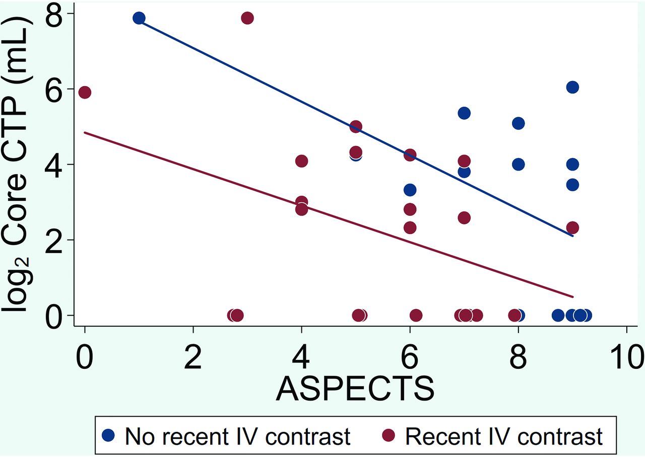
    Scatterplot of CT-ASPECTS versus log-transformed CTP-estimated core infarct for the recent IV contrast group (red) and the contrast-naïve group (blue) with lines of best fit. For the same CT-ASPECTS, the CTP-estimated core infarct is consistently less in the recent IV contrast group with divergence of the lines of best fit toward the lower ASPECTS indicative of more substantial differences between the 2 cohorts and more significant underestimation of estimated core infarct for lower CT-ASPECTS.

  • FIG 2.
    • Download figure
    • Open in new tab
    • Download powerpoint
    FIG 2.

    Scatterplot of CTP-measured core infarct volume versus postoperative core infarct volume as measured by MR imaging or CT for the recent IV contrast group (red) and the contrast-naïve group (blue) with lines of best fit. For the same preoperative CTP-measured core, the postoperative core infarct is greater in the recent IV contrast group, suggesting that recent IV contrast underestimates CTP-measured core infarct volume at presentation.

  • FIG 3.
    • Download figure
    • Open in new tab
    • Download powerpoint
    FIG 3.

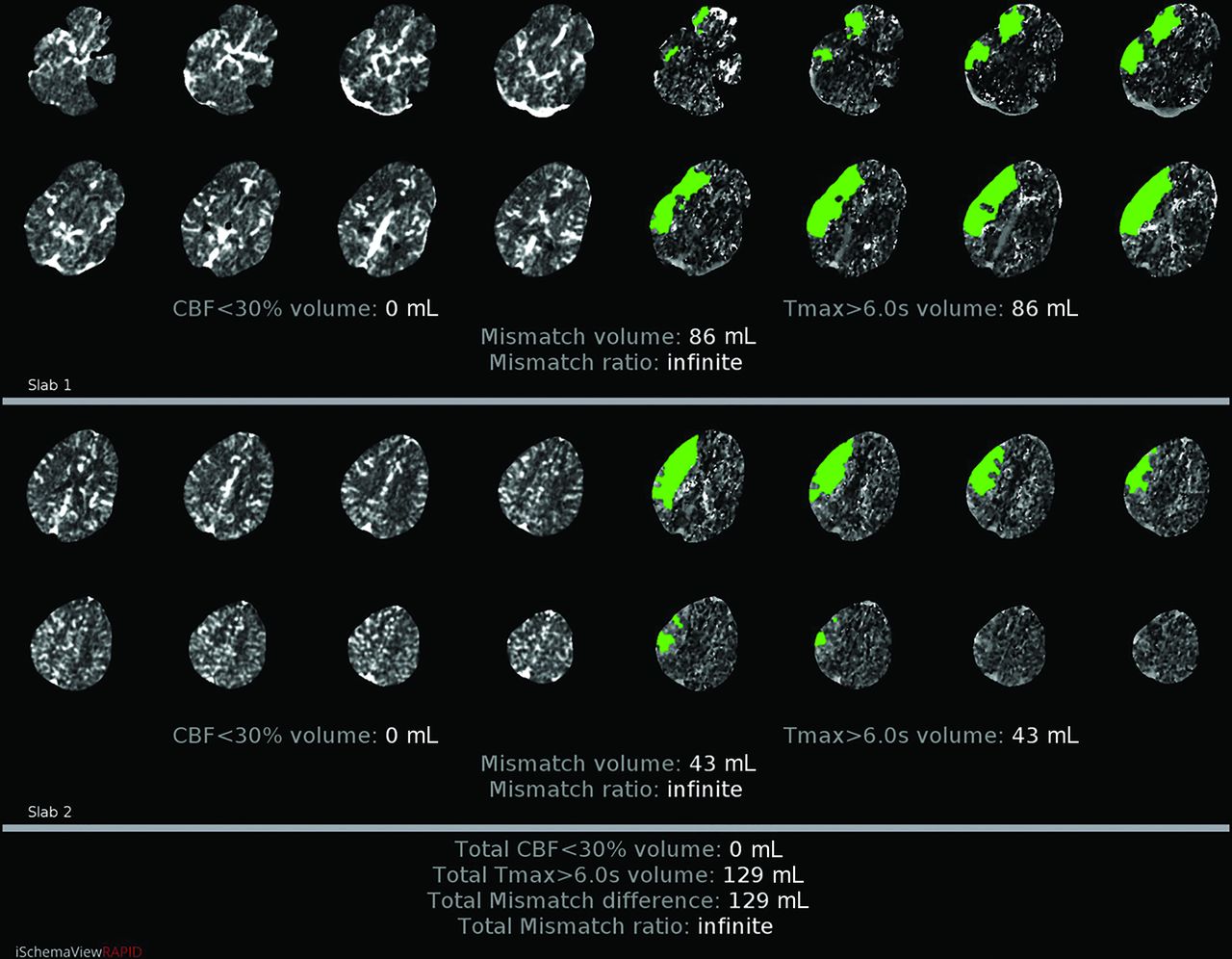
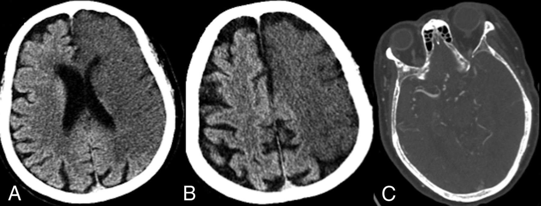
    A 56-year-old man who presented to an outside hospital with a right MCA syndrome and ELVO confirmed on CTA. Last known well was approximately 11 hours before imaging at our institution. A, NCCT demonstrates loss of gray-white matter differentiation, in keeping with acute infarct, involving the right insula as well as the frontal and temporal opercula. B, CTA confirms a right M1 segment (yellow circle) occlusion. C, DWI reveals an extensive area of acute infarct correlating with the areas of hypoattenuation on the NCCT. TICI 3 reperfusion was achieved within 60 minutes of the CTP study, and the MR imaging was obtained later in the day.

  • FIG 4.
    • Download figure
    • Open in new tab
    • Download powerpoint
    FIG 4.

    CTP with RAPID postprocessing from the same patient as in Fig 3 suggests no core infarct (lack of pink color-coding and an rCBF of <30% volume of 0 mL) with a large area of ischemic penumbra (green color-coding with time-to-maximum [Tmax] of >6 seconds of 129 mL).

  • FIG 5.
    • Download figure
    • Open in new tab
    • Download powerpoint
    FIG 5.

    A 67-year-old man who presented directly to our institution (no recent IV contrast) with a left MCA syndrome and last known well approximately 8 hours before imaging. A and B, NCCT demonstrates hypoattenuation involving a large volume of the left MCA and anterior cerebral artery territories. C, CTA demonstrates a internal carotid terminus occlusion with involvement of the carotid terminus and A1 and M1 segments.

  • FIG 6.
    • Download figure
    • Open in new tab
    • Download powerpoint
    FIG 6.

    CTP imaging with RAPID postprocessing from the same patient as in Fig 5 suggests an extensive area of core infarct (pink color-coding and an rCBF of <30% volume of 234 mL) corresponding to the large volume of hypoattenuation on NCCT. Tmax indicates time-to-maximum.

  • FIG 7.
    • Download figure
    • Open in new tab
    • Download powerpoint
    FIG 7.

    The same 56-year-old man with right M1 segment occlusion as presented in Fig 3. A, rCBF map from the RAPID postprocessing of CTP data demonstrates subtle decreased blood flow to the right MCA territory corresponding to the suggested core infarct volume of 0 mL. B, rCBF map from GE Healthcare processing of the CTP data demonstrates much more conspicuous decreased blood flow within the right MCA territory (scale bar = 0–100 mL/g/min).

Tables

  • Figures
    • View popup
    Table 1:

    Baseline characteristics

    CharacteristicRecent IV ContrastaNo Recent IV ContrastaOverallaP Valueb
    Count231538NA
    Age (yr)70.5 ± 16.076.5 ± 18.472.9 ± 17.0.32
    Serum creatinine level (mg/dL)1.08 ± 0.341.12 ± 0.291.10 ± 0.32.78
    Female15 (65%)7 (47%)22 (58%).32
    Time since last known well (hr)12.5 ± 5.69.3 ± 7.811.2 ± 6.6.19
    ASPECTS5.4 ± 2.07.5 ± 2.26.2 ± 2.3.005
    CTP core (median) (range) (mL)4 (0–234)13 (0–208)6 (0–234).28
    Collateral grade.71
     115 (65%)9 (60%)24 (63%)
     26 (26%)3 (20%)9 (24%)
     32 (9%)3 (20%)5 (13%)
    Thrombectomy11 (48%)12 (80%)23 (61%).09
    • Note:—NA indicates not applicable.

    • ↵a Values are No., No. (%), or mean ± SD.

    • ↵b P values are from the Fisher exact test or 2-sample t test with unequal variances.

    • View popup
    Table 2:

    Multivariable linear regression results

    CharacteristicPIa95% Confidence IntervalP Value
    Recent IV contrast0.34(0.12–0.96).04
    ASPECTS0.74(0.59–0.93).01
    Collateral grade (1 = reference group)
     22.27(0.78–6.60).13
     38.46(2.10–34.06).004
    • ↵a Outcome is log2 core CTP.

PreviousNext
Back to top

In this issue

American Journal of Neuroradiology: 41 (12)
American Journal of Neuroradiology
Vol. 41, Issue 12
1 Dec 2020
  • Table of Contents
  • Index by author
  • Complete Issue (PDF)
Advertisement
Print
Download PDF
Email Article

Thank you for your interest in spreading the word on American Journal of Neuroradiology.

NOTE: We only request your email address so that the person you are recommending the page to knows that you wanted them to see it, and that it is not junk mail. We do not capture any email address.

Enter multiple addresses on separate lines or separate them with commas.
Recent Administration of Iodinated Contrast Renders Core Infarct Estimation Inaccurate Using RAPID Software
(Your Name) has sent you a message from American Journal of Neuroradiology
(Your Name) thought you would like to see the American Journal of Neuroradiology web site.
CAPTCHA
This question is for testing whether or not you are a human visitor and to prevent automated spam submissions.
Cite this article
A.Z. Copelan, E.R. Smith, G.T. Drocton, K.H. Narsinh, D. Murph, R.S. Khangura, Z.J. Hartley, A.A. Abla, W.P. Dillon, C.F. Dowd, R.T. Higashida, V.V. Halbach, S.W. Hetts, D.L. Cooke, K. Keenan, J. Nelson, D. Mccoy, M. Ciano, M.R. Amans
Recent Administration of Iodinated Contrast Renders Core Infarct Estimation Inaccurate Using RAPID Software
American Journal of Neuroradiology Dec 2020, 41 (12) 2235-2242; DOI: 10.3174/ajnr.A6908

Citation Manager Formats

  • BibTeX
  • Bookends
  • EasyBib
  • EndNote (tagged)
  • EndNote 8 (xml)
  • Medlars
  • Mendeley
  • Papers
  • RefWorks Tagged
  • Ref Manager
  • RIS
  • Zotero
0 Responses
Respond to this article
Share
Bookmark this article
Recent Administration of Iodinated Contrast Renders Core Infarct Estimation Inaccurate Using RAPID Software
A.Z. Copelan, E.R. Smith, G.T. Drocton, K.H. Narsinh, D. Murph, R.S. Khangura, Z.J. Hartley, A.A. Abla, W.P. Dillon, C.F. Dowd, R.T. Higashida, V.V. Halbach, S.W. Hetts, D.L. Cooke, K. Keenan, J. Nelson, D. Mccoy, M. Ciano, M.R. Amans
American Journal of Neuroradiology Dec 2020, 41 (12) 2235-2242; DOI: 10.3174/ajnr.A6908
del.icio.us logo Twitter logo Facebook logo Mendeley logo
  • Tweet Widget
  • Facebook Like
  • Google Plus One
Purchase

Jump to section

  • Article
    • Abstract
    • ABBREVIATIONS:
    • MATERIALS AND METHODS
    • RESULTS
    • DISCUSSION
    • CONCLUSIONS
    • Footnotes
    • References
  • Figures & Data
  • Info & Metrics
  • Responses
  • References
  • PDF

Related Articles

  • No related articles found.
  • PubMed
  • Google Scholar

Cited By...

  • Evaluation of time-resolved whole brain flat panel detector perfusion imaging using RAPID ANGIO in patients with acute stroke: comparison with CT perfusion imaging
  • Perfusion Scotoma: A Potential Core Underestimation in CT Perfusion in the Delayed Time Window in Patients with Acute Ischemic Stroke
  • Contrast Bolus Interference in a Multimodal CT Stroke Protocol
  • Do Prior Iodine Contrast Injections Affect Cerebral Blood Flow Measurement on CT Perfusion Studies of Patients with Large-Vessel Occlusions?
  • Reply:
  • Acute Stroke Imaging in Transfer Patients Who Received Recent Intravenous Iodinated Contrast at an Outside Facility: An Unrecognized Problem That Deserves More Attention
  • Crossref (15)
  • Google Scholar

This article has been cited by the following articles in journals that are participating in Crossref Cited-by Linking.

  • The R-AI-DIOLOGY checklist: a practical checklist for evaluation of artificial intelligence tools in clinical neuroradiology
    Sven Haller, Sofie Van Cauter, Christian Federau, Dennis M. Hedderich, Myriam Edjlali
    Neuroradiology 2022 64 5
  • Angiography suite cone‐beam CT perfusion for selection of thrombectomy patients: A pilot study
    Santiago Ortega‐Gutierrez, Darko Quispe‐Orozco, Sebastian Schafer, Mudassir Farooqui, Cynthia B Zevallos, Sudeepta Dandapat, Alan Mendez‐Ruiz, Beverly Aagaard‐Kienitz, Nils Petersen, Colin P. Derdeyn
    Journal of Neuroimaging 2022 32 3
  • Evaluation of time-resolved whole brain flat panel detector perfusion imaging using RAPID ANGIO in patients with acute stroke: comparison with CT perfusion imaging
    Christoph C Kurmann, Johannes Kaesmacher, Daniel L Cooke, Marios Psychogios, Johannes Weber, Demetrius K Lopes, Gregory W Albers, Pasquale Mordasini
    Journal of NeuroInterventional Surgery 2023 15 4
  • Perfusion Scotoma: A Potential Core Underestimation in CT Perfusion in the Delayed Time Window in Patients with Acute Ischemic Stroke
    K. Abrams, G. Dabus
    American Journal of Neuroradiology 2022 43 6
  • Combined Use of X-ray Angiography and Intraprocedural MRI Enables Tissue-based Decision Making Regarding Revascularization during Acute Ischemic Stroke Intervention
    Kazim H. Narsinh, Bridget F. Kilbride, Kerstin Mueller, Daniel Murph, Alexander Copelan, Jonathan Massachi, Jeffrey Vitt, Chung-Huan Sun, Himanshu Bhat, Matthew R. Amans, Christopher F. Dowd, Van V. Halbach, Randall T. Higashida, Terilyn Moore, Mark W. Wilson, Daniel L. Cooke, Steven W. Hetts
    Radiology 2021 299 1
  • Contrast Bolus Interference in a Multimodal CT Stroke Protocol
    E. Kellner, A. Rau, T. Demerath, M. Reisert, H. Urbach
    American Journal of Neuroradiology 2021
  • Emerging role of artificial intelligence in stroke imaging
    Giuseppe Corrias, Andrea Mazzotta, Marta Melis, Filippo Cademartiri, Qi Yang, Jasjit S. Suri, Luca Saba
    Expert Review of Neurotherapeutics 2021 21 7
  • Acute Ischemic Stroke in the Clinic and the Laboratory: Targets for Translational Research
    Bart Franx, Rick M. Dijkhuizen, Diederik W.J. Dippel
    Neuroscience 2024 550
  • Acute Stroke Imaging in Transfer Patients Who Received Recent Intravenous Iodinated Contrast at an Outside Facility: An Unrecognized Problem That Deserves More Attention
    E. Gulko, S. Ali, J. Schefflein, H. Mehta, W. Gomes
    American Journal of Neuroradiology 2021 42 6
  • Application of Multimodal CT Combined with RAPID Software in Acute Ischemic Stroke
    容 罗
    Advances in Clinical Medicine 2022 12 01

More in this TOC Section

Adult Brain

  • Diagnostic Neuroradiology of Monoclonal Antibodies
  • Segmentation of Brain Metastases with BLAST
  • NCCT vs. MRI for Brain Atrophy in Acute Stroke
Show more Adult Brain

Functional

  • Glutaric Aciduria Type 1: DK vs. Conventional MRI
  • Kurtosis and Epileptogenic Tubers: A Pilot Study
  • WM Neuroaxonal Loss in Type 1 Diabetes
Show more Functional

Similar Articles

Advertisement

Indexed Content

  • Current Issue
  • Accepted Manuscripts
  • Article Preview
  • Past Issues
  • Editorials
  • Editors Choice
  • Fellow Journal Club
  • Letters to the Editor

Cases

  • Case Collection
  • Archive - Case of the Week
  • Archive - Case of the Month
  • Archive - Classic Case

Special Collections

  • Special Collections

Resources

  • News and Updates
  • Turn around Times
  • Submit a Manuscript
  • Author Policies
  • Manuscript Submission Guidelines
  • Evidence-Based Medicine Level Guide
  • Publishing Checklists
  • Graphical Abstract Preparation
  • Imaging Protocol Submission
  • Submit a Case
  • Become a Reviewer/Academy of Reviewers
  • Get Peer Review Credit from Publons

Multimedia

  • AJNR Podcast
  • AJNR SCANtastic
  • Video Articles

About Us

  • About AJNR
  • Editorial Board
  • Not an AJNR Subscriber? Join Now
  • Alerts
  • Feedback
  • Advertise with us
  • Librarian Resources
  • Permissions
  • Terms and Conditions

American Society of Neuroradiology

  • Not an ASNR Member? Join Now

© 2025 by the American Society of Neuroradiology All rights, including for text and data mining, AI training, and similar technologies, are reserved.
Print ISSN: 0195-6108 Online ISSN: 1936-959X

Powered by HighWire