Advertisement

Main menu

  • Home
  • Content
    • Current Issue
    • Accepted Manuscripts
    • Article Preview
    • Past Issue Archive
    • Video Articles
    • AJNR Case Collection
    • Case of the Week Archive
    • Case of the Month Archive
    • Classic Case Archive
  • Special Collections
    • AJNR Awards
    • Low-Field MRI
    • Alzheimer Disease
    • ASNR Foundation Special Collection
    • Photon-Counting CT
    • View All
  • Multimedia
    • AJNR Podcasts
    • AJNR SCANtastic
    • Trainee Corner
    • MRI Safety Corner
    • Imaging Protocols
  • For Authors
    • Submit a Manuscript
    • Submit a Video Article
    • Submit an eLetter to the Editor/Response
    • Manuscript Submission Guidelines
    • Statistical Tips
    • Fast Publishing of Accepted Manuscripts
    • Graphical Abstract Preparation
    • Imaging Protocol Submission
    • Author Policies
  • About Us
    • About AJNR
    • Editorial Board
    • Editorial Board Alumni
  • More
    • Become a Reviewer/Academy of Reviewers
    • Subscribers
    • Permissions
    • Alerts
    • Feedback
    • Advertisers
    • ASNR Home

User menu

  • Alerts
  • Log in

Search

  • Advanced search
American Journal of Neuroradiology

American Journal of Neuroradiology

ASHNR American Society of Functional Neuroradiology ASHNR American Society of Pediatric Neuroradiology ASSR
  • Alerts
  • Log in

Advanced Search

  • Home
  • Content
    • Current Issue
    • Accepted Manuscripts
    • Article Preview
    • Past Issue Archive
    • Video Articles
    • AJNR Case Collection
    • Case of the Week Archive
    • Case of the Month Archive
    • Classic Case Archive
  • Special Collections
    • AJNR Awards
    • Low-Field MRI
    • Alzheimer Disease
    • ASNR Foundation Special Collection
    • Photon-Counting CT
    • View All
  • Multimedia
    • AJNR Podcasts
    • AJNR SCANtastic
    • Trainee Corner
    • MRI Safety Corner
    • Imaging Protocols
  • For Authors
    • Submit a Manuscript
    • Submit a Video Article
    • Submit an eLetter to the Editor/Response
    • Manuscript Submission Guidelines
    • Statistical Tips
    • Fast Publishing of Accepted Manuscripts
    • Graphical Abstract Preparation
    • Imaging Protocol Submission
    • Author Policies
  • About Us
    • About AJNR
    • Editorial Board
    • Editorial Board Alumni
  • More
    • Become a Reviewer/Academy of Reviewers
    • Subscribers
    • Permissions
    • Alerts
    • Feedback
    • Advertisers
    • ASNR Home
  • Follow AJNR on Twitter
  • Visit AJNR on Facebook
  • Follow AJNR on Instagram
  • Join AJNR on LinkedIn
  • RSS Feeds

AJNR Awards, New Junior Editors, and more. Read the latest AJNR updates

Research ArticleNeurointervention

Endovascular Cerebral Venous Sinus Imaging with Optical Coherence Tomography

C.R. Pasarikovski, J.C. Ku, J. Keith, J. Ramjist, Y. Dobashi, S.M. Priola, L. da Costa and V.X.D. Yang
American Journal of Neuroradiology December 2020, 41 (12) 2292-2297; DOI: https://doi.org/10.3174/ajnr.A6909
C.R. Pasarikovski
aFrom the Division of Neurosurgery (C.R.P., J.C.K.), Department of Surgery, University of Toronto, Toronto, Ontario, Canada
  • Find this author on Google Scholar
  • Find this author on PubMed
  • Search for this author on this site
  • ORCID record for C.R. Pasarikovski
J.C. Ku
aFrom the Division of Neurosurgery (C.R.P., J.C.K.), Department of Surgery, University of Toronto, Toronto, Ontario, Canada
  • Find this author on Google Scholar
  • Find this author on PubMed
  • Search for this author on this site
  • ORCID record for J.C. Ku
J. Keith
bDepartment of Anatomic Pathology (J.K.), Sunnybrook Health Sciences Center, Toronto, Ontario, Canada
  • Find this author on Google Scholar
  • Find this author on PubMed
  • Search for this author on this site
  • ORCID record for J. Keith
J. Ramjist
cDivision of Neurosurgery (J.R., Y.D., L.d.C., V.X.D.Y.), Sunnybrook Hospital, University of Toronto, Toronto, Ontario, Canada
  • Find this author on Google Scholar
  • Find this author on PubMed
  • Search for this author on this site
  • ORCID record for J. Ramjist
Y. Dobashi
cDivision of Neurosurgery (J.R., Y.D., L.d.C., V.X.D.Y.), Sunnybrook Hospital, University of Toronto, Toronto, Ontario, Canada
  • Find this author on Google Scholar
  • Find this author on PubMed
  • Search for this author on this site
  • ORCID record for Y. Dobashi
S.M. Priola
dDivision of Neurosurgery (S.M.P.), Department of Surgery, Health Sciences North, Sudbury, Ontario, Canada
  • Find this author on Google Scholar
  • Find this author on PubMed
  • Search for this author on this site
  • ORCID record for S.M. Priola
L. da Costa
cDivision of Neurosurgery (J.R., Y.D., L.d.C., V.X.D.Y.), Sunnybrook Hospital, University of Toronto, Toronto, Ontario, Canada
  • Find this author on Google Scholar
  • Find this author on PubMed
  • Search for this author on this site
  • ORCID record for L. da Costa
V.X.D. Yang
cDivision of Neurosurgery (J.R., Y.D., L.d.C., V.X.D.Y.), Sunnybrook Hospital, University of Toronto, Toronto, Ontario, Canada
eHurvitz Brain Sciences Research Program (V.X.D.Y.), Sunnybrook Research Institute, Sunnybrook Health Sciences Centre, University of Toronto, Toronto, Ontario, Canada
  • Find this author on Google Scholar
  • Find this author on PubMed
  • Search for this author on this site
  • ORCID record for V.X.D. Yang
  • Article
  • Figures & Data
  • Info & Metrics
  • Responses
  • References
  • PDF
Loading

Article Figures & Data

Figures

  • FIG 1.
    • Download figure
    • Open in new tab
    • Download powerpoint
    FIG 1.

    Swine cerebral venous sinus. A, Combined arterial and venous diagnostic angiography injections in a lateral view via a guide catheter tip in the ascending pharyngeal artery (green arrow) and the internal jugular vein (red arrow), revealing the connection between the internal jugular vein and sigmoid sinus (blue arrows). B, OCT catheter in the superior sagittal sinus. The distal catheter marker (yellow arrow), lens marker (green arrow), and optical fiber (red arrows) are visible in the superior sagittal sinus.

  • FIG 2.
    • Download figure
    • Open in new tab
    • Download powerpoint
    FIG 2.

    Cross-sectional OCT imaging within the superior sagittal sinus. Large draining cortical veins can be observed entering the sinus (light blue arrow) with multiple adjacent dural arteries visible (green arrows), along with adjacent cortical veins outside the sinus lumen (yellow arrows). Small red thrombi (red arrows) are also visible in certain sections, either free-floating or attached to the sinus wall. The white asterisk is the OCT lens, and the yellow asterisk is the artifact from the wire. White bars = 2 mm.

  • FIG 3.
    • Download figure
    • Open in new tab
    • Download powerpoint
    FIG 3.

    Corresponding structural and Doppler OCT imaging. A, Two separate structural images reveal large draining cortical veins (light blue and green arrows) along with dural arteries (yellow arrows) and their corresponding Doppler imaging with a phase-shift color map demonstrating flow in the sinus and dural arteries (B). The white asterisk is the OCT lens, and the yellow asterisk is the artifact from the wire. White bars = 2mm.

  • FIG 4.
    • Download figure
    • Open in new tab
    • Download powerpoint
    FIG 4.

    OCT imaging and corresponding histologic cross-section. A, OCT shows 2 dural arteries (green arrows) and a dural venule (yellow arrow). B, Histologic cross-section with H&E staining. The sinus is collapsed, and the lumen is outlined with red arrows, along with adjacent arterioles (green arrows) and a venule (yellow arrow). The white asterisk is the OCT lens, and the yellow asterisk is the artifact from the wire. White bars = 2 mm. Black scale bar = 500 µm.

  • FIG 5.
    • Download figure
    • Open in new tab
    • Download powerpoint
    FIG 5.

    Bland-Altman plots comparing optical coherence tomography and histology. A, Sinus maximum diameter mean difference of 1.96 [SD, –1.96] mm and a mean of 0.21  [SD, 3.62, −3.19] mm, and 10% (2/20) of points outside the 95% confidence interval. B, Dural arteriole diameter with a mean difference of −8 [SD, 140, −157] µm, and 10% (4/40) of points outside the 95% confidence interval. C, Venule diameter with a mean difference of −15 [SD, 380, −411] µm, and 8% (3/35) of points outside the 95% confidence interval.
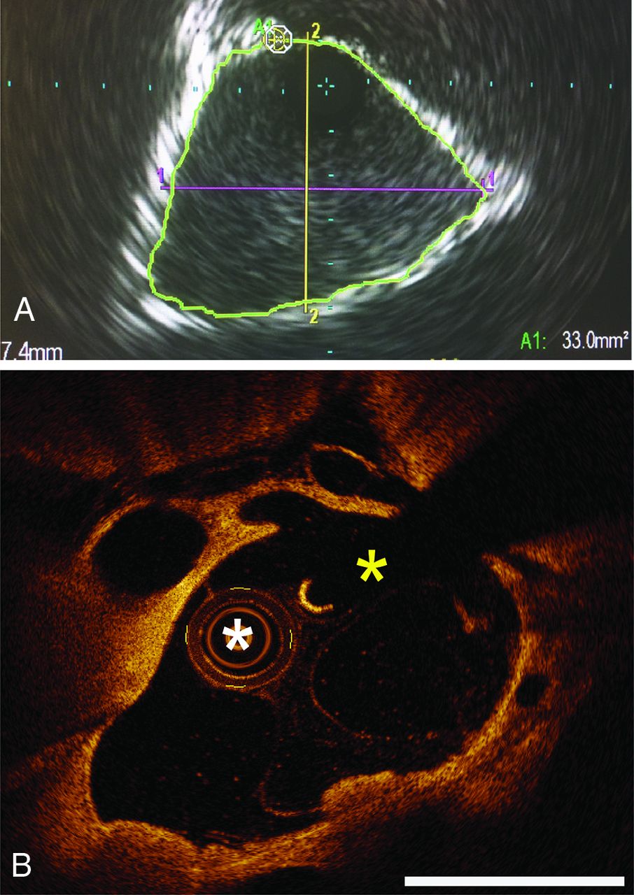
  • FIG 6.
    • Download figure
    • Open in new tab
    • Download powerpoint
    FIG 6.

    IVUS and OCT. A, IVUS of human superior sagittal sinus to determine the maximal diameter and circumference (A), and swine structural OCT imaging of the superior sagittal sinus (B). Although direct anatomic comparison cannot be made, OCT can clearly provide improved visualization of the sinus lumen and adjacent vessels. The white asterisk is the OCT lens, and the yellow asterisk is the artifact from the wire. White bars = 2 mm. Adapted with permission from Boddu et al.5

  • FIG 7.
    • Download figure
    • Open in new tab
    • Download powerpoint
    FIG 7.

    A dural arteriovenous fistula imaged and treated using novel OCT technology. A, The OCT device is navigated into the dural venous sinus and through a transparent dual-lumen balloon (II), optical imaging is undertaken (III) to identify the exact spatial position of arterial feeders (I) into the dural venous sinus. B, Laser ablation of the arterial feeders is accomplished under image guidance through the saline-filled balloon. The arterial feeders (I) previously identified now undergo image-guided laser ablation (III) through the transparent dual lumen balloon (II). Adapted from Pasarikovski et al.21

PreviousNext
Back to top

In this issue

American Journal of Neuroradiology: 41 (12)
American Journal of Neuroradiology
Vol. 41, Issue 12
1 Dec 2020
  • Table of Contents
  • Index by author
  • Complete Issue (PDF)
Advertisement
Print
Download PDF
Email Article

Thank you for your interest in spreading the word on American Journal of Neuroradiology.

NOTE: We only request your email address so that the person you are recommending the page to knows that you wanted them to see it, and that it is not junk mail. We do not capture any email address.

Enter multiple addresses on separate lines or separate them with commas.
Endovascular Cerebral Venous Sinus Imaging with Optical Coherence Tomography
(Your Name) has sent you a message from American Journal of Neuroradiology
(Your Name) thought you would like to see the American Journal of Neuroradiology web site.
CAPTCHA
This question is for testing whether or not you are a human visitor and to prevent automated spam submissions.
Cite this article
C.R. Pasarikovski, J.C. Ku, J. Keith, J. Ramjist, Y. Dobashi, S.M. Priola, L. da Costa, V.X.D. Yang
Endovascular Cerebral Venous Sinus Imaging with Optical Coherence Tomography
American Journal of Neuroradiology Dec 2020, 41 (12) 2292-2297; DOI: 10.3174/ajnr.A6909

Citation Manager Formats

  • BibTeX
  • Bookends
  • EasyBib
  • EndNote (tagged)
  • EndNote 8 (xml)
  • Medlars
  • Mendeley
  • Papers
  • RefWorks Tagged
  • Ref Manager
  • RIS
  • Zotero
0 Responses
Respond to this article
Share
Bookmark this article
Endovascular Cerebral Venous Sinus Imaging with Optical Coherence Tomography
C.R. Pasarikovski, J.C. Ku, J. Keith, J. Ramjist, Y. Dobashi, S.M. Priola, L. da Costa, V.X.D. Yang
American Journal of Neuroradiology Dec 2020, 41 (12) 2292-2297; DOI: 10.3174/ajnr.A6909
del.icio.us logo Twitter logo Facebook logo Mendeley logo
  • Tweet Widget
  • Facebook Like
  • Google Plus One
Purchase

Jump to section

  • Article
    • Abstract
    • ABBREVIATIONS:
    • MATERIALS AND METHODS
    • RESULTS
    • DISCUSSION
    • CONCLUSIONS
    • Footnotes
    • References
  • Figures & Data
  • Info & Metrics
  • Responses
  • References
  • PDF

Related Articles

  • No related articles found.
  • PubMed
  • Google Scholar

Cited By...

  • No citing articles found.
  • Crossref
  • Google Scholar

This article has not yet been cited by articles in journals that are participating in Crossref Cited-by Linking.

More in this TOC Section

  • A Retrospective Study in Tentorial DAVFs
  • Proximal Protection Devices for Carotid Stenting
  • Rescue Reentry in Carotid Near-Occlusion
Show more NEUROINTERVENTION

Similar Articles

Advertisement

Indexed Content

  • Current Issue
  • Accepted Manuscripts
  • Article Preview
  • Past Issues
  • Editorials
  • Editor's Choice
  • Fellows' Journal Club
  • Letters to the Editor
  • Video Articles

Cases

  • Case Collection
  • Archive - Case of the Week
  • Archive - Case of the Month
  • Archive - Classic Case

More from AJNR

  • Trainee Corner
  • Imaging Protocols
  • MRI Safety Corner
  • Book Reviews

Multimedia

  • AJNR Podcasts
  • AJNR Scantastics

Resources

  • Turnaround Time
  • Submit a Manuscript
  • Submit a Video Article
  • Submit an eLetter to the Editor/Response
  • Manuscript Submission Guidelines
  • Statistical Tips
  • Fast Publishing of Accepted Manuscripts
  • Graphical Abstract Preparation
  • Imaging Protocol Submission
  • Evidence-Based Medicine Level Guide
  • Publishing Checklists
  • Author Policies
  • Become a Reviewer/Academy of Reviewers
  • News and Updates

About Us

  • About AJNR
  • Editorial Board
  • Editorial Board Alumni
  • Alerts
  • Permissions
  • Not an AJNR Subscriber? Join Now
  • Advertise with Us
  • Librarian Resources
  • Feedback
  • Terms and Conditions
  • AJNR Editorial Board Alumni

American Society of Neuroradiology

  • Not an ASNR Member? Join Now

© 2025 by the American Society of Neuroradiology All rights, including for text and data mining, AI training, and similar technologies, are reserved.
Print ISSN: 0195-6108 Online ISSN: 1936-959X

Powered by HighWire