
Article Figures & Data
Figures
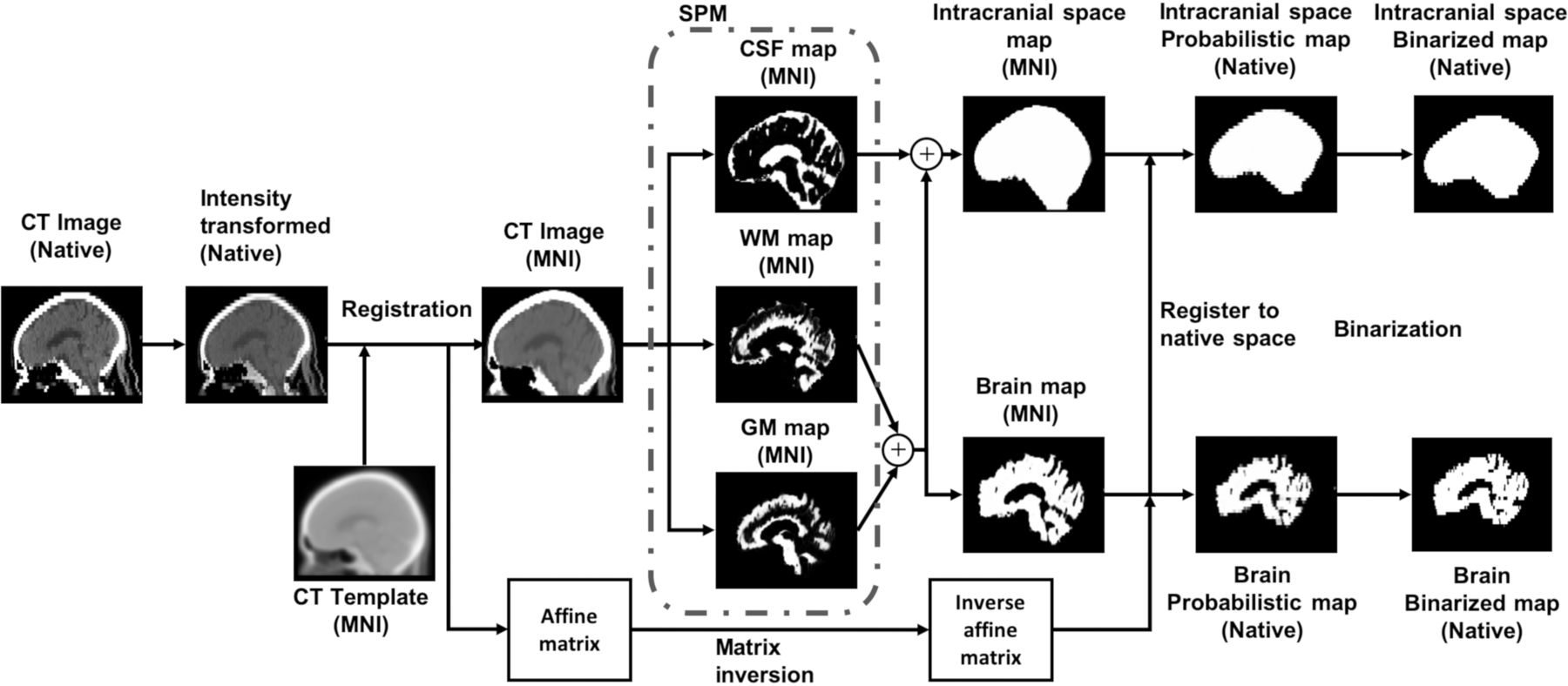
- FIG 1.
CTSeg pipeline for intracranial space and brain parenchyma segmentation from head CT images. Within parentheses is the 3D coordinate space of the image. MNI indicates Montreal Neurological Institute.
- FIG 2.
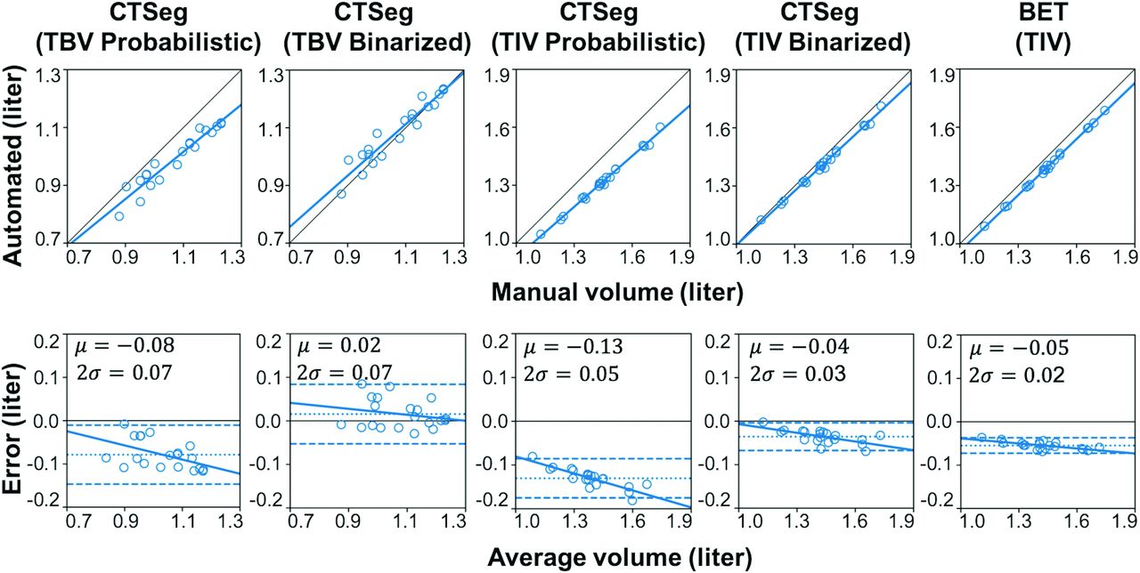
Scatterplots (upper row) of automated-versus-manual volume estimates and the linear fit between automated and manual volumes (thick line) and the line of equality (thin line). Bland-Altman plots (lower row) with automated-minus-manual volume differences on the y-axis and the average of automated and manual volumes on the x-axis. Mean difference ±2 SDs is represented by dotted and dashed horizontal lines, respectively.
- FIG 3.
Scatterplot of %TBV (left) estimated using CTSeg versus age. Scatterplot of TBV versus TIV (right).


Tables
- Table 1:
Comparison of automated TBV and TIV estimates with manual ground truth estimatesa
Parameter/Method %Difference Pearson’s r (P Value) ICC (P Value) Bootstrap Mean ICC (95% CI) TBV CTSeg probabilistic –7.22±2.98 0.96 (<.001) 0.74 (<.001) 0.727 (0.724–0.730) CTSeg binarized 1.58±3.46 0.95 (<.001) 0.94 (<.001) 0.937 (0.935–0.939) TIV CTSeg-probabilistic –12.15±1.44 0.99 (<.001) 0.71 (<.001) 0.685 (0.680–0.689) CTSeg binarized –3.28±1.36 0.99 (<.001) 0.97 (<.001) 0.962 (0.961–0.963) BET –5.12±0.667 0.99 (<.001) 0.94 (<.001) 0.930 (0.928–0.932) ↵a %Difference is reported as means.


