Advertisement

Main menu

  • Home
  • Content
    • Current Issue
    • Accepted Manuscripts
    • Article Preview
    • Past Issue Archive
    • Video Articles
    • AJNR Case Collection
    • Case of the Week Archive
    • Case of the Month Archive
    • Classic Case Archive
  • Special Collections
    • AJNR Awards
    • Low-Field MRI
    • Alzheimer Disease
    • ASNR Foundation Special Collection
    • Photon-Counting CT
    • View All
  • Multimedia
    • AJNR Podcasts
    • AJNR SCANtastic
    • Trainee Corner
    • MRI Safety Corner
    • Imaging Protocols
  • For Authors
    • Submit a Manuscript
    • Submit a Video Article
    • Submit an eLetter to the Editor/Response
    • Manuscript Submission Guidelines
    • Statistical Tips
    • Fast Publishing of Accepted Manuscripts
    • Graphical Abstract Preparation
    • Imaging Protocol Submission
    • Author Policies
  • About Us
    • About AJNR
    • Editorial Board
    • Editorial Board Alumni
  • More
    • Become a Reviewer/Academy of Reviewers
    • Subscribers
    • Permissions
    • Alerts
    • Feedback
    • Advertisers
    • ASNR Home

User menu

  • Alerts
  • Log in

Search

  • Advanced search
American Journal of Neuroradiology

American Journal of Neuroradiology

ASHNR American Society of Functional Neuroradiology ASHNR American Society of Pediatric Neuroradiology ASSR
  • Alerts
  • Log in

Advanced Search

  • Home
  • Content
    • Current Issue
    • Accepted Manuscripts
    • Article Preview
    • Past Issue Archive
    • Video Articles
    • AJNR Case Collection
    • Case of the Week Archive
    • Case of the Month Archive
    • Classic Case Archive
  • Special Collections
    • AJNR Awards
    • Low-Field MRI
    • Alzheimer Disease
    • ASNR Foundation Special Collection
    • Photon-Counting CT
    • View All
  • Multimedia
    • AJNR Podcasts
    • AJNR SCANtastic
    • Trainee Corner
    • MRI Safety Corner
    • Imaging Protocols
  • For Authors
    • Submit a Manuscript
    • Submit a Video Article
    • Submit an eLetter to the Editor/Response
    • Manuscript Submission Guidelines
    • Statistical Tips
    • Fast Publishing of Accepted Manuscripts
    • Graphical Abstract Preparation
    • Imaging Protocol Submission
    • Author Policies
  • About Us
    • About AJNR
    • Editorial Board
    • Editorial Board Alumni
  • More
    • Become a Reviewer/Academy of Reviewers
    • Subscribers
    • Permissions
    • Alerts
    • Feedback
    • Advertisers
    • ASNR Home
  • Follow AJNR on Twitter
  • Visit AJNR on Facebook
  • Follow AJNR on Instagram
  • Join AJNR on LinkedIn
  • RSS Feeds

AJNR Awards, New Junior Editors, and more. Read the latest AJNR updates

Research ArticleAdult Brain

Effects of Susceptibility Artifacts on Perfusion MRI in Patients with Primary Brain Tumor: A Comparison of Arterial Spin-Labeling versus DSC

H. Maral, E. Ertekin, Ö. Tunçyürek and Y. Özsunar
American Journal of Neuroradiology February 2020, 41 (2) 255-261; DOI: https://doi.org/10.3174/ajnr.A6384
H. Maral
aFrom the Department of Radiology (H.M.), Dr. Siyami Ersek Thoracic and Cardiovascular Surgery Training and Research Hospital, Istanbul, Turkey
  • Find this author on Google Scholar
  • Find this author on PubMed
  • Search for this author on this site
  • ORCID record for H. Maral
E. Ertekin
bDepartment of Radiology (E.E., Ö.T., Y.Ö.), Aydın Adnan Menderes University Faculty of Medicine, Aydın, Turkey
  • Find this author on Google Scholar
  • Find this author on PubMed
  • Search for this author on this site
  • ORCID record for E. Ertekin
Ö. Tunçyürek
bDepartment of Radiology (E.E., Ö.T., Y.Ö.), Aydın Adnan Menderes University Faculty of Medicine, Aydın, Turkey
cDepartment of Radiology (Ö.T.), Near East University Faculty of Medicine, Nicosia, Cyprus.
  • Find this author on Google Scholar
  • Find this author on PubMed
  • Search for this author on this site
  • ORCID record for Ö. Tunçyürek
Y. Özsunar
bDepartment of Radiology (E.E., Ö.T., Y.Ö.), Aydın Adnan Menderes University Faculty of Medicine, Aydın, Turkey
  • Find this author on Google Scholar
  • Find this author on PubMed
  • Search for this author on this site
  • ORCID record for Y. Özsunar
  • Article
  • Figures & Data
  • Info & Metrics
  • Responses
  • References
  • PDF
Loading

Article Figures & Data

Figures

  • Tables
  • FIG 1.
    • Download figure
    • Open in new tab
    • Download powerpoint
    FIG 1.

    ROIs were located in the tumor and white matter of the contralateral hemisphere, and the measurements are seen on mpASL (A) and DSC (B) perfusion maps.

  • FIG 2.
    • Download figure
    • Open in new tab
    • Download powerpoint
    FIG 2.

    Anaplastic astrocytoma in a 44-year-old female patient. There is a mass in the T2-weighted (B) sequence showing a signal increase in the right thalamus. Also, there is a slight expansion in both T2-weighted (B) and the postcontrast T1-weighted (A) sequences. There is no significant enhancement in the postcontrast T1-weighted (A) MR image. There are no significant increases in perfusion in the mass localization derived from the DSC (C) perfusion color map (DSC rrCBF = 1.1). In the mpASL perfusion map (D), the perfusion increase in the mass localization can be seen clearly (ASL rrCBF = 3.3) (arrow).
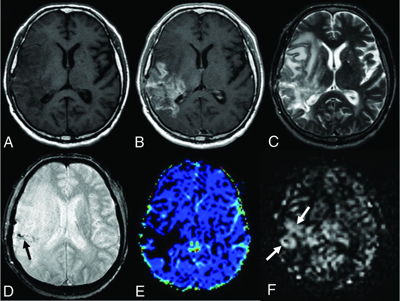
  • FIG 3.
    • Download figure
    • Open in new tab
    • Download powerpoint
    FIG 3.

    A 49-year-old male patient who underwent an operation and received chemoradiotherapy due to a glioblastoma in the temporoparietal region 1 year ago. T1-weighted (A) and T2-weighted (C) MR images show postoperative changes and alterations in the signal intensities in the right temporoparietal region. The postcontrast image (B) shows wide-scale enhancement in the site of the operation. The T2*-weighted image (D) shows hypointensities secondary to old bleeding residues (black arrow). The DSC perfusion map (E) shows a large perfusion signal loss caused by leakage artifacts in the contrasted areas (DSC rrCBF = 1.3). In the mpASL perfusion map (F), there is a significant increase in perfusion at the site of the operation (white arrows) (ASL rrCBF = 3.5). The patient was diagnosed with a recurrent glioblastoma after the second operation.

  • FIG 4.
    • Download figure
    • Open in new tab
    • Download powerpoint
    FIG 4.

    The ROC analysis curve. The blue line shows mpASL perfusion, and the green and the brown lines represent the DSC perfusion MR imaging technique for rrCBF and rrCBV, respectively.
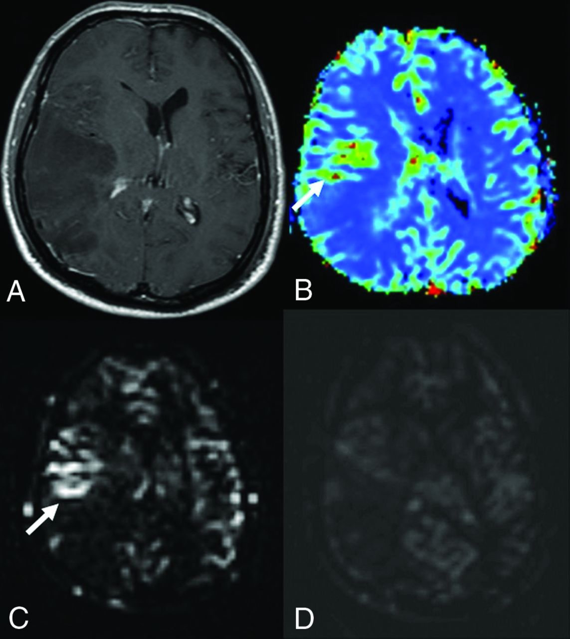
  • FIG 5.
    • Download figure
    • Open in new tab
    • Download powerpoint
    FIG 5.

    Anaplastic astrocytoma in a 41-year-old male patient. Postcontrast T1-weighted (A) image shows a wide nonenhancing mass lesion in the right temporoparietal region. There was no diffusion restriction (not shown here). The DSC (B) and mpASL (C) maps show significantly hyperperfused areas within the mass (arrows) (DSC rrCBF = 2.3, ASL rrCBF = 4.6). The spASL (D) map shows no increases in perfusion. It is also remarkable that the signal-to-noise ratio of spASL is lower than that of mpASL.

Tables

  • Figures
    • View popup
    Table 1:

    Perfusion MR imaging parameters

    Sequence ParametersspASLmpASLDSC
    Acquired orientationAxialAxialAxial
    TR (ms)40002501800
    TE (ms)252040
    Flip angle70°35°75°
    FOV (mm2)240240224
    Matrix (pixel)68 × 6868 × 6888 × 88
    Thickness/gap (mm)6/0.66/0.65/0
    Pulse delay times (ms)1200300, 550, 800, 1050, 1300, 1550, 1800, 2050–
    Label thickness/gap (mm)100/20130/20–
    Acquisition time4 min 8 sec4 min 8 sec1 min 40 sec
    • Note:—–indicates “no labelling” and “no pulse delay time”; DSC, dynamic susceptibility contrast; FOV, field-of-view; mpASL, multi phase arterial spin-labeling; spASL, single phase arterial spin-labeling; TE, echo time; TR, repetition time.

    • View popup
    Table 2:

    Histopathologic and follow-up diagnosis

    No.Percentage
    Histopathologic diagnosis3844.2
    Benign lesions1315.1
     Diffuse astrocytoma78.1
     WHO grade II oligodendroglioma22.3
     DNET22.3
     Cerebellar astrocytoma11.2
     Ganglioglioma11.2
    Malignant lesions2529.1
     Glioblastoma1416.3
     Anaplastic astrocytoma78.1
     Anaplastic oligondendroglioma22.3
     Gliosarcoma22.3
    Follow-up diagnosis4855.8
     Benign lesions3237.2
     Malignant lesions1618.6
    Total86100
    • Note:—DNET indicate dysembryoplastic neuroepithelial tumors.

    • View popup
    Table 3:

    Perfusion parameters of benign and malignant tumors on the quantitative evaluation

    Benign Tumors Mean (± SD)Malignant Tumors Mean (± SD)PaSensitivity (%)Specificity (%)Accuracy (%)
    All lesions (n = 86)b
     mpASL rrCBF1.19 (0.59)2.84 (1.44)<.00185.484.484.9
     DSC rrCBF1.19 (0.69)2.93 (1.57)<.00182.980.081.45
     DSC rrCBV1.20 (0.62)3.07 (1.43)<.00168.397.883.05
    Group 1 (n = 38)c
     mpASL rrCBF1.24 (0.47)3.06 (1.45)<.00194.480.087.2
     DSC rrCBF1.33 (0.83)3.13 (1.80)<.00188.975.081.95
     DSC rrCBV1.32 (0.67)3.22 (1.43)<.00166.710083.35
    Group 2 (n = 48)d
     mpASL rrCBF1.16 (0.68)2.67 (1.45)<.00178.388.083.15
     DSC rrCBF1.08 (0.54)2.77 (1.39)<.00178.384.081.15
     DSC rrCBV1.10 (0.58)2.96 (1.45)<.00169.696.082.8
    • ↵a Mann-Whitney U test.

    • ↵b Benign (n = 45), malignant (n = 41).

    • ↵c Benign (n = 20), malignant (n = 18).

    • ↵d Benign (n = 25), malignant (n = 23).

PreviousNext
Back to top

In this issue

American Journal of Neuroradiology: 41 (2)
American Journal of Neuroradiology
Vol. 41, Issue 2
1 Feb 2020
  • Table of Contents
  • Index by author
  • Complete Issue (PDF)
Advertisement
Print
Download PDF
Email Article

Thank you for your interest in spreading the word on American Journal of Neuroradiology.

NOTE: We only request your email address so that the person you are recommending the page to knows that you wanted them to see it, and that it is not junk mail. We do not capture any email address.

Enter multiple addresses on separate lines or separate them with commas.
Effects of Susceptibility Artifacts on Perfusion MRI in Patients with Primary Brain Tumor: A Comparison of Arterial Spin-Labeling versus DSC
(Your Name) has sent you a message from American Journal of Neuroradiology
(Your Name) thought you would like to see the American Journal of Neuroradiology web site.
CAPTCHA
This question is for testing whether or not you are a human visitor and to prevent automated spam submissions.
Cite this article
H. Maral, E. Ertekin, Ö. Tunçyürek, Y. Özsunar
Effects of Susceptibility Artifacts on Perfusion MRI in Patients with Primary Brain Tumor: A Comparison of Arterial Spin-Labeling versus DSC
American Journal of Neuroradiology Feb 2020, 41 (2) 255-261; DOI: 10.3174/ajnr.A6384

Citation Manager Formats

  • BibTeX
  • Bookends
  • EasyBib
  • EndNote (tagged)
  • EndNote 8 (xml)
  • Medlars
  • Mendeley
  • Papers
  • RefWorks Tagged
  • Ref Manager
  • RIS
  • Zotero
0 Responses
Respond to this article
Share
Bookmark this article
Effects of Susceptibility Artifacts on Perfusion MRI in Patients with Primary Brain Tumor: A Comparison of Arterial Spin-Labeling versus DSC
H. Maral, E. Ertekin, Ö. Tunçyürek, Y. Özsunar
American Journal of Neuroradiology Feb 2020, 41 (2) 255-261; DOI: 10.3174/ajnr.A6384
del.icio.us logo Twitter logo Facebook logo Mendeley logo
  • Tweet Widget
  • Facebook Like
  • Google Plus One
Purchase

Jump to section

  • Article
    • Abstract
    • ABBREVIATIONS:
    • MATERIALS AND METHODS
    • RESULTS
    • DISCUSSION
    • CONCLUSIONS
    • References
  • Figures & Data
  • Info & Metrics
  • Responses
  • References
  • PDF

Related Articles

  • No related articles found.
  • PubMed
  • Google Scholar

Cited By...

  • No citing articles found.
  • Crossref
  • Google Scholar

This article has not yet been cited by articles in journals that are participating in Crossref Cited-by Linking.

More in this TOC Section

  • Diagnostic Neuroradiology of Monoclonal Antibodies
  • Clinical Outcomes After Chiari I Decompression
  • Segmentation of Brain Metastases with BLAST
Show more Adult Brain

Similar Articles

Advertisement

Indexed Content

  • Current Issue
  • Accepted Manuscripts
  • Article Preview
  • Past Issues
  • Editorials
  • Editor's Choice
  • Fellows' Journal Club
  • Letters to the Editor
  • Video Articles

Cases

  • Case Collection
  • Archive - Case of the Week
  • Archive - Case of the Month
  • Archive - Classic Case

More from AJNR

  • Trainee Corner
  • Imaging Protocols
  • MRI Safety Corner
  • Book Reviews

Multimedia

  • AJNR Podcasts
  • AJNR Scantastics

Resources

  • Turnaround Time
  • Submit a Manuscript
  • Submit a Video Article
  • Submit an eLetter to the Editor/Response
  • Manuscript Submission Guidelines
  • Statistical Tips
  • Fast Publishing of Accepted Manuscripts
  • Graphical Abstract Preparation
  • Imaging Protocol Submission
  • Evidence-Based Medicine Level Guide
  • Publishing Checklists
  • Author Policies
  • Become a Reviewer/Academy of Reviewers
  • News and Updates

About Us

  • About AJNR
  • Editorial Board
  • Editorial Board Alumni
  • Alerts
  • Permissions
  • Not an AJNR Subscriber? Join Now
  • Advertise with Us
  • Librarian Resources
  • Feedback
  • Terms and Conditions
  • AJNR Editorial Board Alumni

American Society of Neuroradiology

  • Not an ASNR Member? Join Now

© 2025 by the American Society of Neuroradiology All rights, including for text and data mining, AI training, and similar technologies, are reserved.
Print ISSN: 0195-6108 Online ISSN: 1936-959X

Powered by HighWire