Advertisement

Main menu

  • Home
  • Content
    • Current Issue
    • Accepted Manuscripts
    • Article Preview
    • Past Issue Archive
    • Video Articles
    • AJNR Case Collection
    • Case of the Week Archive
    • Case of the Month Archive
    • Classic Case Archive
  • Special Collections
    • AJNR Awards
    • Low-Field MRI
    • Alzheimer Disease
    • ASNR Foundation Special Collection
    • Photon-Counting CT
    • View All
  • Multimedia
    • AJNR Podcasts
    • AJNR SCANtastic
    • Trainee Corner
    • MRI Safety Corner
    • Imaging Protocols
  • For Authors
    • Submit a Manuscript
    • Submit a Video Article
    • Submit an eLetter to the Editor/Response
    • Manuscript Submission Guidelines
    • Statistical Tips
    • Fast Publishing of Accepted Manuscripts
    • Graphical Abstract Preparation
    • Imaging Protocol Submission
    • Author Policies
  • About Us
    • About AJNR
    • Editorial Board
    • Editorial Board Alumni
  • More
    • Become a Reviewer/Academy of Reviewers
    • Subscribers
    • Permissions
    • Alerts
    • Feedback
    • Advertisers
    • ASNR Home

User menu

  • Alerts
  • Log in

Search

  • Advanced search
American Journal of Neuroradiology

American Journal of Neuroradiology

ASHNR American Society of Functional Neuroradiology ASHNR American Society of Pediatric Neuroradiology ASSR
  • Alerts
  • Log in

Advanced Search

  • Home
  • Content
    • Current Issue
    • Accepted Manuscripts
    • Article Preview
    • Past Issue Archive
    • Video Articles
    • AJNR Case Collection
    • Case of the Week Archive
    • Case of the Month Archive
    • Classic Case Archive
  • Special Collections
    • AJNR Awards
    • Low-Field MRI
    • Alzheimer Disease
    • ASNR Foundation Special Collection
    • Photon-Counting CT
    • View All
  • Multimedia
    • AJNR Podcasts
    • AJNR SCANtastic
    • Trainee Corner
    • MRI Safety Corner
    • Imaging Protocols
  • For Authors
    • Submit a Manuscript
    • Submit a Video Article
    • Submit an eLetter to the Editor/Response
    • Manuscript Submission Guidelines
    • Statistical Tips
    • Fast Publishing of Accepted Manuscripts
    • Graphical Abstract Preparation
    • Imaging Protocol Submission
    • Author Policies
  • About Us
    • About AJNR
    • Editorial Board
    • Editorial Board Alumni
  • More
    • Become a Reviewer/Academy of Reviewers
    • Subscribers
    • Permissions
    • Alerts
    • Feedback
    • Advertisers
    • ASNR Home
  • Follow AJNR on Twitter
  • Visit AJNR on Facebook
  • Follow AJNR on Instagram
  • Join AJNR on LinkedIn
  • RSS Feeds

AJNR Awards, New Junior Editors, and more. Read the latest AJNR updates

Research ArticlePediatric Neuroimaging

Thalamic Massa Intermedia in Children with and without Midline Brain Malformations

M.T. Whitehead and N. Najim
American Journal of Neuroradiology April 2020, 41 (4) 729-735; DOI: https://doi.org/10.3174/ajnr.A6446
M.T. Whitehead
aFrom the Department of Radiology (M.T.W., N.N.), Children’s National Hospital, Washington, DC
bThe George Washington University Hospital (M.T.W.), Washington, DC.
  • Find this author on Google Scholar
  • Find this author on PubMed
  • Search for this author on this site
  • ORCID record for M.T. Whitehead
N. Najim
aFrom the Department of Radiology (M.T.W., N.N.), Children’s National Hospital, Washington, DC
  • Find this author on Google Scholar
  • Find this author on PubMed
  • Search for this author on this site
  • ORCID record for N. Najim
  • Article
  • Figures & Data
  • Supplemental
  • Info & Metrics
  • Responses
  • References
  • PDF
Loading

Article Figures & Data

Figures

  • Tables
  • Fig 1.
    • Download figure
    • Open in new tab
    • Download powerpoint
    Fig 1.

    Sagittal midline T1WI from a brain MR imaging with normal findings in a 17-year-old adolescent girl with retroauricular pain depicting the normal thalamic MI centered in the anterior/superior portion of the third ventricle (arrow).

  • Fig 2.
    • Download figure
    • Open in new tab
    • Download powerpoint
    Fig 2.

    Sagittal midline T1WI from a brain MR imaging with normal findings in an 11-year-old girl with headache, showing a normal MI with part of its superior and posterior margin volume averaged with the medial thalami in a patient with a small third ventricle (large arrow). This appearance has the potential to compromise MI evaluation both qualitatively and quantitatively. However, a faint marginal distinction is often seen when carefully analyzed (small arrow).
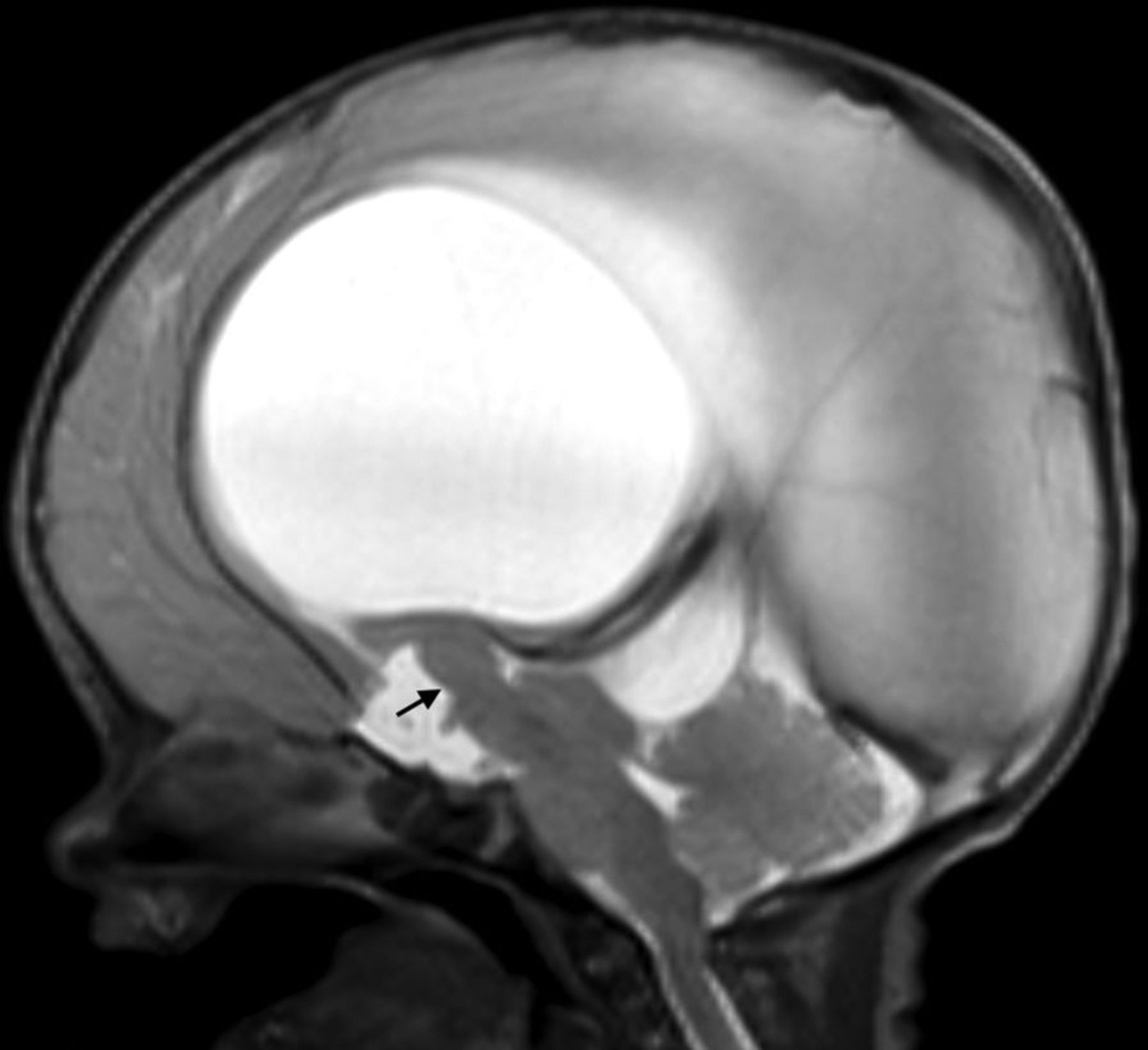
  • Fig 3.
    • Download figure
    • Open in new tab
    • Download powerpoint
    Fig 3.

    Sagittal midline T1WI demonstrating MI absence in association with multiple additional midline abnormalities, including marked enlargement of the fourth ventricle/posterior fossa and under-rotation of a hypoplastic/dysplastic vermis (Dandy-Walker malformation), hypoplasia of the anterior commissure (arrow), pontine hypoplasia, and agenesis of the corpus callosum.

  • Fig 4.
    • Download figure
    • Open in new tab
    • Download powerpoint
    Fig 4.

    Sagittal midline T1WI showing MI absence in association with multiple additional midline abnormalities in a patient with septopreoptic holoprosencephaly. Additional findings include agenesis of the corpus callosum, interhypothalamic adhesion (small straight arrow), tectal dysplasia (small curved arrow), and hypoplasia of the vermis (long straight arrow).

  • Fig 5.
    • Download figure
    • Open in new tab
    • Download powerpoint
    Fig 5.

    Sagittal midline T2WI from a patient with diencephalic-mesencephalic junction dysplasia showing a thickened thalamic massa intermedia connected to the midbrain (arrow). The midbrain is dysplastic with associated aqueductal stenosis and consequent hydrocephalus.

  • Fig 6.
    • Download figure
    • Open in new tab
    • Download powerpoint
    Fig 6.

    Sagittal midline T1WI depicting MI thinning/hypoplasia in a patient with Aicardi syndrome (arrow). Other findings include callosal dysgenesis and a pericallosal arachnoid cyst (star).

Tables

  • Figures
  • Demographic and structural variables in healthy and midline abnormality groups

    Healthy Group (Avg ± SD)Midline Group (Avg ± SD)Total (Avg ± SD)P
    Age (yr)11.7 ± 5.34.1 ± 5.57.9 ± 6.6.001
    Sexn = 55 f (52%)n = 52 f (50%)n = 107 f (51%).78
    MI presentn = 92 (88%)n = 62 (60%)n = 154 (74%).001
     Morphologyn = 89 (n); 3(T)n = 45 (n); 10(T); 7(t)n = 134 (n); 13(T); 7(t).001
     tr (mm)1.5 ± 0.22 ± 0.21.7 ± 1.002
     ap (mm)7.3 ± 2.36.8 ± 3.97 ± 3.1.144
     cc (mm)6.7 ± 5.66.1 ± 3.56.4 ± 4.9.202
     Area (mm2)42.9 ± 23.944.2 ± 48.943.2 ± 35.9.458
     Locationn = 84 (a/s); 6 (p/s); 2 (p/i)n = 34 (a/s); 14 (p/s); 3 (p/i); 1 (a/i); 10 (all)n = 118 (a/s); 20 (p/s); 5 (p/i); 1 (a/i); 10 (all).001
    CC presentn = 105 (100%)n = 70 (68%)n = 175 (84%).001
     Morphologyn = 105 (n)n = 13 (n); 6 (h); 46 (h/d); 5 (d)n = 118 (n); 6 (h); 46 (h/); (5d).001
     Length (mm)68.2 ± 7.639.9 ± 21.557 ± 20.2.001
     Genu (mm)10.5 ± 27.4 ± 109.3 ± 6.6.007
     Body (mm)5.7 ± 1.23.5 ± 2.24.9 ± 1.9.001
     Splen (mm)10.3 ± 24.7 ± 2.98.4 ± 3.6.001
     Area (mm2)546 ± 129214 ± 199415 ± 228.001
    AC presentn = 105 (100%)n = 101 (98%)n = 206 (99%).151
     Morphologyn = 105 (n)n = 41 (n); 53 (h); 7 (T)n = 146 (n); 53 (h); 7 (T).001
     tr (mm)1.8 ± 0.43.9 ± 1.52.8 ± 1.5.001
     ap (mm)2.6 ± 0.71.7 ± 1.12.2 ± 1.001
     cc (mm)3.5 ± 12.2 ± 1.32.9 ± 1.3.001
     Area (mm2)8.1 ± 3.84.4 ± 5.16.3 ± 4.9.001
    Septum pellucidumn = 96 (n); 9 (c)n = 13 (n); 7 (c); 47 (a); 36 (h)n = 109 (n); 16 (c); 47 (a); 36 (h).001
    Fornixn = 105 (n)n = 36 (n); 25 (a); 42 (h)n = 141 (n); 25 (a); 42 (h).001
    Hypothalamusn = 105 (n)n = 84 (n); 19 (IHA)n = 189 (n); 19 (IHA).001
    Hippocampusn = 87 (n); 18 (u)n = 11 (n); 88 (u); 2 (d); 2 (mts)n = 98 (n); 106 (u); 2 (d); 2 (mts).001
    Brain stemn = 105 (n)n = 67 (n); 18 (h); 18 (d)n = 172 (n); 18 (h); 18 (d).001
    Vermisn = 105 (n)n = 77 (n); 15 (h); 11 (d)n = 182 (n); 15 (h); 11 (d).001
    3rd Ventricle (mm)2.8 ± 0.95.5 ± 3.34.1 ± 2.8.001
    • Note:—Avg indicates average; tr, transverse diameter; ap, anteroposterior diameter; cc, craniocaudal diameter; CC, corpus callosum; Splen, splenium; AC, anterior commissure; n, normal; T, thick; t, thin; a/s, anterior/superior quadrant of 3rd ventricle; a/i, anterior-inferior; p/s, posterior-superior; p/i, posterior-inferior; all, all quadrants; h, hypoplastic; h/d, hypogenetic ± dysgenetic; d, dysgenetic/dysplastic; c, cavum; a, absent; IHA, interhypothalamic adhesion; u, under-rotated; mts, hippocampal sclerosis; f, female.

PreviousNext
Back to top

In this issue

American Journal of Neuroradiology: 41 (4)
American Journal of Neuroradiology
Vol. 41, Issue 4
1 Apr 2020
  • Table of Contents
  • Index by author
  • Complete Issue (PDF)
Advertisement
Print
Download PDF
Email Article

Thank you for your interest in spreading the word on American Journal of Neuroradiology.

NOTE: We only request your email address so that the person you are recommending the page to knows that you wanted them to see it, and that it is not junk mail. We do not capture any email address.

Enter multiple addresses on separate lines or separate them with commas.
Thalamic Massa Intermedia in Children with and without Midline Brain Malformations
(Your Name) has sent you a message from American Journal of Neuroradiology
(Your Name) thought you would like to see the American Journal of Neuroradiology web site.
CAPTCHA
This question is for testing whether or not you are a human visitor and to prevent automated spam submissions.
Cite this article
M.T. Whitehead, N. Najim
Thalamic Massa Intermedia in Children with and without Midline Brain Malformations
American Journal of Neuroradiology Apr 2020, 41 (4) 729-735; DOI: 10.3174/ajnr.A6446

Citation Manager Formats

  • BibTeX
  • Bookends
  • EasyBib
  • EndNote (tagged)
  • EndNote 8 (xml)
  • Medlars
  • Mendeley
  • Papers
  • RefWorks Tagged
  • Ref Manager
  • RIS
  • Zotero
0 Responses
Respond to this article
Share
Bookmark this article
Thalamic Massa Intermedia in Children with and without Midline Brain Malformations
M.T. Whitehead, N. Najim
American Journal of Neuroradiology Apr 2020, 41 (4) 729-735; DOI: 10.3174/ajnr.A6446
del.icio.us logo Twitter logo Facebook logo Mendeley logo
  • Tweet Widget
  • Facebook Like
  • Google Plus One
Purchase

Jump to section

  • Article
    • Abstract
    • ABBREVIATION:
    • MATERIALS AND METHODS
    • RESULTS
    • DISCUSSION
    • CONCLUSIONS
    • References
  • Figures & Data
  • Supplemental
  • Info & Metrics
  • Responses
  • References
  • PDF

Related Articles

  • PubMed
  • Google Scholar

Cited By...

  • Characterizing massa intermedia morphology in schizophrenia: associations with aging, neuropsychological functioning, and atypical hippocampal development
  • Sotos Syndrome: Deep Neuroimaging Phenotyping Reveals a High Prevalence of Malformations of Cortical Development
  • A large cohort study (n = 591) on the impact of the presence or absence of the interthalamic adhesion: cognitive, neuroimaging, and genetic results
  • Third Ventricle Diameter Is Inversely Related to Thalamic Massa Intermedia Thickness in Hydrocephalus Caused by Congenital Aqueductal Stenosis
  • Crossref
  • Google Scholar

This article has not yet been cited by articles in journals that are participating in Crossref Cited-by Linking.

More in this TOC Section

  • FRACTURE MR in Congenital Vertebral Anomalies
  • Comparing MRI Perfusion in Pediatric Brain Tumors
  • Sodium MRI in Pediatric Brain Tumors
Show more Pediatric Neuroimaging

Similar Articles

Advertisement

Indexed Content

  • Current Issue
  • Accepted Manuscripts
  • Article Preview
  • Past Issues
  • Editorials
  • Editor's Choice
  • Fellows' Journal Club
  • Letters to the Editor
  • Video Articles

Cases

  • Case Collection
  • Archive - Case of the Week
  • Archive - Case of the Month
  • Archive - Classic Case

More from AJNR

  • Trainee Corner
  • Imaging Protocols
  • MRI Safety Corner
  • Book Reviews

Multimedia

  • AJNR Podcasts
  • AJNR Scantastics

Resources

  • Turnaround Time
  • Submit a Manuscript
  • Submit a Video Article
  • Submit an eLetter to the Editor/Response
  • Manuscript Submission Guidelines
  • Statistical Tips
  • Fast Publishing of Accepted Manuscripts
  • Graphical Abstract Preparation
  • Imaging Protocol Submission
  • Evidence-Based Medicine Level Guide
  • Publishing Checklists
  • Author Policies
  • Become a Reviewer/Academy of Reviewers
  • News and Updates

About Us

  • About AJNR
  • Editorial Board
  • Editorial Board Alumni
  • Alerts
  • Permissions
  • Not an AJNR Subscriber? Join Now
  • Advertise with Us
  • Librarian Resources
  • Feedback
  • Terms and Conditions
  • AJNR Editorial Board Alumni

American Society of Neuroradiology

  • Not an ASNR Member? Join Now

© 2025 by the American Society of Neuroradiology All rights, including for text and data mining, AI training, and similar technologies, are reserved.
Print ISSN: 0195-6108 Online ISSN: 1936-959X

Powered by HighWire