
Article Figures & Data
Figures
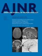
- Fig 1.
Brain MR imaging of patient 15. Brain stem images on T2WI (A), FLAIR (B), and DIR (C). There is a fluffy structure on T2WI (A, black arrows) and high intensity of the hypoplastic pontine surface on the FLAIR image (B, white arrows). High signal intensity along the brain stem surface on the DIR image is recognized more clearly than on the T2WI or FLAIR image (C, white arrows). T2WIs of the left temporal lobe (D–F) show lissencephalic and the abnormally located cortical superficial vessels running through the superficial layer of the lissencephalic cortex (white arrows). Microhemorrhages were seen in the cerebellar cyst on T2WI (G) and SWI (H). More microhemorrhages are recognizable on the SWI than on T2WI (white arrowheads). Cerebral SWI (I) shows hypoplasia of a superficial cerebral vein in both frontal lobes (white arrows) and dilated and tortuous subependymal veins (white circle).
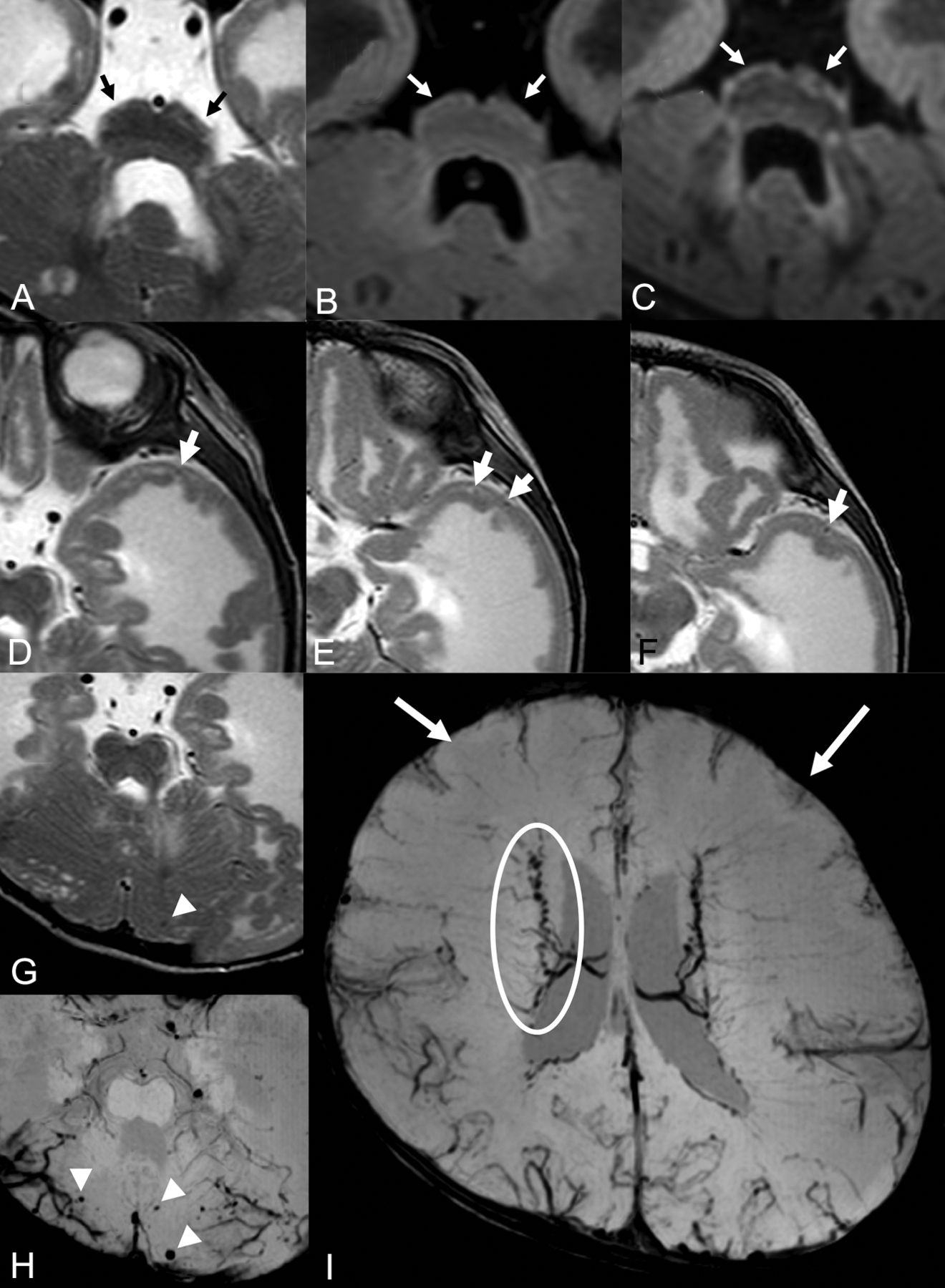
- Fig 2.
Brain stem (A and B) and left temporal (C–E) MRIs of patient 20. A brain stem T2WI (A) shows a fluffy structure and high intensity of the hypoplastic pontine surface (black arrows). The FLAIR image (B) detects high intensity of the pontine surface (white arrows). Left temporal T2WIs (C–E) show lissencephaly, which contains abnormally located superficial cortical vessels running through and beneath the cortex (white arrows).
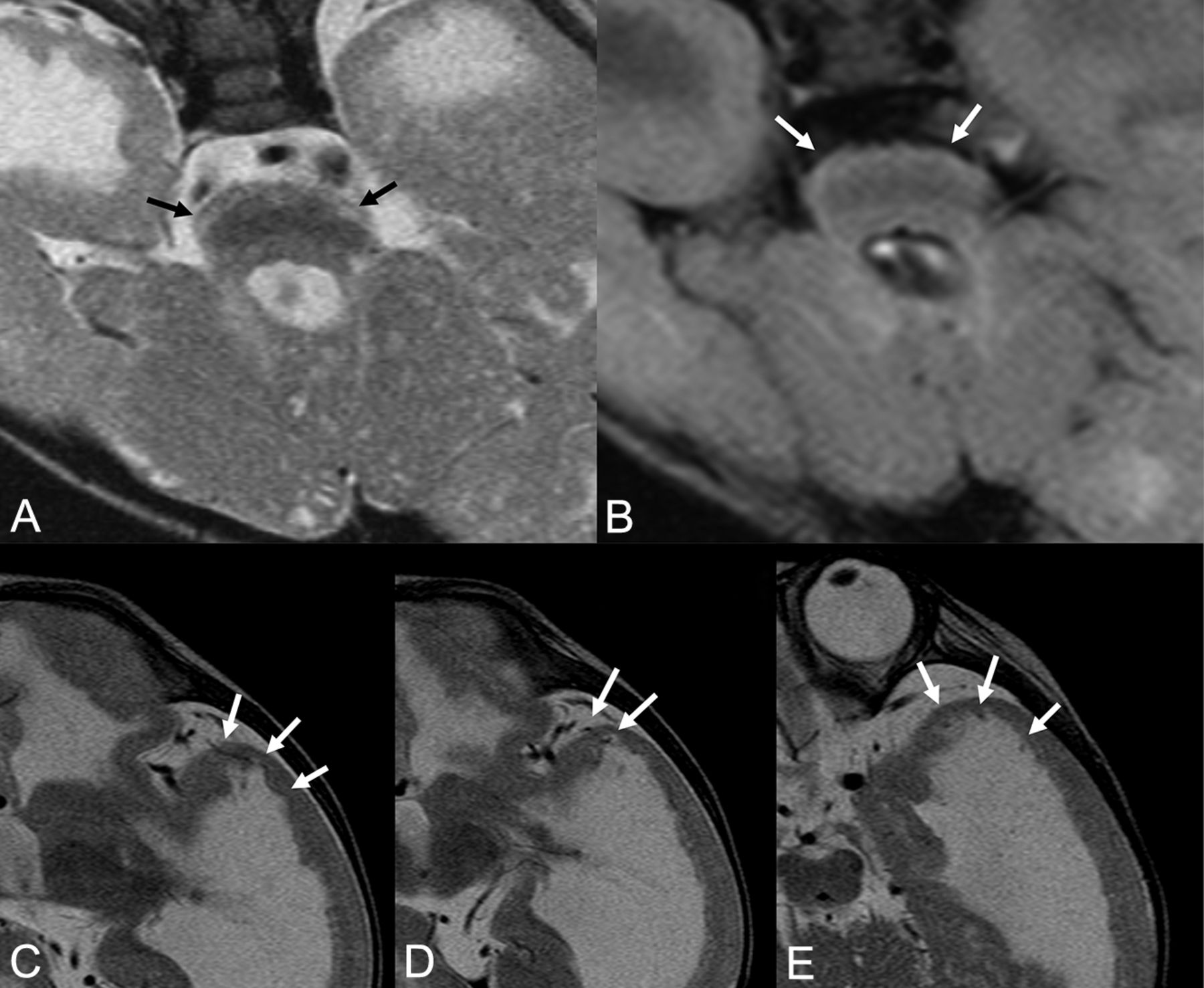
- Fig 3.
Cerebral MRIs of patient 13. T2WIs (A and B) show polymicrogyria and diffuse high signal in the white matter. SWIs (C and D) reveal hypoplasia of superficial cerebral veins in the frontal lobe (white arrows) and meandering subependymal veins (white circles).
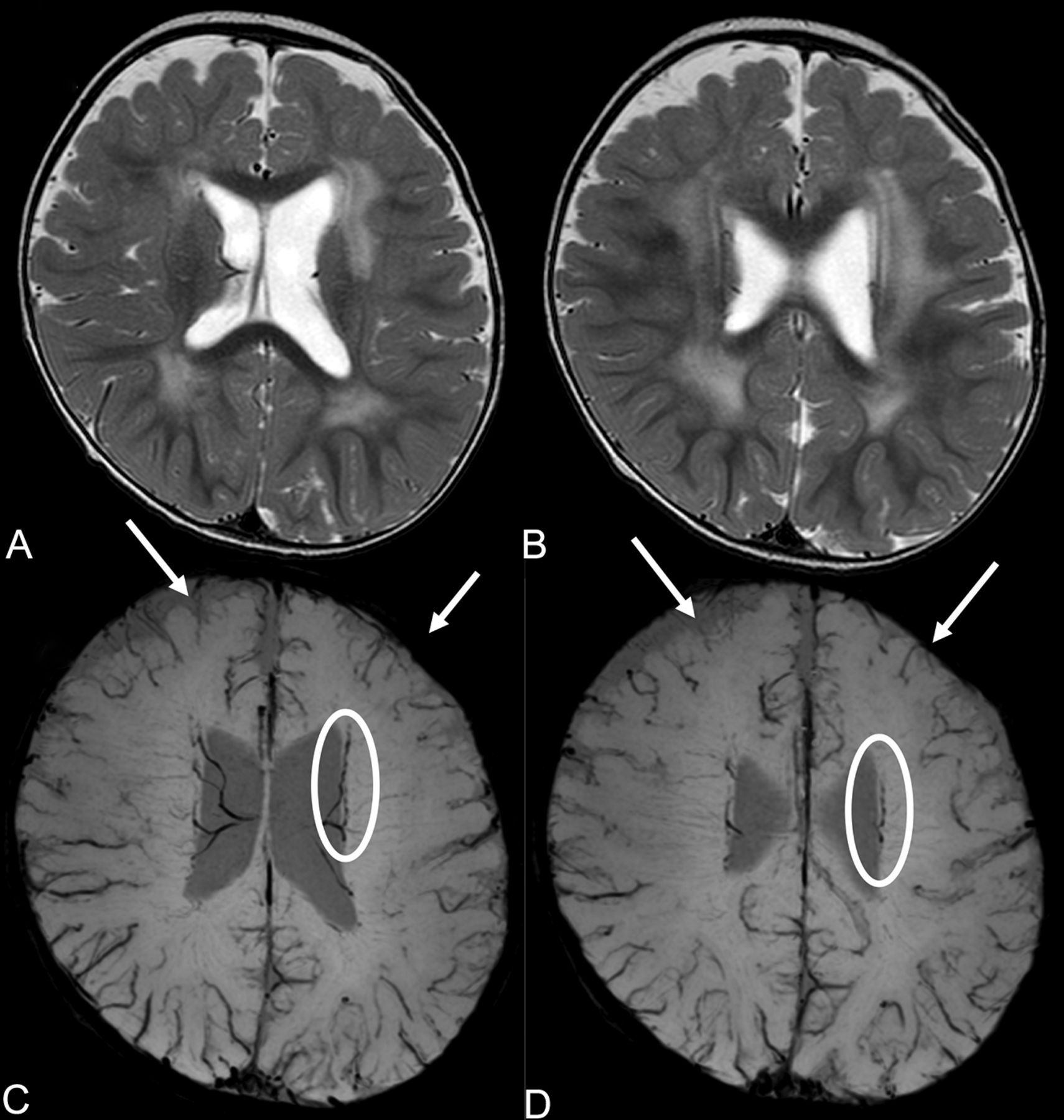
- Fig 4.
Cerebral MRIs of patient 25. There are hemorrhages in the deep and subcortical white matter of the atrophic left frontal lobe (white arrows) on T2WIs. The location of the hemorrhage in this patient is different from that of the subependymal hemorrhage in a premature neonate.


Jump to section
Related Articles
Cited By...
- No citing articles found.