
Article Figures & Data
Figures
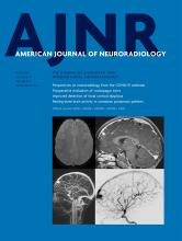
- Fig 1.
The difference between conventional T1-weighted sagittal imaging with a head coil (A) and MC-MR imaging T1-weighted sagittal imaging (B). In this patient, a lens prosthesis (white arrow) inserted during cataract surgery is demonstrated.
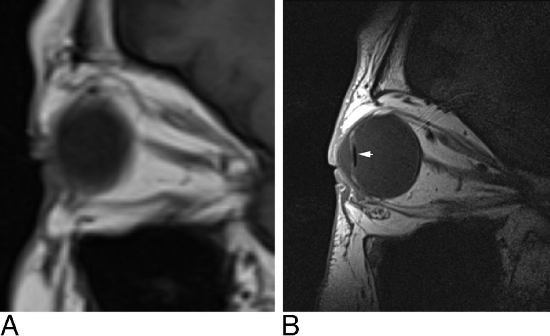
- Fig 2.
A, MC-MR imaging T1-weighted axial image demonstrates a sphenoid wing meningioma, engulfing the optic nerve as well as the medial (MR) and lateral rectus (LR) muscles. MC-MR imaging demonstrates widening of the superior orbital fissure (double-headed arrow), but beyond the orbital apex, the signal-to-noise ratio is low. B, Head coil T1-weighted fat-saturation postcontrast axial image demonstrates the lesion surrounding the left internal carotid artery (white arrow), infiltrating the left cavernous sinus and extending past the midline (dotted white line) in the intercavernous sinus.
- Fig 3.
Correct patient and coil positioning. Photographs of the author (N.W.D.) showing final patient and coil positioning in the imaging magnet with headphones wedged against head coil for immobilization (A) and coil positioning over the orbit with extensive taping (B).
- Fig 4.
Persistence pays off with movement artifacts. A, T2-weighted axial image of the orbit with image degradation due to movement artifacts. B, The same patient and same imaging protocol, after ensuring patient comfort.
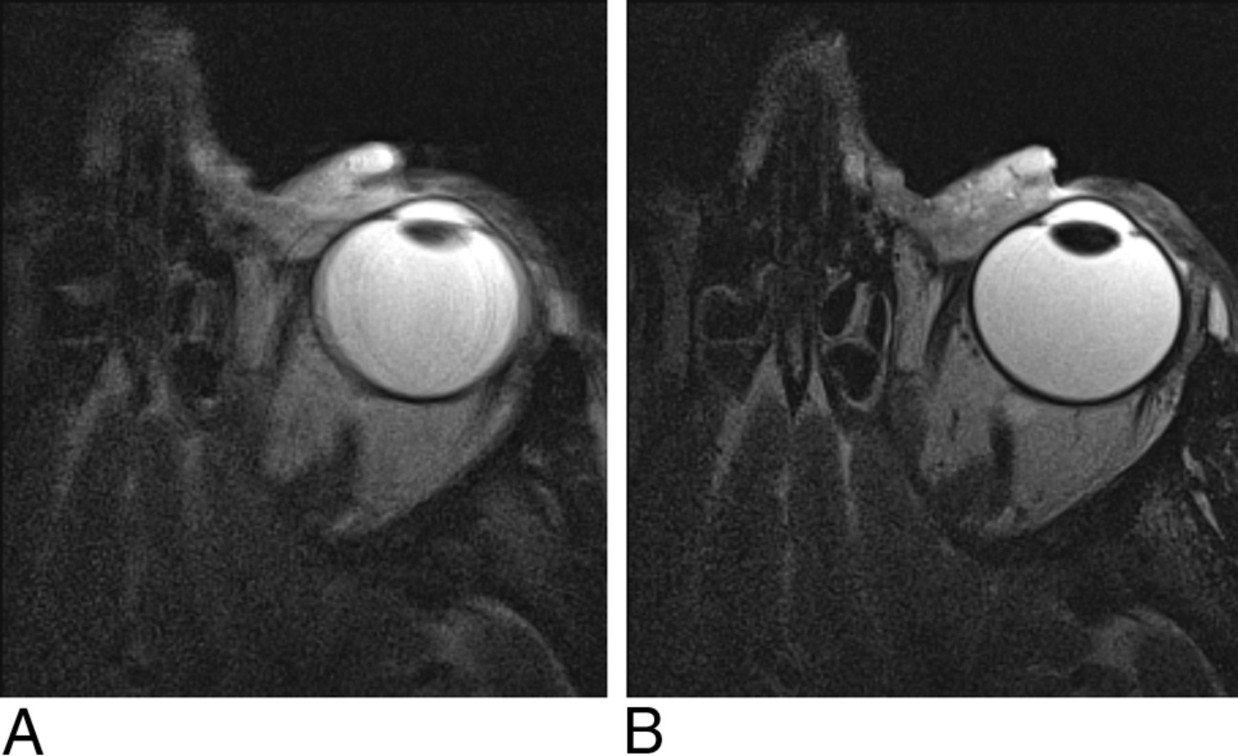
- Fig 5.
MC-MR imaging T2-weighted sagittal image degraded by metallic artifacts from mascara.
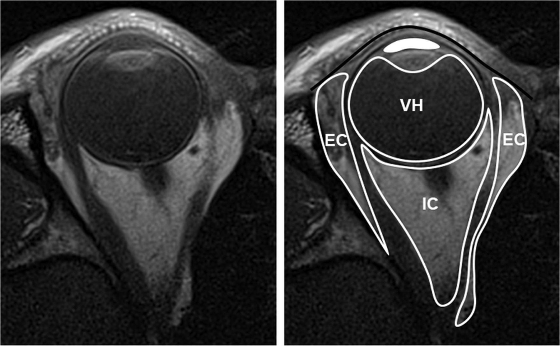
- Fig 6.
Compartmental anatomy shown by MC-MR imaging T1-weighted axial image, original on the left and annotated on the right. The solid black line indicates the orbital septum, defined by native high signal of the tarsal plate; EC, extraconal space, external to the extraocular muscles; IC, intraconal space, inside the extraocular muscles; VH, vitreous humor, behind the lens; solid white fill, aqueous humor, anterior to the lens and ciliary muscles.
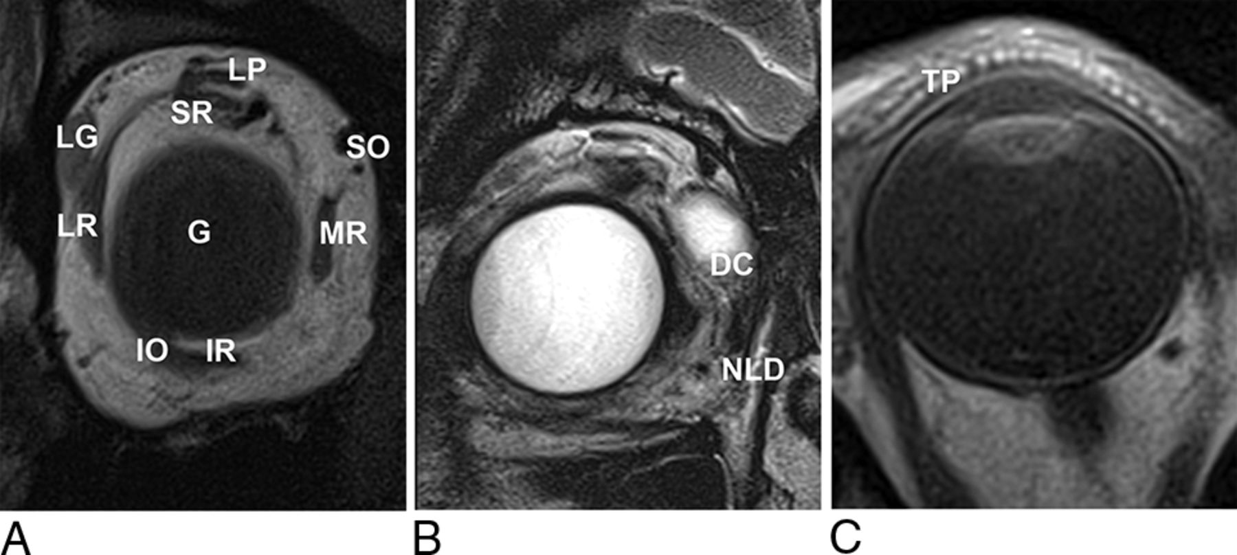
- Fig 7.
A, T1-weighted coronal MC-MR image. LG indicates the lacrimal gland, positioned superolaterally in the orbit, lying directly inferior to the orbital rim; LP, levator palpebrae superioris muscle; SR, superior rectus muscle; SO, superior oblique tendon; MR, medial rectus muscle; IR, inferior rectus muscle; IO, inferior oblique muscle; LR, lateral rectus muscle; G, globe. B, T2-weighted coronal MC-MR image. In this patient, a protruding dermoid cyst (DC) caused epiphora, filling the nasolacrimal duct (NLD) and allowing its demonstration with high signal on T2-weighted imaging. C, T1-weighted axial MC-MR image illustrates the high-signal dots of the meibomian glands within the tarsal plate (TP).
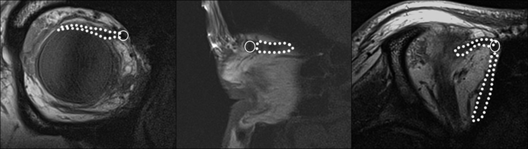
- Fig 8.
T1-weighted MC-MR images in the coronal, sagittal, and axial planes demonstrating the course of the superior oblique muscle and tendon (dotted line) through the trochlea (circle).
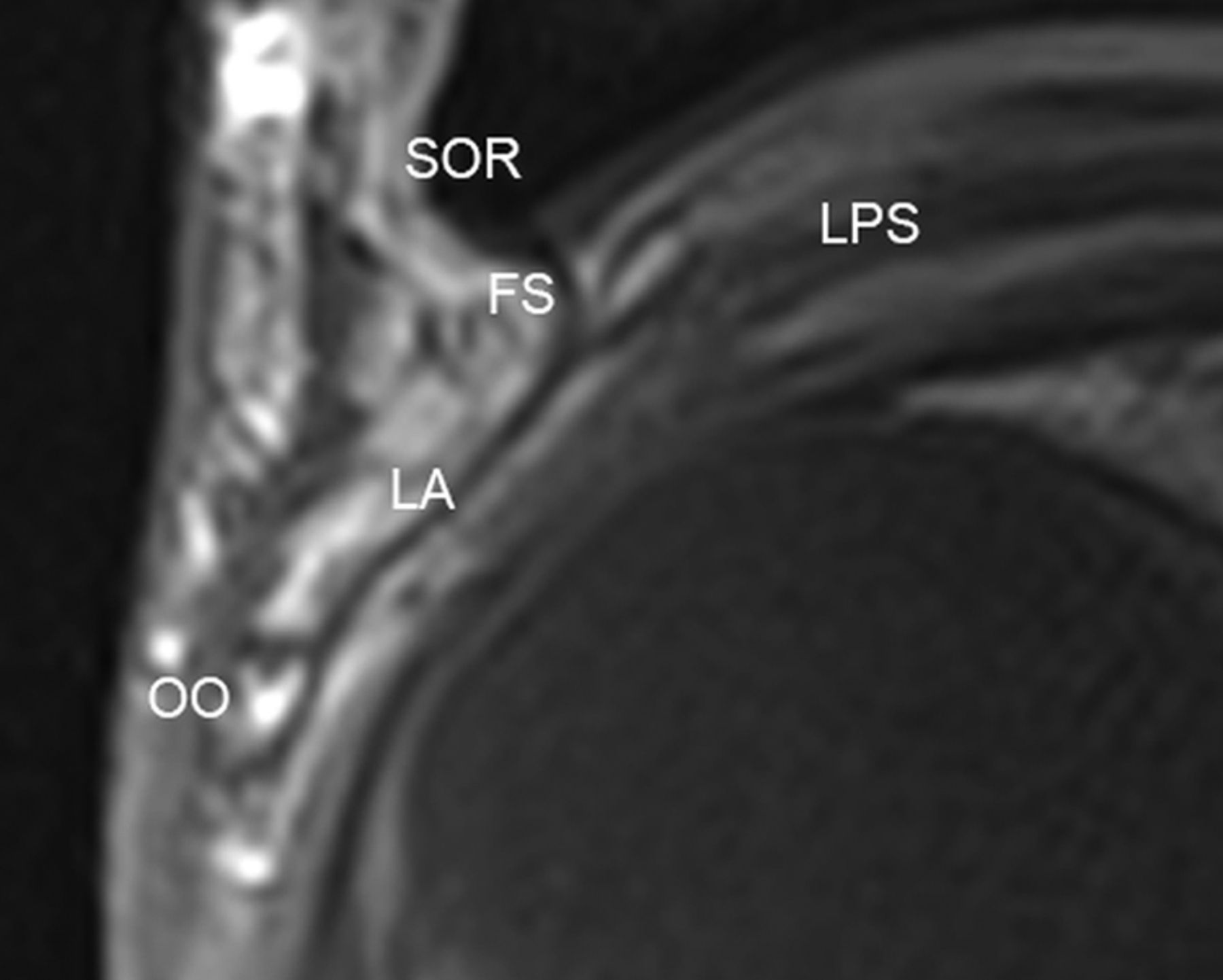
- Fig 9.
The levator aponeurosis shown on MC-MR imaging T1-weighted sagittal image. SOR indicates superior orbital rim; LPS, levator palpebrae superioris; FS, fibrous orbital septum; LA, levator aponeurosis; OO, orbicularis oculi.
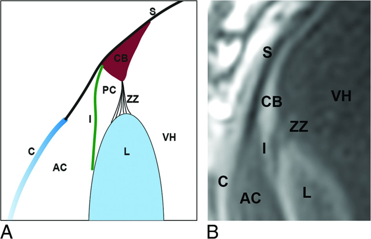
- Fig 10.
Diagrammatic sagittal representation of anterior segment anatomy (A) and its depiction on T1-weighted sagittal MC-MR imaging (B). S indicates sclera; CB, ciliary body; ZZ, zonule of Zinn; VH, vitreous humor; I, Iris; AC, anterior chamber; PC, posterior chamber; L, lens; C, cornea.


Tables
Sequence parameters
Parameters for MC-MR Imaging Parameter T1-Weighted TSE T2-Weighted TSE TR (ms) 400 4000 TE (ms) 15 123 Signals averaged 2 3 FOV (mm) 80 × 70 80 × 70 Martrix 224 × 256 224 × 256 Section thickness (mm) 1 1.5 Turbo factor 3 13 Voxel size (mm) 0.3 × 0.3 × 1.5 0.3 × 0.3 × 1.5 Acquisition time 5 min, 42 sec 7 min, 6 sec


Jump to section
Related Articles
Cited By...
- A-eye: Automated 3D Segmentation of Healthy Human Eye and Orbit Structures and Axial Length Extraction
- Eye-Opening Advances: Automated 3D Segmentation, Key Biomarkers Extraction, and the First Large-Scale MRI Eye Atlas
- Radioanatomic Characteristics of the Posteromedial Intraconal Space: Implications for Endoscopic Resection of Orbital Lesions