
Article Figures & Data
Figures
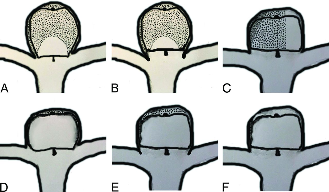
- FIG 1.
Different types of contrast-in-WEB phenomenon. Areas with black spots are thrombosed. A, Partial opacification of the WEB without opacification of the neck or aneurysm. B, Partial opacification of the WEB and opacification of the neck (neck remnant). C, Partial opacification of the WEB and aneurysm dome (aneurysm remnant). D, Complete opacification of the WEB without opacification of the neck or aneurysm. E, Complete opacification of the WEB and opacification of the neck (neck remnant). F, Complete opacification of the WEB and opacification of the aneurysm (aneurysm remnant).
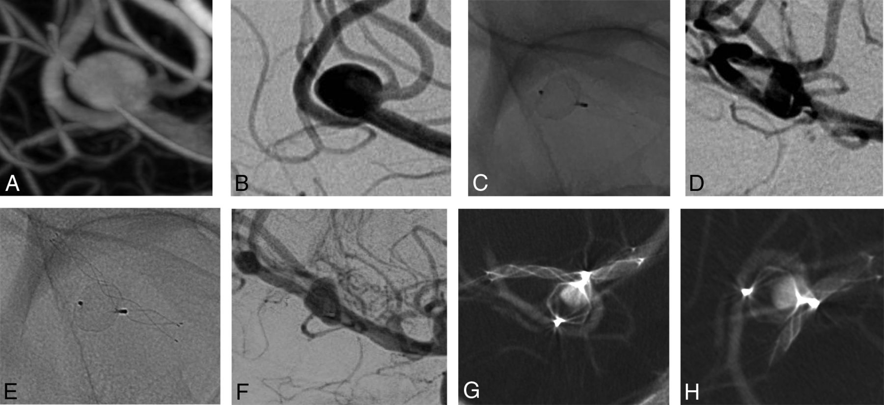
- FIG 2.
3D (A) and 2D (B) DSA show a basilar artery aneurysm. Nonsubtraction (C) and subtraction (D) images of the first DSA follow-up after 1 year show partial filling of the WEB with contrast. A similar finding is observed at 2 years (E and F). VasoCT in 2 different views (G and H) confirms a partial opacification of the WEB and no opacification of the aneurysm.
- FIG 3.
3D (A) and 2D (B) DSA show an anterior communicating artery aneurysm. Nonsubtraction (C) and subtraction (D) images of the first DSA follow-up after 6 months and of the latest DSA follow-up after 4 years (E and F) show complete filling of the WEB with contrast. VasoCT in 2 different views (G and H) shows opacification of the WEB and no opacification of the aneurysm.
- FIG 4.
3D (A) and 2D (B) DSA show an MCA aneurysm. Nonsubtraction (C) and subtraction (D) images of the first DSA follow-up after 6 months and of the latest DSA follow-up after 2 years (E and F) show partial opacification of the WEB and the aneurysm sac, confirmed by VasoCT in 2 different views (G and H).
- FIG 5.
3D (A) and 2D (B) DSA show an MCA aneurysm. Nonsubtraction (C) and subtraction (D) images of the first DSA follow-up after 6 months and of the latest DSA follow-up after 1 year (E and F) show complete opacification of the WEB and the aneurysm dome, confirmed by VasoCT in 2 different views (G and H).


Tables
No. Location Sac Width (mm) Sac Height (mm) Neck Width (mm) Dome-to-Neck Ratio Ruptured Statusa WEB Deployed 1 AcomA 6.0 4.7 4.5 1.3 0 SL 7-3 2 BA 3.4 5.0 3.2 1.1 0 SL 5-3 3 MCA 7.2 5.9 5.5 1.3 0 SL 8-3 4 MCA 6.7 4.0 6.0 1.1 0 SL 7-3 5 MCA 5.3 3.9 4.4 1.2 0 SL 6-3 6 MCA 3.2 4.5 3.3 1.0 0 SL 4.5-3 7 MCA 5.2 5.0 4.4 1.2 0 SL 7-3 8 AcomA 3.7 4.1 3.0 1.2 1 SL 4.5-2 Note:—AcomA indicates anterior communicating artery; BA, basilar artery.
↵a 0, Unruptured; 1, ruptured.
No. Height Postoperation (mm) Height after 6 Months (mm) Height after 1 Year (mm) Height after 2 Years (mm) Height after 3 Years (mm) Height after 4 Years (mm) 1 6.2 5.8 5.8 5.9 5.6 5.8 2 3.6 – 3.4 3.6 – – 3 7.8 7.9 7.6 7.7 – – 4 5.5 5.9 5.1 – – – 5 5.4 6.0 5.8 – – – 6 4.1 4.2 4.1 – – – 7 6.0 6.0 – – – – 8 3.7 3.7 3.8 – – – Note:—– indicates no imaging was performed at this date.


