
Article Figures & Data
Figures
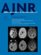
- Fig 1.
Chiari type I malformation versus SIH. A, Sagittal T1WI demonstrates a Chiari type I malformation with descent of cerebellar tonsils 1.4 cm below the foramen magnum (black arrow). Note the typical “peglike” morphology. The prepontine cistern is preserved, and the pons maintains a rounded ventral morphology (white arrow). B, Sagittal T1WI in a patient with severe brain sag due to SIH shows mild descent of the cerebellar tonsils at the foramen magnum (black arrow), but with normal morphology. The pons is flattened (white arrow) with effacement of the prepontine cistern. There is also profound effacement of the subarachnoid spaces in the posterior fossa.
- Fig 2.
“Acquired” Chiari malformation showing findings of CSF hypotension. A, Sagittal T1WI demonstrates a normal appearance of the posterior fossa of a 6-year-old patient with a ventriculoperitoneal shunt. B, Six years later, imaging demonstrates findings of overshunting resulting in CSF hypotension. This has previously (mistakenly) been called “acquired Chiari malformation.” In addition to descent of the tonsils, there is also effacement of the subarachnoid spaces, flattening of the pons, descent of the brain stem, and a decrease in the volume of the lateral and fourth ventricles, all of which indicate reduced CSF volume.
- Fig 3.
Bilateral subdural hematomas as an initial presentation in SIH. A, Sagittal T1WI shows a subtle decrease in the mamillopontine distance (white arrow). Otherwise, the typical stigmata of SIH are not present. B, Coronal postcontrast T1WI demonstrates large bilateral subdural hematomas with mass effect on the cerebral hemispheres. A hyperdynamic CT myelogram showed a leak in the thoracic spine, and a targeted blood patch was performed. The subdural hematomas subsequently required evacuation due to cerebral compression. All of the patient’s symptoms resolved, and the hematomas did not recur.
- Fig 4.
IgG4-related pachymeningitis. A, Coronal contrast-enhanced T1WI from a 53-year-old man with IgG4-related disease with Castleman-like features shows abnormal pachymeningeal enhancement (arrows). B, Follow-up imaging after steroid and rituximab (Rituxan) treatment demonstrates interval near resolution of the pachymeningeal enhancement.
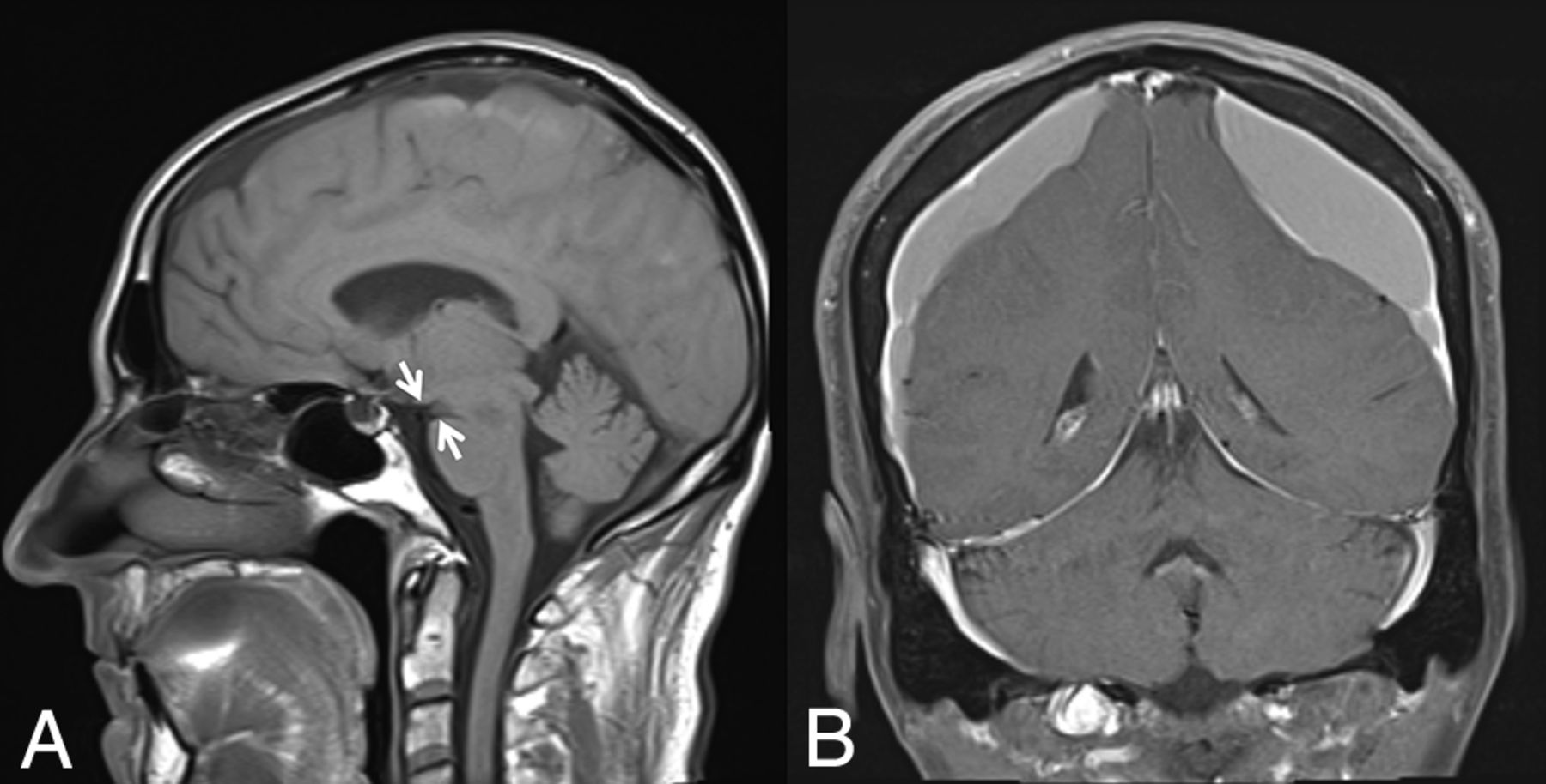
- Fig 5.
Autonomic failure presenting as orthostatic headache. Beat-to-beat blood pressure (black) and heart rate (green) of an 80-year-old woman during a tilt-table test show a sustained drop in blood pressure during head-up tilt (gray shading). The lack of heart rate acceleration suggests neurogenic orthostatic hypotension. When her mean blood pressure fell below 85 mm Hg, she developed posterior head and neck tightness (coat hanger pain) and lightheadedness. Thus, pure autonomic failure was initially misdiagnosed as orthostatic headache.
- Fig 6.
SIH presenting with venous sinus thrombosis. A 43-year-old man presented with sudden onset of headaches, dizziness, and nausea clinically suspected to represent migraines. A, Contrast-enhanced T1-weighted image demonstrates diffuse, smooth, nonspecific thin pachymeningeal enhancement (arrows). The patient presented to the emergency department 3 days later with a marked increase in his headache and sudden onset of right lower-extremity weakness. D, Unenhanced coronal head CT demonstrates high attenuation and expansile content within the superior sagittal sinus and adjacent cortical veins, compatible with acute dural venous sinus and cortical vein thrombosis (selected clot highlighted by arrows). Sagittal MIP (B) and coronal (E) images from a contrast-enhanced CT venogram confirm a long-segment acute, expansile thrombus as a filling defect within the superior sagittal sinus, as well as multiple bilateral left-greater-than-right superior cortical veins (arrows). While he was being treated for sinus thrombosis, he reported that his headaches were orthostatic in nature. His condition continued to deteriorate with progression of the thrombosis despite treatment and parenchymal hemorrhage. Axial (C) and sagittal (F) T2WI demonstrates a ventral extradural fluid collection. A hyperdynamic CT myelogram was performed, and a leak was found at a spiculated osteophyte at T1-2.
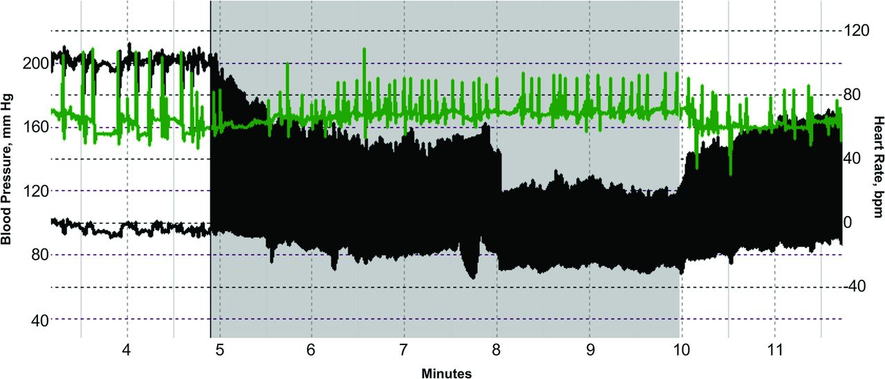
- Fig 7.
SIH presenting as pituitary apoplexy. Sagittal T1WI (A) and coronal postcontrast T1WI (B) images demonstrate a hyperintense lesion filling the sella and extending into the suprasellar cistern. The patient was diagnosed with pituitary apoplexy of a macroadenoma or a Rathke cleft cyst and a Chiari I malformation. However, the MR imaging actually demonstrates the expected intracranial stigmata of an SIH, including severe brain sag and dural enhancement. Despite extensive evaluation, the source of the leak was not found. The patient was treated with a nontargeted blood patch. His headaches and imaging findings (C and D) resolved.
- Fig 8.
SIH presenting as superficial siderosis. A 37-year-old man who presented with episodes of dizziness and loss of consciousness was found to have diffuse posterior fossa–predominant superficial siderosis on axial SWI (A and B). Subsequent imaging of the spine demonstrated a ventral extradural fluid collection (curved arrow, C; straight arrows, D). A subsequent hyperdynamic CT myelogram identified the source of the CSF leak, which was surgically repaired.


Tables
- Table 1:
Distinguishing features of alternative diagnoses that occasionally mimic spontaneous intracranial hypotension
Distinguishing Features SIH Radiologic mimickers Chiari type I malformation Cerebellar tonsils inferiorly pointed
Midbrain descent absentNormal cerebellar tonsil shape
Midbrain descent presentSubdural fluid collections Usually unilateral Usually bilateral
Brain sag and focal dural enhancementConditions with dural thickening (IgG4-related disease, neurosarcoidosis, tuberculosis, autoimmune diseases, infectious diseases) Focal or diffuse
May have leptomeningeal involvement, skull base prominence, hypertrophic pachymeningitis
Usually systemic symptoms and involvement of other organsDiffuse, non-nodular dural thickening and enhancement Clinical mimickers POTS Increased heart rate with minimal change in blood pressure on standing from a seated or supine position Stable heart rate with postural changes Orthostatic hypotension Autonomic failure, medication effect, hypovolemia
Fall in systolic (20 mm Hg) and/or diastolic (10 mm Hg) blood pressure on standing from a seated or supine positionStable blood pressure with postural changes Cervicogenic headache Headache with neck pain that worsens with cervical motion, relieved with medication Pain is typically centered in the head and not worsened by cervical motion or improved with medications Vestibular migraine Vertigo, unilateral headache, nystagmus, presence of aura, history of migraine Hearing changes and tinnitus more than vertigo or nystagmus Coexisting Condition Pathogenesis Cerebral venous sinus thrombosis Compensatory venous engorgement and stasis secondary to decrease in intracranial CSF volume Frontotemporal dementia Brain sag, obstruction of venous outflow, and swelling of the diencephalon may precipitate behavior and personality changes Pituitary enlargement or apoplexy Compensatory enlargement and congestion of hypophyseal veins may cause pituitary engorgement and predisposition to apoplexy POTS Prolonged supine deconditioning secondary to spinal CSF leak Superficial siderosis Venous traction at the skull base may cause microhemorrhages, or bleeding may occur at the site of the dural defect


Jump to section
Related Articles
Cited By...
- Spinal CSF Leaks: The Neuroradiologist Transforming Care
- Skull Base CSF Leaks: Potential Underlying Pathophysiology and Evaluation of Brain MR Imaging Findings Associated with Spontaneous Intracranial Hypotension
- Natural history of spontaneous intracranial hypotension: a clinical and imaging study
- Likelihood of Discovering a CSF Leak Based on Intracranial MRI Findings in Patients without a Spinal Longitudinal Extradural Collection: A New Probabilistic Scoring System
- The Monro-Kellie Doctrine: A Review and Call for Revision
- Case of spontaneous intracranial hypotension: clinical, neuroimaging and treatment approach