
Article Figures & Data
Figures
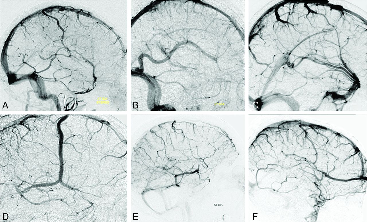
- Fig 1.
Illustrative variations of superficial venous drainage. A, Balanced pattern. B, Dominant Labbe. C, Dominant superficial Sylvian veins. D, Dominant Rolandic. E, Dominant basal vein with variant drainage. F, Dominant anterior frontal vein, a treacherous variation in cases of surgical sacrifice of a distal superior sagittal sinus.
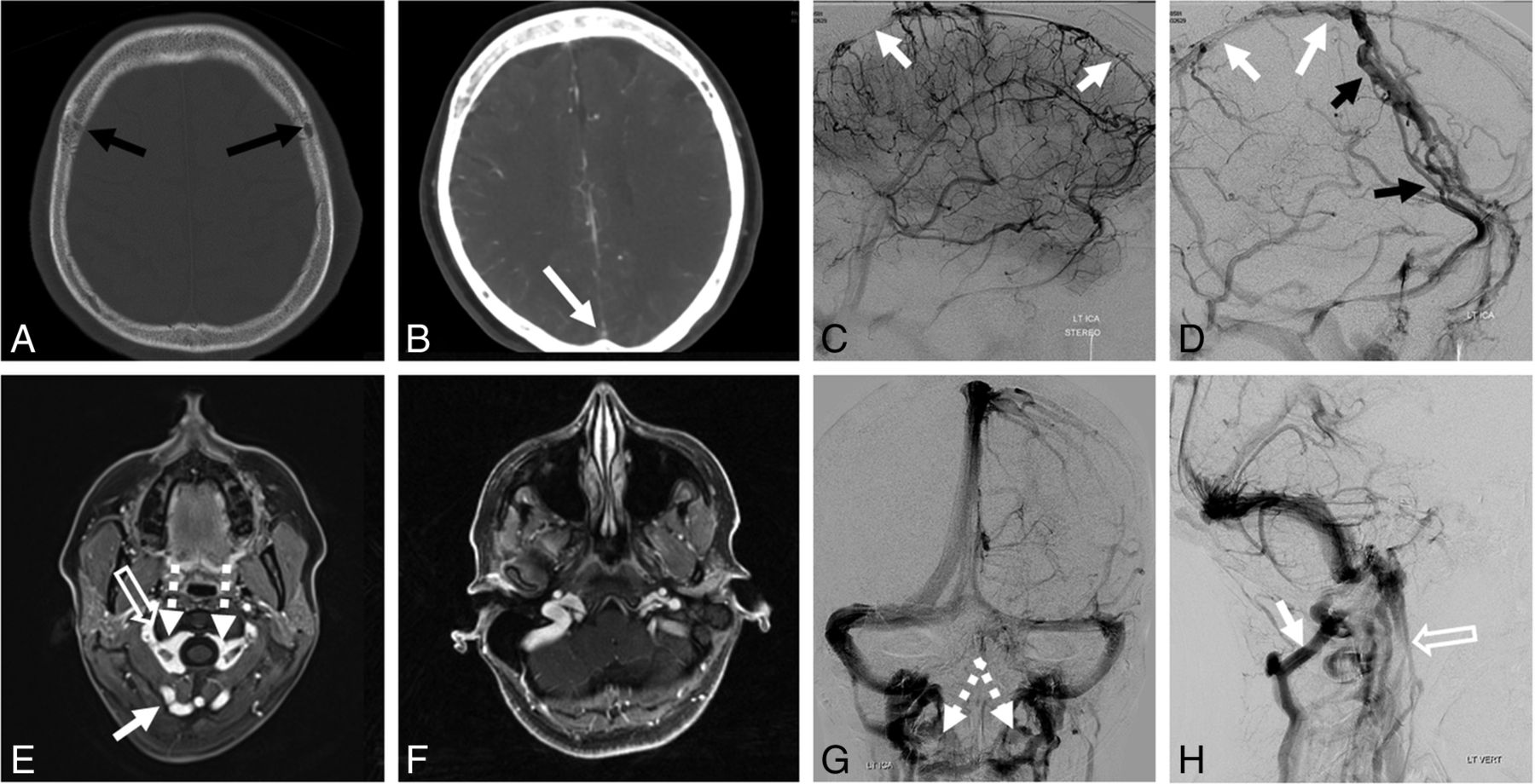
- Fig 2.
Variations of venous drainage. A–D, Hypoplastic superior sagittal sinus (white arrows) is associated with prominent diploic veins (black arrows). E–H, C1 lateral mass region stenoses of the jugular system (open arrows) are associated with compensatory prominence of condylar/suboccipital veins (white arrows), draining in large part into the vertebral venous plexus (dashed arrows). This configuration is further modified in nonrecumbent positions.
- Fig 3.
The variety of dural venous channels. Any part of the dura can be a venous channel, supra- or infratentorial. There is nothing particularly unique about a tentorial sinus except for the increased frequency of dural venous channels there. 1) Dural venous channel (tentorial sinus group) receiving the basal vein; 2) Dural venous channel (tentorial sinus group) receiving the inferior cerebellar vein; 3) Dural venous channel receiving the vein of Labbe; 4) Dural venous channel (falcine sinus group) receiving a parieto-occipital vein; 5) Dural venous channel (parietal) receiving either a medial or lateral parietal vein such as the vein of Trolard; 6) Dural venous channel (venous lake) receiving the Rolandic/Trolard group of veins; 7) Dural venous channel receiving the superficial Sylvian group of veins. Together with the Labbe dural channel (3 above), these drain into the sigmoid sinus.
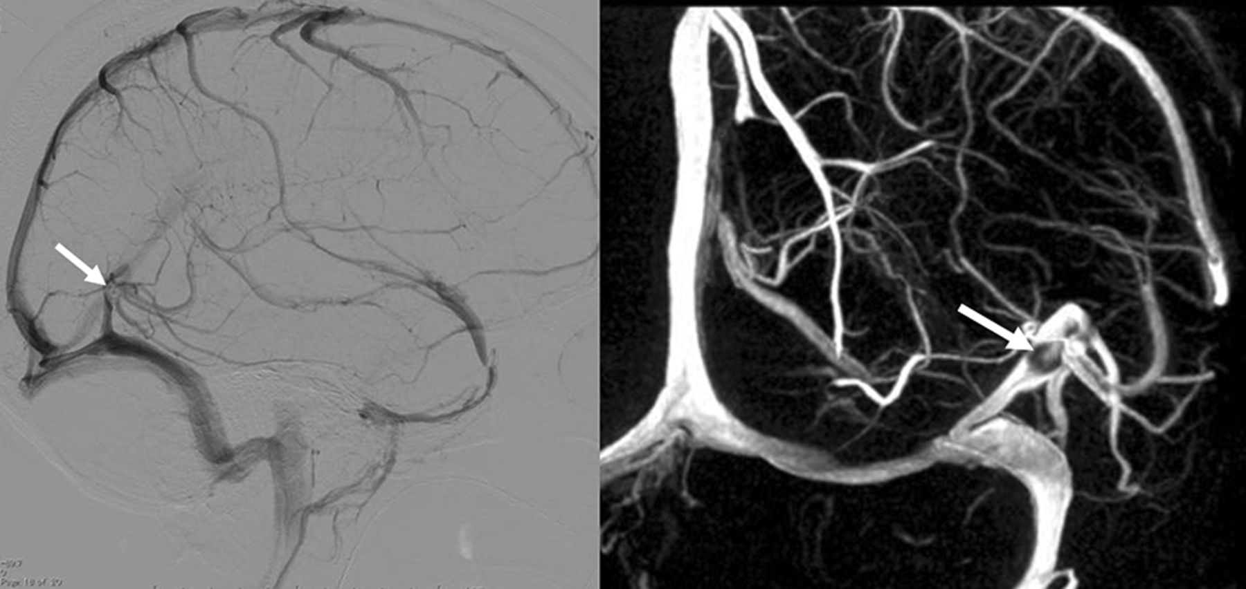
- Fig 4.
Typical dural venous channel (white arrows) draining a large vein of Labbe (white arrowheads). A and B, Angiographic images. C and D, Postcontrast T1 axial volumetric source images. E and F, Volume-rendered reconstructions of the above axial T1 post-contrast dataset.
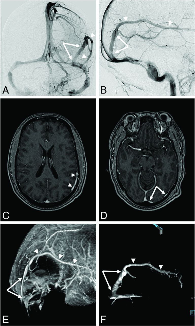
- Fig 5.
Mirror image of dural venous channels. Right ICA (A and B), left ICA (C and D), and vertebral (E and F) injections. On the right, the posterior temporal (white arrowhead) vein drains into a dural venous channel (white arrow). The inferior temporal veins drain into a separate dural venous channel (dashed arrow). The 2 channels form a common dural channel (open arrow) draining into the proximal transverse sinus. A mirror image appearance is seen on the left. C and D, Corresponding black arrows. E and F, Vertebral injection opacifies the same dural venous channels (white and black arrows), collecting posterior temporal and occipital veins. Also seen are several dural venous channels in the tentorium cerebelli, also known as tentorial sinuses (curved arrows, ballpoint arrow). G and H, Left anterior oblique views of the left ICA injection profile dural venous channels seen in C and D. Notice the flattened appearance and location of channels lateral to parenchymal veins and corresponding indentations on the inner surface of skull due to flow-related remodeling.
- Fig 6.
Dural venous channel with an associated arachnoid granulation (arrows)—another characteristic of a dural structure.
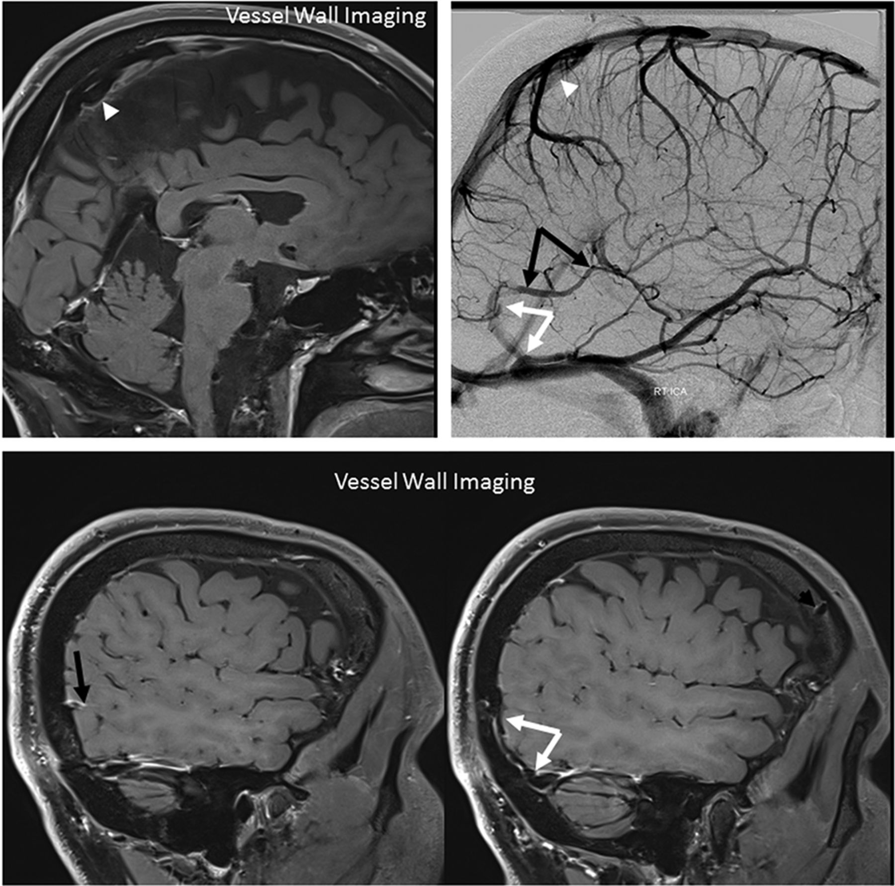
- Fig 7.
Vessel wall imaging of dural venous channels in the falx cerebri (white arrowheads), collecting the Trolard group, and lateral tentorium (white arrows), collecting the Labbe group (black arrows).
- Fig 8.
A, Classic dural fistula with an arteriovenous connection directly involving a dural (sigmoid in this case) sinus. B, Dural fistula with an arteriovenous connection involving a dural venous channel segment of the Labbe (1) and posterior cerebellar (2) veins. Thrombosis or restriction of antegrade outflow (black circles) into a draining sinus results in retrograde congestion of the cortical venous system. These seemingly “direct” fistulas draining exclusively into a cortical vein are proposed to primarily involve a dural venous channel, explaining the seemingly random dural location of these fistulas.
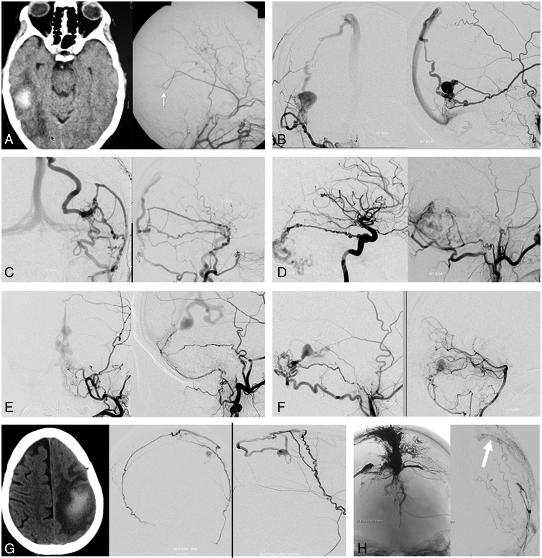
- Fig 9.
Dural fistulas draining directly into a cortical vein instead of a sinus, which may illustrate the concept of a dural venous channel fistula shown in Fig 8. A, Venous congestion hemorrhage due to a fistula (arrow) between the middle meningeal artery and the vein of Labbe. B and C, Fistulas just above the transverse sinus, with typical arterial supply and drainage into the parietal veins. D–F, Tentorial cerebelli fistulas congesting various cerebellar veins. These can be found anywhere on the tentorium, including the midline/vermian region (E). G, A small parasagittal fistula between the artery of the superior sagittal sinus supplied by both middle meningeal arteries and the vein of Trolard. Despite the small fistula size, the resulting Trolard congestion precipitated a large venous infarct. H, Complex vertex/frontoparietal dural fistula. Despite the presence of extensive Onyx (Covidien) and n-BCA embolic material in the sinus and adjacent venous lakes, there remains a parasagittal fistula between the middle meningeal artery and the Rolandic vein (white arrow). The fistula is clearly lateral to the sinus, illustrating the shunt location within the parasagittal venous lake. Multiple similar fistulas in this case have already been closed at this stage.


Tables
Supratentorial dural venous channel characteristics, based on review of 100 consecutive angiograms
No. % Patients with supratentorial dural venous channels 26 26 No. of supratentorial dural venous channels 29 29 Average dural venous channel length (mm) 20 No. of dural venous channels on right side 19 66 Dural venous channel, female patients 20 77 Overall No. of females in angiographic sample 63 63 Location of supratentorial dural venous channela Posterior temporal 19 66 Parietal 4 14 Occipital 6 21 ↵a Excludes “dural venous lakes” and vertex falx cerebri channels.


Jump to section
Related Articles
Cited By...
- Principles, techniques and applications of high resolution cone beam CT angiography in the neuroangio suite
- Cerebral venous anatomy: implications for the neurointerventionalist
- Cerebral venous anatomy: implications for the neurointerventionalist
- Principles, techniques and applications of high resolution cone beam CT angiography in the neuroangio suite
- The So-Called Cranial Dural Channels and Their Relationship with the Bridging Veins