Advertisement

Main menu

  • Home
  • Content
    • Current Issue
    • Accepted Manuscripts
    • Article Preview
    • Past Issue Archive
    • Video Articles
    • AJNR Case Collection
    • Case of the Week Archive
    • Case of the Month Archive
    • Classic Case Archive
  • Special Collections
    • AJNR Awards
    • Low-Field MRI
    • Alzheimer Disease
    • ASNR Foundation Special Collection
    • Photon-Counting CT
    • View All
  • Multimedia
    • AJNR Podcasts
    • AJNR SCANtastic
    • Trainee Corner
    • MRI Safety Corner
    • Imaging Protocols
  • For Authors
    • Submit a Manuscript
    • Submit a Video Article
    • Submit an eLetter to the Editor/Response
    • Manuscript Submission Guidelines
    • Statistical Tips
    • Fast Publishing of Accepted Manuscripts
    • Graphical Abstract Preparation
    • Imaging Protocol Submission
    • Author Policies
  • About Us
    • About AJNR
    • Editorial Board
    • Editorial Board Alumni
  • More
    • Become a Reviewer/Academy of Reviewers
    • Subscribers
    • Permissions
    • Alerts
    • Feedback
    • Advertisers
    • ASNR Home

User menu

  • Alerts
  • Log in

Search

  • Advanced search
American Journal of Neuroradiology

American Journal of Neuroradiology

ASHNR American Society of Functional Neuroradiology ASHNR American Society of Pediatric Neuroradiology ASSR
  • Alerts
  • Log in

Advanced Search

  • Home
  • Content
    • Current Issue
    • Accepted Manuscripts
    • Article Preview
    • Past Issue Archive
    • Video Articles
    • AJNR Case Collection
    • Case of the Week Archive
    • Case of the Month Archive
    • Classic Case Archive
  • Special Collections
    • AJNR Awards
    • Low-Field MRI
    • Alzheimer Disease
    • ASNR Foundation Special Collection
    • Photon-Counting CT
    • View All
  • Multimedia
    • AJNR Podcasts
    • AJNR SCANtastic
    • Trainee Corner
    • MRI Safety Corner
    • Imaging Protocols
  • For Authors
    • Submit a Manuscript
    • Submit a Video Article
    • Submit an eLetter to the Editor/Response
    • Manuscript Submission Guidelines
    • Statistical Tips
    • Fast Publishing of Accepted Manuscripts
    • Graphical Abstract Preparation
    • Imaging Protocol Submission
    • Author Policies
  • About Us
    • About AJNR
    • Editorial Board
    • Editorial Board Alumni
  • More
    • Become a Reviewer/Academy of Reviewers
    • Subscribers
    • Permissions
    • Alerts
    • Feedback
    • Advertisers
    • ASNR Home
  • Follow AJNR on Twitter
  • Visit AJNR on Facebook
  • Follow AJNR on Instagram
  • Join AJNR on LinkedIn
  • RSS Feeds

AJNR Awards, New Junior Editors, and more. Read the latest AJNR updates

Research ArticleHead and Neck Imaging

Indentation and Transverse Diameter of the Meckel Cave: Imaging Markers to Diagnose Idiopathic Intracranial Hypertension

A. Kamali, K.C. Sullivan, F. Rahmani, A. Gandhi, A. Aein, O. Arevalo, P. Rabiei, S.J. Choi, X. Zhang, R.E. Gabr and R.F. Riascos
American Journal of Neuroradiology August 2020, 41 (8) 1487-1494; DOI: https://doi.org/10.3174/ajnr.A6682
A. Kamali
aFrom the Department of Diagnostic Radiology (A.K., A.A., O.A., P.R., X.Z., R.E.G., R.F.R.), University of Texas at Houston, Houston, Texas
  • Find this author on Google Scholar
  • Find this author on PubMed
  • Search for this author on this site
  • ORCID record for A. Kamali
K.C. Sullivan
bUniversity of Texas Medical School Health Science Center Houston (K.C.S., S.J.C.), Houston, Texas
  • Find this author on Google Scholar
  • Find this author on PubMed
  • Search for this author on this site
  • ORCID record for K.C. Sullivan
F. Rahmani
cNeuroimaging Laboratory at Mallinckrodt Institute of Radiology (F.R.), Washington University School of Medicine, St. Louis, Missouri
  • Find this author on Google Scholar
  • Find this author on PubMed
  • Search for this author on this site
  • ORCID record for F. Rahmani
A. Gandhi
dRice University (A.G.), Houston, Texas.
  • Find this author on Google Scholar
  • Find this author on PubMed
  • Search for this author on this site
  • ORCID record for A. Gandhi
A. Aein
aFrom the Department of Diagnostic Radiology (A.K., A.A., O.A., P.R., X.Z., R.E.G., R.F.R.), University of Texas at Houston, Houston, Texas
  • Find this author on Google Scholar
  • Find this author on PubMed
  • Search for this author on this site
  • ORCID record for A. Aein
O. Arevalo
aFrom the Department of Diagnostic Radiology (A.K., A.A., O.A., P.R., X.Z., R.E.G., R.F.R.), University of Texas at Houston, Houston, Texas
  • Find this author on Google Scholar
  • Find this author on PubMed
  • Search for this author on this site
  • ORCID record for O. Arevalo
P. Rabiei
aFrom the Department of Diagnostic Radiology (A.K., A.A., O.A., P.R., X.Z., R.E.G., R.F.R.), University of Texas at Houston, Houston, Texas
  • Find this author on Google Scholar
  • Find this author on PubMed
  • Search for this author on this site
  • ORCID record for P. Rabiei
S.J. Choi
bUniversity of Texas Medical School Health Science Center Houston (K.C.S., S.J.C.), Houston, Texas
  • Find this author on Google Scholar
  • Find this author on PubMed
  • Search for this author on this site
  • ORCID record for S.J. Choi
X. Zhang
aFrom the Department of Diagnostic Radiology (A.K., A.A., O.A., P.R., X.Z., R.E.G., R.F.R.), University of Texas at Houston, Houston, Texas
  • Find this author on Google Scholar
  • Find this author on PubMed
  • Search for this author on this site
  • ORCID record for X. Zhang
R.E. Gabr
aFrom the Department of Diagnostic Radiology (A.K., A.A., O.A., P.R., X.Z., R.E.G., R.F.R.), University of Texas at Houston, Houston, Texas
  • Find this author on Google Scholar
  • Find this author on PubMed
  • Search for this author on this site
  • ORCID record for R.E. Gabr
R.F. Riascos
aFrom the Department of Diagnostic Radiology (A.K., A.A., O.A., P.R., X.Z., R.E.G., R.F.R.), University of Texas at Houston, Houston, Texas
  • Find this author on Google Scholar
  • Find this author on PubMed
  • Search for this author on this site
  • ORCID record for R.F. Riascos
  • Article
  • Figures & Data
  • Info & Metrics
  • Responses
  • References
  • PDF
Loading

Article Figures & Data

Figures

  • Tables
  • Fig 1.
    • Download figure
    • Open in new tab
    • Download powerpoint
    Fig 1.

    Two coronal T2-weighted images of the Meckel cave in a healthy subject (A) versus a patient with IIH (B). The white arrows in A represent the curvature of the Meckel cave and no indentation in a healthy subject. The white arrow in B demonstrates an acute angle of indentation and a bilobed appearance of the Meckel cave in a patient with IIH. The 2-way arrows in B demonstrate the craniocaudal diameter (along the oblique axis of the right MC) and transverse diameter of the left MC (perpendicular to both walls in the widest segment of the MC on the coronal plane).

  • Fig 2.
    • Download figure
    • Open in new tab
    • Download powerpoint
    Fig 2.

    Coronal T2 view of a dilated MC in a patient with IIH. The black arrows represent the indentation of the Meckel cave. The white arrow shows the dilated cisternal space of the V3 segment of the left trigeminal nerve and points to the foramen ovale where the V3 nerve exits the intracranial space at the skull base. Dilation of this cisternal space may contribute to the indented appearance of the MC in patients with IIH.

  • Fig 3.
    • Download figure
    • Open in new tab
    • Download powerpoint
    Fig 3.

    The appearance of the Meckel cave in a healthy person (A) versus a patient with IIH (B) on the axial T2 planes. The 2-way arrows represent the wall-to-wall measurement of the Meckel cave in the AP (A) and transverse (B) diameters.
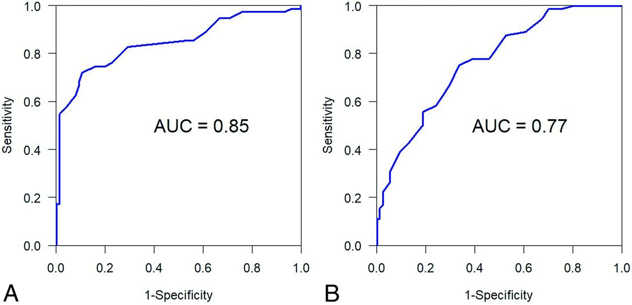
  • Fig 4.
    • Download figure
    • Open in new tab
    • Download powerpoint
    Fig 4.

    Figs 4–7. The AUC was used to evaluate the overall differentiability of a marker to identify those with IIH from healthy controls. The optimal cutoff point for the marker was obtained by evaluating the Youden index. On the basis of the optimal cutoff point, we reported estimates of sensitivity, specificity, positive predictive value, and negative predictive value, together with their 95% Wald confidence intervals. Maximum transverse values. A, coronal data. B, axial data.

  • Fig 5.
    • Download figure
    • Open in new tab
    • Download powerpoint
    Fig 5.

    Right transverse values. A, Coronal data. B, Axial data.
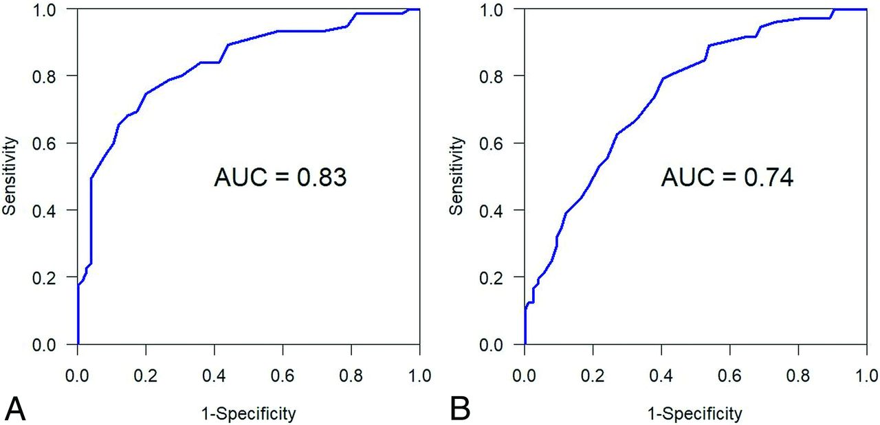
  • Fig 6.
    • Download figure
    • Open in new tab
    • Download powerpoint
    Fig 6.

    Left transverse values. A, Coronal data. B, Axial data.

  • Fig 7.
    • Download figure
    • Open in new tab
    • Download powerpoint
    Fig 7.

    Receiver operating characteristic curves for MC volumes: right MC (right), left MC (center), and maximum values (left).

Tables

  • Figures
    • View popup
    Table 1:

    Incidence, sensitivity, and specificity of imaging findings in IIH and control groups

    Imaging SignsIIHControlsSensitivitySpecificity
    Optic nerve sheath dilation63 (84%)12 (16%)8484
    Empty/partial empty sella69 (92%)20 (26%)9274
    Posterior scleral flattening41 (55%)055100
    Papilledema34 (45%)045100
    Bilateral TSS55 (73%)6 (8%)7392
    Enlarged MC56 (75%)11 (14%)7586
    • View popup
    Table 2:

    Demographics for 2 groups

    IIH (n = 75)Healthy Controls (n = 75)P Value
    Age (mean ± SD) (yr)33.84 ± 9.1534.65 ± 9.37.59
    Male sex6 (8%)15 (20%).034
    LP opening pressure (median) (IQR)29 (24–38)Not Available
    • Note:—IIH indicates idiopathic intracranial hypertension patients; LP, lumbar puncture; IQR, interquartile range.

    • View popup
    Table 3:

    Meckel cave indentationa

    YesNoTotal
    IIH571875
    Healthy controls215475
    • ↵a Seventy-six percent sensitivity, 72% specificity, positive predictive value of 73% and negative predictive value of 75% for the diagnosis of IIH.

    • View popup
    Table 4:

    Descriptive statistics in IIH patients (n = 75) and controls (n = 75)a

    VariablePatients with IIHControlsP Value
    Right MC height11.51 ± 1.789.51 ± 1.46<.001
    Left MC height11.93 ± 1.909.53 ± 1.43<.001
    Max MC height12.50 ± 1.929.97 ± 1.51<.001
    Right MC AP12.37 ± 2.439.68 ± 1.43<.001
    Left MC AP12.92 ± 2.709.55 ± 1.47<.001
    Max MC AP13.41 ± 2.4810.13 ± 1.43<.001
    Right MC transverse5.14 ± 1.213.94 ±0.73<.001
    Left MC transverse4.94 ± 0.963.88 ± 0.75<.001
    Max MC transverse5.49 ± 1.114.14 ± 0.72<.001
    Right volume747.3 ± 293368.7 ± 121<.001
    Left volume775.4 ± 278357.2 ± 115<.001
    Max volume938.4 ± 333424.0 ± 131<.001
    • Note:—Max indicates maximum.

    • ↵a Mean ± SD are shown. Summary statistics for anterior-posterior (AP) and transverse diameters of MC on axial T2, and craniocaudal diameter of MC on coronal T2 weighted sequences. Max is larger value between right and left. Volume is product of height, AP, and transverse diameters.

    • View popup
    Table 5:

    AUC estimates for differentiating patients with IIH from controls

    VariableAUC95% CI
    Right MC height0.816(0.748–0.884)
    Left MC height0.852(0.793–0.911)
    Max MC height0.850(0.789–0.910)
    Right MC AP0.830(0.768–0.893)
    Left MC AP0.861(0.804–0.917)
    Max MC AP0.874(0.821–0.927)
    Right MC transverse0.810(0.740–0.879)
    Left MC transverse0.807(0.737–0.877)
    Max MC transverse0.852(0.791–0.913)
    Right volume0.907(0.861–0.954)
    Left volume0.922(0.881–0.964)
    Max volume0.936(0.900–0.972)
    • Note:—Max indicates maximum.

    • View popup
    Table 6:

    Estimated Spearman correlation coefficients (n = 75)a

    Variable 1Variable 2Estimated Spearman Correlation Coefficient95% CIP Value
    Right MC volumeCSF opening pressure−0.104−0.323−0.127.38
    Left MC volumeCSF opening pressure−0.050−0.274−0.179.67
    • ↵a The Spearman correlation coefficient was only −0.10 for right MC volume and CSF pressure, and −0.05 for left MC volume and CSF opening pressure. Correlation coefficients are very close to zero. We may conclude that opening CSF pressure is not correlated with right or left MC volumes.

PreviousNext
Back to top

In this issue

American Journal of Neuroradiology: 41 (8)
American Journal of Neuroradiology
Vol. 41, Issue 8
1 Aug 2020
  • Table of Contents
  • Index by author
  • Complete Issue (PDF)
Advertisement
Print
Download PDF
Email Article

Thank you for your interest in spreading the word on American Journal of Neuroradiology.

NOTE: We only request your email address so that the person you are recommending the page to knows that you wanted them to see it, and that it is not junk mail. We do not capture any email address.

Enter multiple addresses on separate lines or separate them with commas.
Indentation and Transverse Diameter of the Meckel Cave: Imaging Markers to Diagnose Idiopathic Intracranial Hypertension
(Your Name) has sent you a message from American Journal of Neuroradiology
(Your Name) thought you would like to see the American Journal of Neuroradiology web site.
CAPTCHA
This question is for testing whether or not you are a human visitor and to prevent automated spam submissions.
Cite this article
A. Kamali, K.C. Sullivan, F. Rahmani, A. Gandhi, A. Aein, O. Arevalo, P. Rabiei, S.J. Choi, X. Zhang, R.E. Gabr, R.F. Riascos
Indentation and Transverse Diameter of the Meckel Cave: Imaging Markers to Diagnose Idiopathic Intracranial Hypertension
American Journal of Neuroradiology Aug 2020, 41 (8) 1487-1494; DOI: 10.3174/ajnr.A6682

Citation Manager Formats

  • BibTeX
  • Bookends
  • EasyBib
  • EndNote (tagged)
  • EndNote 8 (xml)
  • Medlars
  • Mendeley
  • Papers
  • RefWorks Tagged
  • Ref Manager
  • RIS
  • Zotero
0 Responses
Respond to this article
Share
Bookmark this article
Indentation and Transverse Diameter of the Meckel Cave: Imaging Markers to Diagnose Idiopathic Intracranial Hypertension
A. Kamali, K.C. Sullivan, F. Rahmani, A. Gandhi, A. Aein, O. Arevalo, P. Rabiei, S.J. Choi, X. Zhang, R.E. Gabr, R.F. Riascos
American Journal of Neuroradiology Aug 2020, 41 (8) 1487-1494; DOI: 10.3174/ajnr.A6682
del.icio.us logo Twitter logo Facebook logo Mendeley logo
  • Tweet Widget
  • Facebook Like
  • Google Plus One
Purchase

Jump to section

  • Article
    • Abstract
    • ABBREVIATIONS:
    • MATERIALS AND METHODS
    • RESULTS
    • DISCUSSION
    • CONCLUSIONS
    • Footnotes
    • References
  • Figures & Data
  • Info & Metrics
  • Responses
  • References
  • PDF

Related Articles

  • No related articles found.
  • PubMed
  • Google Scholar

Cited By...

  • No citing articles found.
  • Crossref
  • Google Scholar

This article has not yet been cited by articles in journals that are participating in Crossref Cited-by Linking.

More in this TOC Section

  • Hydrops Herniation into the Semicircular Canals
  • ASL Sensitivity for Head and Neck Paraganglioma
  • Post SRS Peritumoral Hyperintense Signal of VSs
Show more HEAD AND NECK IMAGING

Similar Articles

Advertisement

Indexed Content

  • Current Issue
  • Accepted Manuscripts
  • Article Preview
  • Past Issues
  • Editorials
  • Editor's Choice
  • Fellows' Journal Club
  • Letters to the Editor
  • Video Articles

Cases

  • Case Collection
  • Archive - Case of the Week
  • Archive - Case of the Month
  • Archive - Classic Case

More from AJNR

  • Trainee Corner
  • Imaging Protocols
  • MRI Safety Corner
  • Book Reviews

Multimedia

  • AJNR Podcasts
  • AJNR Scantastics

Resources

  • Turnaround Time
  • Submit a Manuscript
  • Submit a Video Article
  • Submit an eLetter to the Editor/Response
  • Manuscript Submission Guidelines
  • Statistical Tips
  • Fast Publishing of Accepted Manuscripts
  • Graphical Abstract Preparation
  • Imaging Protocol Submission
  • Evidence-Based Medicine Level Guide
  • Publishing Checklists
  • Author Policies
  • Become a Reviewer/Academy of Reviewers
  • News and Updates

About Us

  • About AJNR
  • Editorial Board
  • Editorial Board Alumni
  • Alerts
  • Permissions
  • Not an AJNR Subscriber? Join Now
  • Advertise with Us
  • Librarian Resources
  • Feedback
  • Terms and Conditions
  • AJNR Editorial Board Alumni

American Society of Neuroradiology

  • Not an ASNR Member? Join Now

© 2025 by the American Society of Neuroradiology All rights, including for text and data mining, AI training, and similar technologies, are reserved.
Print ISSN: 0195-6108 Online ISSN: 1936-959X

Powered by HighWire