Advertisement

Main menu

  • Home
  • Content
    • Current Issue
    • Accepted Manuscripts
    • Article Preview
    • Past Issue Archive
    • AJNR Case Collection
    • Case of the Week Archive
    • Classic Case Archive
    • Case of the Month Archive
  • Special Collections
    • Spinal CSF Leak Articles (Jan 2020-June 2024)
    • 2024 AJNR Journal Awards
    • Most Impactful AJNR Articles
  • Multimedia
    • AJNR Podcast
    • AJNR Scantastics
    • Video Articles
  • For Authors
    • Submit a Manuscript
    • Author Policies
    • Fast publishing of Accepted Manuscripts
    • Graphical Abstract Preparation
    • Manuscript Submission Guidelines
    • Imaging Protocol Submission
    • Submit a Case for the Case Collection
  • About Us
    • About AJNR
    • Editorial Board
  • More
    • Become a Reviewer/Academy of Reviewers
    • Subscribers
    • Permissions
    • Alerts
    • Feedback
    • Advertisers
    • ASNR Home
  • Other Publications
    • ajnr

User menu

  • Alerts
  • Log in

Search

  • Advanced search
American Journal of Neuroradiology

American Journal of Neuroradiology

ASHNR American Society of Functional Neuroradiology ASHNR American Society of Pediatric Neuroradiology ASSR
  • Alerts
  • Log in

Advanced Search

  • Home
  • Content
    • Current Issue
    • Accepted Manuscripts
    • Article Preview
    • Past Issue Archive
    • AJNR Case Collection
    • Case of the Week Archive
    • Classic Case Archive
    • Case of the Month Archive
  • Special Collections
    • Spinal CSF Leak Articles (Jan 2020-June 2024)
    • 2024 AJNR Journal Awards
    • Most Impactful AJNR Articles
  • Multimedia
    • AJNR Podcast
    • AJNR Scantastics
    • Video Articles
  • For Authors
    • Submit a Manuscript
    • Author Policies
    • Fast publishing of Accepted Manuscripts
    • Graphical Abstract Preparation
    • Manuscript Submission Guidelines
    • Imaging Protocol Submission
    • Submit a Case for the Case Collection
  • About Us
    • About AJNR
    • Editorial Board
  • More
    • Become a Reviewer/Academy of Reviewers
    • Subscribers
    • Permissions
    • Alerts
    • Feedback
    • Advertisers
    • ASNR Home
  • Follow AJNR on Twitter
  • Visit AJNR on Facebook
  • Follow AJNR on Instagram
  • Join AJNR on LinkedIn
  • RSS Feeds

Welcome to the new AJNR, Updated Hall of Fame, and more. Read the full announcements.


AJNR is seeking candidates for the position of Associate Section Editor, AJNR Case Collection. Read the full announcement.


Research ArticlePediatrics

MRI Head Coil Malfunction Producing Artifacts Mimicking Malformation of Cortical Development in Pediatric Epilepsy Work-Up

N. Kashani, N. Khan, J.M. Ospel and X.-C. Wei
American Journal of Neuroradiology August 2020, 41 (8) 1538-1540; DOI: https://doi.org/10.3174/ajnr.A6639
N. Kashani
aFrom the Department of Radiology (N. Kashani. N. Khan, J.M.O.), University of Calgary, Calgary, Alberta, Canada
  • Find this author on Google Scholar
  • Find this author on PubMed
  • Search for this author on this site
  • ORCID record for N. Kashani
N. Khan
aFrom the Department of Radiology (N. Kashani. N. Khan, J.M.O.), University of Calgary, Calgary, Alberta, Canada
  • Find this author on Google Scholar
  • Find this author on PubMed
  • Search for this author on this site
  • ORCID record for N. Khan
J.M. Ospel
aFrom the Department of Radiology (N. Kashani. N. Khan, J.M.O.), University of Calgary, Calgary, Alberta, Canada
  • Find this author on Google Scholar
  • Find this author on PubMed
  • Search for this author on this site
  • ORCID record for J.M. Ospel
X.-C. Wei
bDiagnostic Imaging (X.-C.W.), Alberta Children’s Hospital, Calgary, Alberta, Canada.
  • Find this author on Google Scholar
  • Find this author on PubMed
  • Search for this author on this site
  • ORCID record for X.-C. Wei
  • Article
  • Figures & Data
  • Supplemental
  • Info & Metrics
  • Responses
  • References
  • PDF
Loading

Article Figures & Data

Figures

  • Fig 1.
    • Download figure
    • Open in new tab
    • Download powerpoint
    Fig 1.

    Picture of a typical multichannel MR head coil (A) shows the positions of the multiple coil elements. A modified screenshot from the operator console of the system (B) shows indicators of the positions of the 4 groups of a 20-channel head coil. Red bars show the position of the individual groups of head coil elements. The coils 1 and 2 are anterior coils, while the coils 3 and 4 are posterior coils.
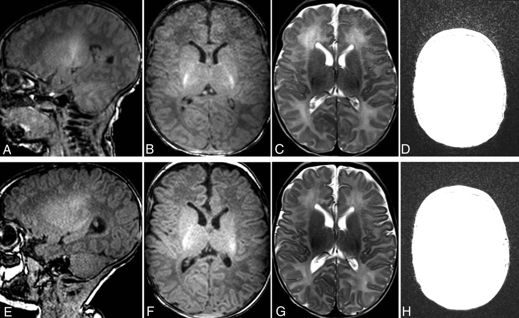
  • Fig 2.
    • Download figure
    • Open in new tab
    • Download powerpoint
    Fig 2.

    MR imaging of the brain of case 1 at presentation (A–D) and 20 days later (E–H). On the sagittal and axial T1WI (A and B) and the axial T2WI (C) of the first scan, the cerebral cortices of the bilateral frontal lobes show apparent irregular surfaces and gray-white matter junctions. Because the irregularity was observed on both T1WI and T2WI and the posterior cerebral cortices have rather smooth surfaces, without knowing the possibility of coil malfunction, we interpreted the findings as possible polymicrogyria. On the sagittal and axial T1WI (E and F) and the axial T2WI (G) of the follow-up scan obtained only 20 days later, the irregularity is no longer present, suggesting that the irregular appearance of the cortex seen on the first scan is artifactual rather than pathologic. When the window setting was adjusted to evaluate the background noise of the axial T1WI of the first (D) and follow-up (H) scan, it is obvious that the first scan has pronounced background noise in the frontal part of the image.

PreviousNext
Back to top

In this issue

American Journal of Neuroradiology: 41 (8)
American Journal of Neuroradiology
Vol. 41, Issue 8
1 Aug 2020
  • Table of Contents
  • Index by author
  • Complete Issue (PDF)
Advertisement
Print
Download PDF
Email Article

Thank you for your interest in spreading the word on American Journal of Neuroradiology.

NOTE: We only request your email address so that the person you are recommending the page to knows that you wanted them to see it, and that it is not junk mail. We do not capture any email address.

Enter multiple addresses on separate lines or separate them with commas.
MRI Head Coil Malfunction Producing Artifacts Mimicking Malformation of Cortical Development in Pediatric Epilepsy Work-Up
(Your Name) has sent you a message from American Journal of Neuroradiology
(Your Name) thought you would like to see the American Journal of Neuroradiology web site.
CAPTCHA
This question is for testing whether or not you are a human visitor and to prevent automated spam submissions.
Cite this article
N. Kashani, N. Khan, J.M. Ospel, X.-C. Wei
MRI Head Coil Malfunction Producing Artifacts Mimicking Malformation of Cortical Development in Pediatric Epilepsy Work-Up
American Journal of Neuroradiology Aug 2020, 41 (8) 1538-1540; DOI: 10.3174/ajnr.A6639

Citation Manager Formats

  • BibTeX
  • Bookends
  • EasyBib
  • EndNote (tagged)
  • EndNote 8 (xml)
  • Medlars
  • Mendeley
  • Papers
  • RefWorks Tagged
  • Ref Manager
  • RIS
  • Zotero
0 Responses
Respond to this article
Share
Bookmark this article
MRI Head Coil Malfunction Producing Artifacts Mimicking Malformation of Cortical Development in Pediatric Epilepsy Work-Up
N. Kashani, N. Khan, J.M. Ospel, X.-C. Wei
American Journal of Neuroradiology Aug 2020, 41 (8) 1538-1540; DOI: 10.3174/ajnr.A6639
del.icio.us logo Twitter logo Facebook logo Mendeley logo
  • Tweet Widget
  • Facebook Like
  • Google Plus One
Purchase

Jump to section

  • Article
    • Abstract
    • DISCUSSION
    • Acknowledgments
    • Footnotes
    • References
  • Figures & Data
  • Supplemental
  • Info & Metrics
  • Responses
  • References
  • PDF

Related Articles

  • PubMed
  • Google Scholar

Cited By...

  • No citing articles found.
  • Crossref
  • Google Scholar

This article has not yet been cited by articles in journals that are participating in Crossref Cited-by Linking.

More in this TOC Section

  • Comparison of Image Quality and Radiation Dose in Pediatric Temporal Bone CT Using Photon-Counting Detector CT and Energy-Integrating Detector CT
  • SyMRI & MR Fingerprinting in Brainstem Myelination
  • Pons&Vermis Localization on Fetal MRI Using U-Net
Show more Pediatrics

Similar Articles

Advertisement

Indexed Content

  • Current Issue
  • Accepted Manuscripts
  • Article Preview
  • Past Issues
  • Editorials
  • Editors Choice
  • Fellow Journal Club
  • Letters to the Editor

Cases

  • Case Collection
  • Archive - Case of the Week
  • Archive - Case of the Month
  • Archive - Classic Case

Special Collections

  • Special Collections

Resources

  • News and Updates
  • Turn around Times
  • Submit a Manuscript
  • Author Policies
  • Manuscript Submission Guidelines
  • Evidence-Based Medicine Level Guide
  • Publishing Checklists
  • Graphical Abstract Preparation
  • Imaging Protocol Submission
  • Submit a Case
  • Become a Reviewer/Academy of Reviewers
  • Get Peer Review Credit from Publons

Multimedia

  • AJNR Podcast
  • AJNR SCANtastic
  • Video Articles

About Us

  • About AJNR
  • Editorial Board
  • Not an AJNR Subscriber? Join Now
  • Alerts
  • Feedback
  • Advertise with us
  • Librarian Resources
  • Permissions
  • Terms and Conditions

American Society of Neuroradiology

  • Not an ASNR Member? Join Now

© 2025 by the American Society of Neuroradiology All rights, including for text and data mining, AI training, and similar technologies, are reserved.
Print ISSN: 0195-6108 Online ISSN: 1936-959X

Powered by HighWire