Advertisement

Main menu

  • Home
  • Content
    • Current Issue
    • Accepted Manuscripts
    • Article Preview
    • Past Issue Archive
    • AJNR Case Collection
    • Case of the Week Archive
    • Classic Case Archive
    • Case of the Month Archive
  • Special Collections
    • Spinal CSF Leak Articles (Jan 2020-June 2024)
    • 2024 AJNR Journal Awards
    • Most Impactful AJNR Articles
  • Multimedia
    • AJNR Podcast
    • AJNR Scantastics
    • Video Articles
  • For Authors
    • Submit a Manuscript
    • Author Policies
    • Fast publishing of Accepted Manuscripts
    • Graphical Abstract Preparation
    • Manuscript Submission Guidelines
    • Imaging Protocol Submission
    • Submit a Case for the Case Collection
  • About Us
    • About AJNR
    • Editorial Board
  • More
    • Become a Reviewer/Academy of Reviewers
    • Subscribers
    • Permissions
    • Alerts
    • Feedback
    • Advertisers
    • ASNR Home
  • Other Publications
    • ajnr

User menu

  • Alerts
  • Log in

Search

  • Advanced search
American Journal of Neuroradiology

American Journal of Neuroradiology

ASHNR American Society of Functional Neuroradiology ASHNR American Society of Pediatric Neuroradiology ASSR
  • Alerts
  • Log in

Advanced Search

  • Home
  • Content
    • Current Issue
    • Accepted Manuscripts
    • Article Preview
    • Past Issue Archive
    • AJNR Case Collection
    • Case of the Week Archive
    • Classic Case Archive
    • Case of the Month Archive
  • Special Collections
    • Spinal CSF Leak Articles (Jan 2020-June 2024)
    • 2024 AJNR Journal Awards
    • Most Impactful AJNR Articles
  • Multimedia
    • AJNR Podcast
    • AJNR Scantastics
    • Video Articles
  • For Authors
    • Submit a Manuscript
    • Author Policies
    • Fast publishing of Accepted Manuscripts
    • Graphical Abstract Preparation
    • Manuscript Submission Guidelines
    • Imaging Protocol Submission
    • Submit a Case for the Case Collection
  • About Us
    • About AJNR
    • Editorial Board
  • More
    • Become a Reviewer/Academy of Reviewers
    • Subscribers
    • Permissions
    • Alerts
    • Feedback
    • Advertisers
    • ASNR Home
  • Follow AJNR on Twitter
  • Visit AJNR on Facebook
  • Follow AJNR on Instagram
  • Join AJNR on LinkedIn
  • RSS Feeds

Welcome to the new AJNR, Updated Hall of Fame, and more. Read the full announcements.


AJNR is seeking candidates for the position of Associate Section Editor, AJNR Case Collection. Read the full announcement.


Getting new auth cookie, if you see this message a lot, tell someone!
Research ArticleAdult Brain
Open Access

MR Susceptibility Imaging with a Short TE (MR-SISET): A Clinically Feasible Technique to Resolve Thalamic Nuclei

S. Chung, P. Storey, T.M. Shepherd and Y.W. Lui
American Journal of Neuroradiology September 2020, 41 (9) 1629-1631; DOI: https://doi.org/10.3174/ajnr.A6683
S. Chung
aFrom the Center for Advanced Imaging Innovation and Research (CAI2R), and Bernard and Irene Schwartz Center for Biomedical Imaging, Department of Radiology, New York University Grossman School of Medicine, New York, New York.
  • Find this author on Google Scholar
  • Find this author on PubMed
  • Search for this author on this site
  • ORCID record for S. Chung
P. Storey
aFrom the Center for Advanced Imaging Innovation and Research (CAI2R), and Bernard and Irene Schwartz Center for Biomedical Imaging, Department of Radiology, New York University Grossman School of Medicine, New York, New York.
  • Find this author on Google Scholar
  • Find this author on PubMed
  • Search for this author on this site
  • ORCID record for P. Storey
T.M. Shepherd
aFrom the Center for Advanced Imaging Innovation and Research (CAI2R), and Bernard and Irene Schwartz Center for Biomedical Imaging, Department of Radiology, New York University Grossman School of Medicine, New York, New York.
  • Find this author on Google Scholar
  • Find this author on PubMed
  • Search for this author on this site
  • ORCID record for T.M. Shepherd
Y.W. Lui
aFrom the Center for Advanced Imaging Innovation and Research (CAI2R), and Bernard and Irene Schwartz Center for Biomedical Imaging, Department of Radiology, New York University Grossman School of Medicine, New York, New York.
  • Find this author on Google Scholar
  • Find this author on PubMed
  • Search for this author on this site
  • ORCID record for Y.W. Lui
  • Article
  • Figures & Data
  • Supplemental
  • Info & Metrics
  • Responses
  • References
  • PDF
Loading

Abstract

SUMMARY: The thalamus consists of several functionally distinct nuclei, some of which serve as targets for functional neurosurgery. Visualization of such nuclei is a major challenge due to their low signal contrast on conventional imaging. We introduce MR susceptibility imaging with a short TE, leveraging susceptibility differences among thalamic nuclei, to automatically delineate 15 thalamic subregions. The technique has the potential to enable direct targeting of thalamic nuclei for functional neurosurgical guidance.

ABBREVIATIONS:

MR-SISET
MR susceptibility imaging with a short TE
STh
subthalmic
CM
centromedian

The thalamus plays a critical role as a central relay hub and comprises functionally specific nuclei. However, it appears relatively homogeneous in structure on conventional imaging (Fig 1A). This appearance limits the ability of physicians to accurately target specific nuclei in functional neurosurgical therapies, such as the ventral intermediate/lateral and subthalamic (STh) nuclei for the treatment of essential tremor and tremor-dominant Parkinson disease1 and the centromedian (CM) nucleus for the treatment of refractory epilepsy2 and Tourette syndrome.3 Neurosurgical outcome is critically dependent on accurate targeting; however, current clinical practice relies on indirect localization based on standard coordinates because there is no reliable, noninvasive means currently available to resolve individual nuclei. Indirect targeting is suboptimal due to anatomic variation across individuals, which may lead to inaccurate targeting with potential implications for clinical outcome. Histology-based atlases may improve accuracy4 but are subject to limitations such as the typically small number of subjects on which they are based and registration errors when mapping from atlas to patient. DTI tractography techniques aimed at delineating thalamic nuclei are problematic when white matter tracts are difficult to track.5

FIG 1.
  • Download figure
  • Open in new tab
  • Download powerpoint
FIG 1.

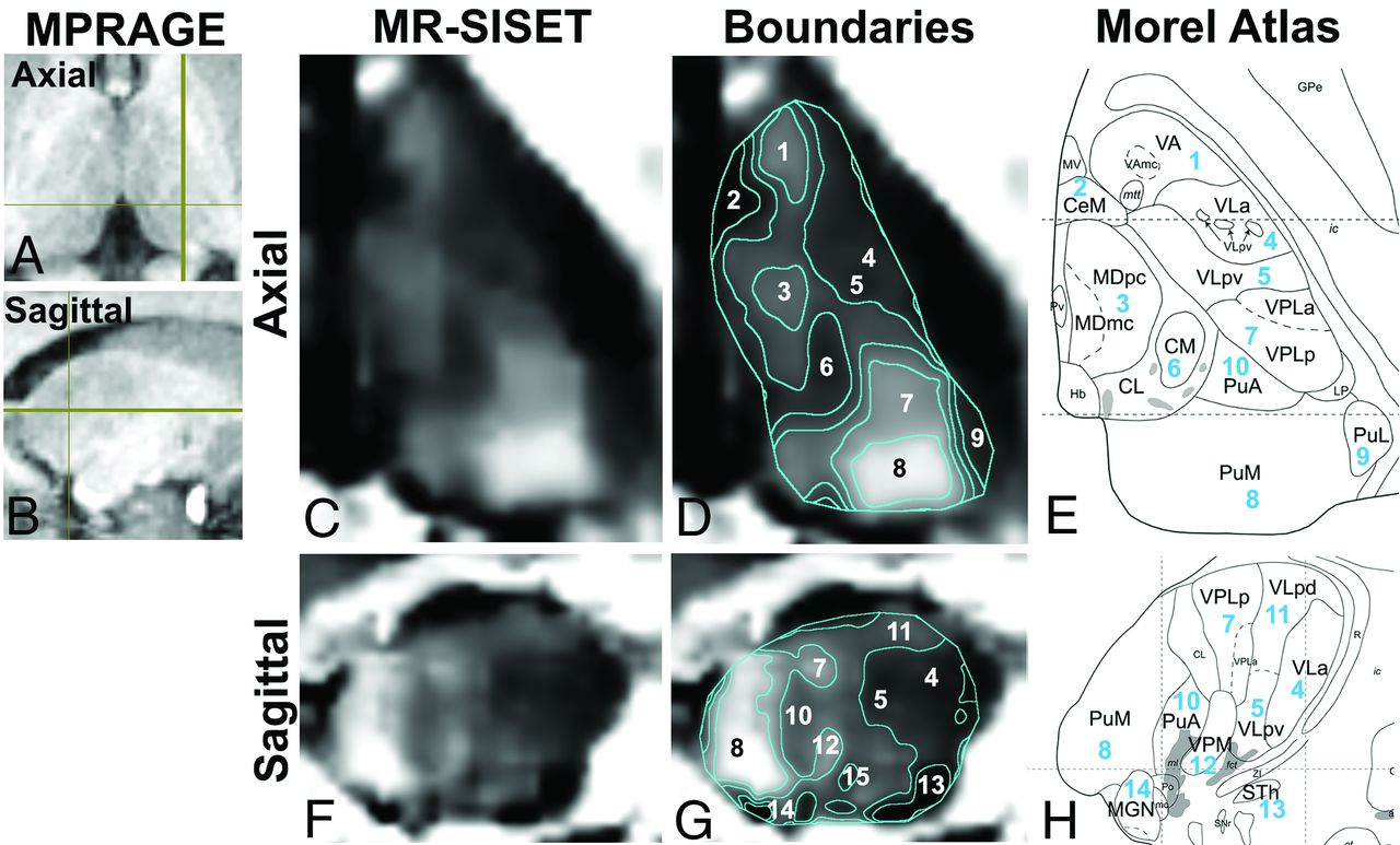
MPRAGE images are shown in the axial (A) and sagittal planes (B), indicating section location. Axial images: C, MR-SISET map. D, Boundaries of 15 thalamic nuclei derived from K-means clustering. E, Corresponding Morel atlas labels are shown. F–H, Sagittal images (1-VA, 2-CeM, 3-MD, 4-VLa, 5-VLpv, 6-CM, 7-VPL, 8-PuM, 9-PuL, 10-PuA, 11-VLpd, 12-VPM, 13-STh, 14-MGN, 15-VM (not shown in H). Reprinted with permission from Morel, 2007.6

The thalamus is a deep gray matter structure that contains a considerable amount of WM. In the thalamus, iron content, degree of WM myelination,6 and WM fiber orientation7 may all contribute to susceptibility differences within the structure. There are recent reports showing the potential of susceptibility imaging for delineating thalamic nuclei at 7T;8,9 however, clinically practical protocols at 3T are lacking. Here, we introduce a novel method, leveraging short TEs, termed MR susceptibility imaging with a short TE (MR-SISET), to bring out differences in contrast among thalamic nuclei and provide an automated method to delineate the intrathalamic subregions using K-means clustering.

Technique

The study was approved by the local institutional review board, and all subjects provided informed consent. Ten healthy controls were included in the study spanning a broad age range (23–65 years of age; 3 men) to assess intersubject variation due to age-related myelination changes. MR-SISET was performed on 3T MR imaging scanners (Magnetom Skyra/Prisma [6/4 subjects]; Siemens) using a 3D multiecho gradient-echo sequence: FOV = 220 ×170 × 75 mm3, matrix = 176 × 136 × 60, 1.25-mm isotropic resolution, flip angle = 22°, TR = 92 ms, 20 multiple TE = 1.90–45.98 ms with echo spacing of 2.32 ms, bandwidth = 840 Hz/pixel. MR-SISET maps were generated using the MEDI toolbox10 with a relatively high regularization parameter (λ= 2000) related to data fidelity and sharpness to optimize image contrast. Automatic delineation was performed using a K-means algorithm with 6 different intensity clusters (Matlab R2019b; MathWorks). Subregions were visually inspected and compared against known thalamic nuclei as delineated in the Morel atlas.4 Susceptibility contrast relative to that of the CM nucleus was calculated for each subregion.

RESULTS

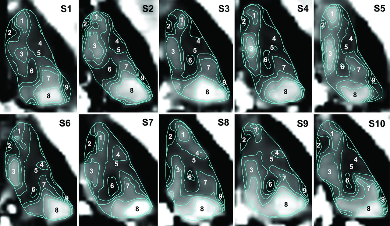
MR-SISET maps showed clear internal heterogeneity with 15 distinct nuclei showing a high degree of signal contrast with neighboring nuclei (Fig 1). There were clear similarities between subjects with several subregions that were consistently segmented and appeared to correspond to the following thalamic nuclei on the Morel atlas (Fig 2 and On-line Figure): CM (1), central medial (CeM, 0.92 ± 0.08), lateral pulvinar (PuL, 0.87 ± 0.09), STh (0.62 ± 0.17), and ventral medial (VM 0.95 ± 0.08) regions were consistently darker than their neighbors, while ventral anterior (VA, 1.32 ± 0.09), mediodorsal (MD, 1.41 ± 0.2), ventral posterior lateral (VPL, 1.29 ± 0.11), ventral posterior medial (VPM, 1.29 ± 0.13), and medial pulvinar (PuM, 1.79 ± 0.31) regions were brighter than their surroundings (contrast ratios are shown in parentheses). The ventral lateral anterior (VLa, 1.10 ± 0.1) and ventral lateral posterior, ventral division (VLpv, 1.04 ± 0.06) regions were not easily separable. The anterior pulvinar (PuA, 1.17 ± 0.13), ventral lateral posterior, dorsal division (VLpd, 1.17 ± 0.15), and medial geniculate (MGN, 1.18 ± 0.19) regions had similar contrast. The terms are defined and contrast ratios are summarized in the On-line Table.

FIG 2.
  • Download figure
  • Open in new tab
  • Download powerpoint
FIG 2.

MR-SISET maps with corresponding boundaries for all 10 subjects, showing clear similarities between subjects with several subregions that were consistently segmented. S indicates subject.

DISCUSSION

MR-SISET delineates 15 distinct thalamic subregions at 3T, as a result of a greater sensitivity to myelin content and WM packing geometry, supported by previous works.11 A limitation of this study is the lack of ground truth. Instead, indirect comparisons have been performed against the Morel atlas, which is based on histology, though from a small number of subjects. The regions segmented using MR-SISET do appear to correspond well to nuclei delineated in the Morel atlas. Further work is warranted in patients with relevant pathology to assess the potential utility in patients undergoing functional neurosurgery.

CONCLUSIONS

MR-SISET enables subject-specific delineation of intrathalamic structures. Thus far, magnetic susceptibility–based techniques have not been widely exploited for resolving intrathalamic structures, particularly at 3T. Our preliminary results suggest that MR-SISET has the potential to aid in more personalized, direct visualization of thalamic nuclei for functional neurosurgery.

Footnotes

  • This work was supported by the National Institutes of Health R01 NS039135-11, R21 NS090349, P41 EB017183; and the Leon Lowenstein Foundation.

  • Disclosures: Sohae Chung—RELATED: Grant: National Institutes of Health R01 NS039135-11, R21 NS090349, P41 EB017183.* Pippa Storey—RELATED: Grant: National Institutes of Health.* Timothy M. Shepherd—UNRELATED: Stock/Stock Options: Microstructure Imaging, Comments: founder and equity for startup, no money has been paid to me or my institution. Yvonne W. Lui—RELATED: Grant: National Institutes of Health*; UNRELATED: Grants/Grants Pending: Leon Lowenstein Foundation, Department of Defense.* *Money paid to the institution.

Indicates open access to non-subscribers at www.ajnr.org

References

  1. 1.↵
    1. Benabid AL,
    2. Pollak P,
    3. Gervason C, et al
    . Long-term suppression of tremor by chronic stimulation of the ventral intermediate thalamic nucleus. Lancet 1991;337:403–06 doi:10.1016/0140-6736(91)91175-t pmid:1671433
    CrossRefPubMed
  2. 2.↵
    1. Son BC,
    2. Shon YM,
    3. Choi JG, et al
    . Clinical outcome of patients with deep brain stimulation of the centromedian thalamic nucleus for refractory epilepsy and location of the active contacts. Stereotact Funct Neurosurg 2016;94:187–97 doi:10.1159/000446611 pmid:27434073
    CrossRefPubMed
  3. 3.↵
    1. Vandewalle V,
    2. van der Linden C,
    3. Groenewegen HJ, et al
    . Stereotactic treatment of Gilles de la Tourette syndrome by high frequency stimulation of thalamus. Lancet 1999;353:724 doi:10.1016/s0140-6736(98)05964-9 pmid:10073521
    CrossRefPubMed
  4. 4.↵
    1. Morel A,
    2. Magnin M,
    3. Jeanmonod D
    . Multiarchitectonic and stereotactic atlas of the human thalamus. J Comp Neurol 1997;387:588–630 doi:10.1002/(SICI)1096-9861(19971103)387:4<588::AID-CNE8>3.0.CO;2-Z pmid:9373015
    CrossRefPubMed
  5. 5.↵
    1. Sammartino F,
    2. Krishna V,
    3. King NK, et al
    . Tractography-based ventral intermediate nucleus targeting: novel methodology and intraoperative validation. Mov Disord 2016;31:1217–25 doi:10.1002/mds.26633
    CrossRefPubMed
  6. 6.↵
    1. Morel A
    . Stereotactic Atlas of the Human Thalamus and Basal Ganglia. Informa Healthcare; 2007:13–53
  7. 7.↵
    1. Lancione M,
    2. Tosetti M,
    3. Donatelli G, et al
    . The impact of white matter fiber orientation in single-acquisition quantitative susceptibility mapping. NMR Biomed 2017;30:doi:10.1002/nbm.3798 doi:10.1002/nbm.3798 pmid:28902421
    CrossRefPubMed
  8. 8.↵
    1. Abosch A,
    2. Yacoub E,
    3. Ugurbil K, et al
    . An assessment of current brain targets for deep brain stimulation surgery with susceptibility-weighted imaging at 7 Tesla. Neurosurgery 2010;67:1745–56; discussion 1756 doi:10.1227/NEU.0b013e3181f74105 pmid:21107206
    CrossRefPubMed
  9. 9.↵
    1. Deistung A,
    2. Schafer A,
    3. Schweser F, et al
    . Toward in vivo histology: a comparison of quantitative susceptibility mapping (QSM) with magnitude-, phase-, and R2*-imaging at ultra-high magnetic field strength. Neuroimage 2013;65:299–314 doi:10.1016/j.neuroimage.2012.09.055 pmid:23036448
    CrossRefPubMed
  10. 10.↵
    1. Liu T,
    2. Xu W,
    3. Spincemaille P, et al
    . Accuracy of the morphology enabled dipole inversion (MEDI) algorithm for quantitative susceptibility mapping in MRI. IEEE Trans Med Imaging 2012;31:816–24 doi:10.1109/TMI.2011.2182523 pmid:22231170
    CrossRefPubMed
  11. 11.↵
    1. Sood S,
    2. Urriola J,
    3. Reutens D, et al
    . Echo time-dependent quantitative susceptibility mapping contains information on tissue properties. Magn Reson Med 2017;77:1946–58 doi:10.1002/mrm.26281 pmid:27221590
    CrossRefPubMed
  • Received November 13, 2019.
  • Accepted after revision May 11, 2020.
  • © 2020 by American Journal of Neuroradiology
View Abstract
PreviousNext
Back to top

In this issue

American Journal of Neuroradiology: 41 (9)
American Journal of Neuroradiology
Vol. 41, Issue 9
1 Sep 2020
  • Table of Contents
  • Index by author
  • Complete Issue (PDF)
Advertisement
Print
Download PDF
Email Article

Thank you for your interest in spreading the word on American Journal of Neuroradiology.

NOTE: We only request your email address so that the person you are recommending the page to knows that you wanted them to see it, and that it is not junk mail. We do not capture any email address.

Enter multiple addresses on separate lines or separate them with commas.
MR Susceptibility Imaging with a Short TE (MR-SISET): A Clinically Feasible Technique to Resolve Thalamic Nuclei
(Your Name) has sent you a message from American Journal of Neuroradiology
(Your Name) thought you would like to see the American Journal of Neuroradiology web site.
CAPTCHA
This question is for testing whether or not you are a human visitor and to prevent automated spam submissions.
Cite this article
S. Chung, P. Storey, T.M. Shepherd, Y.W. Lui
MR Susceptibility Imaging with a Short TE (MR-SISET): A Clinically Feasible Technique to Resolve Thalamic Nuclei
American Journal of Neuroradiology Sep 2020, 41 (9) 1629-1631; DOI: 10.3174/ajnr.A6683

Citation Manager Formats

  • BibTeX
  • Bookends
  • EasyBib
  • EndNote (tagged)
  • EndNote 8 (xml)
  • Medlars
  • Mendeley
  • Papers
  • RefWorks Tagged
  • Ref Manager
  • RIS
  • Zotero
0 Responses
Respond to this article
Share
Bookmark this article
MR Susceptibility Imaging with a Short TE (MR-SISET): A Clinically Feasible Technique to Resolve Thalamic Nuclei
S. Chung, P. Storey, T.M. Shepherd, Y.W. Lui
American Journal of Neuroradiology Sep 2020, 41 (9) 1629-1631; DOI: 10.3174/ajnr.A6683
del.icio.us logo Twitter logo Facebook logo Mendeley logo
  • Tweet Widget
  • Facebook Like
  • Google Plus One
Purchase

Jump to section

  • Article
    • Abstract
    • ABBREVIATIONS:
    • RESULTS
    • DISCUSSION
    • CONCLUSIONS
    • Footnotes
    • References
  • Figures & Data
  • Supplemental
  • Info & Metrics
  • Responses
  • References
  • PDF

Related Articles

  • PubMed
  • Google Scholar

Cited By...

  • No citing articles found.
  • Crossref
  • Google Scholar

This article has not yet been cited by articles in journals that are participating in Crossref Cited-by Linking.

More in this TOC Section

  • Diagnostic Neuroradiology of Monoclonal Antibodies
  • Segmentation of Brain Metastases with BLAST
  • NCCT vs. MRI for Brain Atrophy in Acute Stroke
Show more Adult Brain

Similar Articles

Advertisement

Indexed Content

  • Current Issue
  • Accepted Manuscripts
  • Article Preview
  • Past Issues
  • Editorials
  • Editors Choice
  • Fellow Journal Club
  • Letters to the Editor

Cases

  • Case Collection
  • Archive - Case of the Week
  • Archive - Case of the Month
  • Archive - Classic Case

Special Collections

  • Special Collections

Resources

  • News and Updates
  • Turn around Times
  • Submit a Manuscript
  • Author Policies
  • Manuscript Submission Guidelines
  • Evidence-Based Medicine Level Guide
  • Publishing Checklists
  • Graphical Abstract Preparation
  • Imaging Protocol Submission
  • Submit a Case
  • Become a Reviewer/Academy of Reviewers
  • Get Peer Review Credit from Publons

Multimedia

  • AJNR Podcast
  • AJNR SCANtastic
  • Video Articles

About Us

  • About AJNR
  • Editorial Board
  • Not an AJNR Subscriber? Join Now
  • Alerts
  • Feedback
  • Advertise with us
  • Librarian Resources
  • Permissions
  • Terms and Conditions

American Society of Neuroradiology

  • Not an ASNR Member? Join Now

© 2025 by the American Society of Neuroradiology All rights, including for text and data mining, AI training, and similar technologies, are reserved.
Print ISSN: 0195-6108 Online ISSN: 1936-959X

Powered by HighWire