
Article Figures & Data
Figures
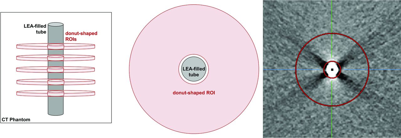
- FIG 1.
Schematic illustration of the donut-shaped ROI created by the MITK with an inner radius of 6 mm and an outer radius of 26 mm. The tube filled with the LEA of interest was placed centrally with a radius equal to the radius of the donut hole. For each tube, the measurement was performed with an ROI in 5 different positions according to the length of the LEA cast.
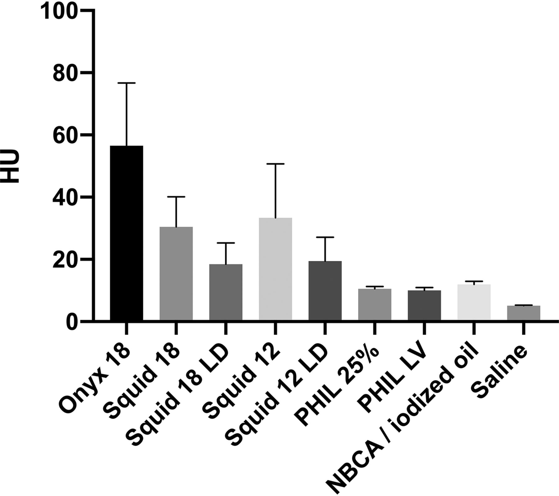
- FIG 2.
Illustration of the results of the quantitative image analysis. Different degrees of artifacts were observed among all study groups. Post hoc testing showed differences among the different types of LEAs (eg, Squid 18 versus PHIL 25% and Squid 12 versus PHIL LV). Bars indicate mean; whiskers, SD.
- FIG 3.
Representative CT images in the axial plane in a standard brain window with a width of 80 and a length of 40. Note the more severe artifacts for the EVOH-based LEAs (Onyx and Squid) and the relatively low degree of artifacts for LEAs that used iodine as a radiopaque component (PHIL and n-BCA/iodized oil).
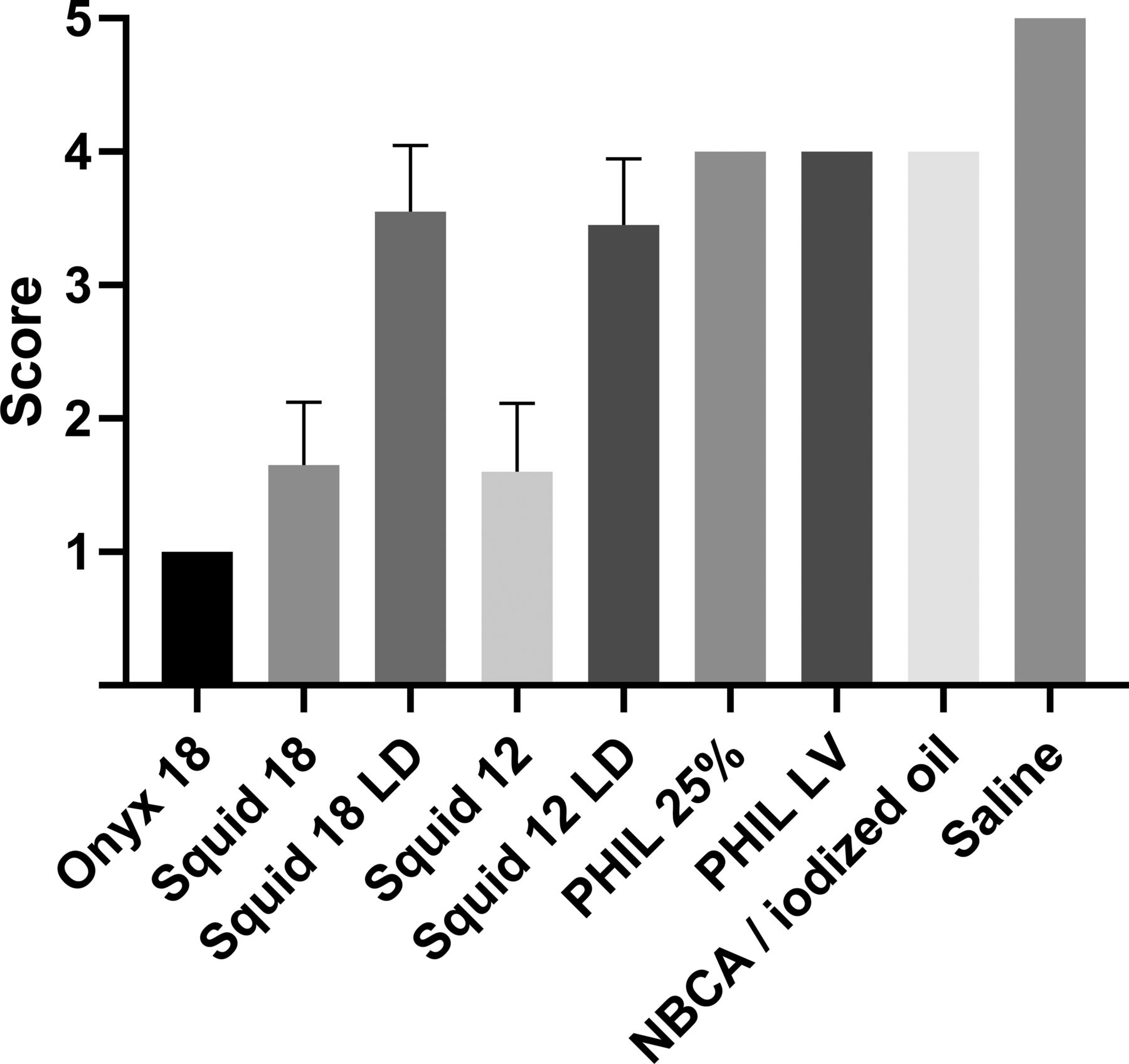
- FIG 4.
Illustration of the results of the qualitative image analysis using a 5-point scale. Different degrees of artifacts were observed among all study groups using the Dunn test for multiple comparisons with statistical hypothesis testing. Bars indicate mean; whiskers, SD. Five-point scale: 1) severe artifacts, 2) marked artifacts, 3) moderate artifacts, 4) minor artifacts, and 5) no artifacts.


Tables
Liquid Embolic Agent SD of Donut-Shaped ROI P Valuea Onyx 18 56.6 ± 20.1 HU Squid 18 30.3 ± 9.7 HU Squid 18 LD 18.5 ± 6.8 HU Squid 12 33.4 ± 17.3 HU Squid 12 LD 19.4 ± 7.7 HU P < .001 PHIL 25% 10.6 ± 0.8 HU PHIL LV 10.1 ± 0.9 HU n-BCA/iodized oil 11.9 ± 1.0 HU Saline 5.1 ± 0.1 HU ↵a Kruskal-Wallis test; for the P values of the post hoc analysis, see Table 2.
Liquid Embolic Agent Onyx 18 Squid 18 Squid 18 LD Squid 12 Squid 12 LD PHIL 25% PHIL LV n-BCA/Iodized Oil Saline P < .001a P < .001a P < .001a P < .001a P < .001a P = .001a P = .034a P < .001a n-BCA/iodized oil P < .001a P < .001a P = .106 P < .001a P = .468 P = .987 P = .096 PHIL LV NA NA NA P < .001 NA P > .999 PHIL 25% P < .001a P < .001a NA NA NA Squid 12 LD NA NA P > .999 P = .013a Squid 12 NA P > .999 NA Squid 18 LD NA P = .057 Squid 18 P = .118 Note:—NA indicates no P value available because the Dunn test was only performed for corresponding LEA variants.
↵a Statistical significance.
Liquid Embolic Agent Onyx 18 Squid 18 Squid 18 LD Squid 12 Squid 12 LD PHIL 25% PHIL LV n-BCA/Iodized Oil Saline P < .001a P < .001a P = .022a P < .001a P = .009a P = .665 P = .665 P = .665 n-BCA/iodized oil P < .001a P = .004a P > .999 P = .003a P > .999 P > .999 P > .999 PHIL LV NA NA NA P = .003a NA P > .999 PHIL 25% P < .001a P = .004a NA NA NA Squid 12 LD NA NA P > .999 P = .311 Squid 12 NA P > .999 NA Squid 18 LD NA P = .201 Squid 18 P > .999 Note:—NA indicates no P value available because the Dunn test was performed for only corresponding LEA variants.
↵a Statistical significance.


