
Article Figures & Data
Figures
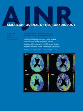
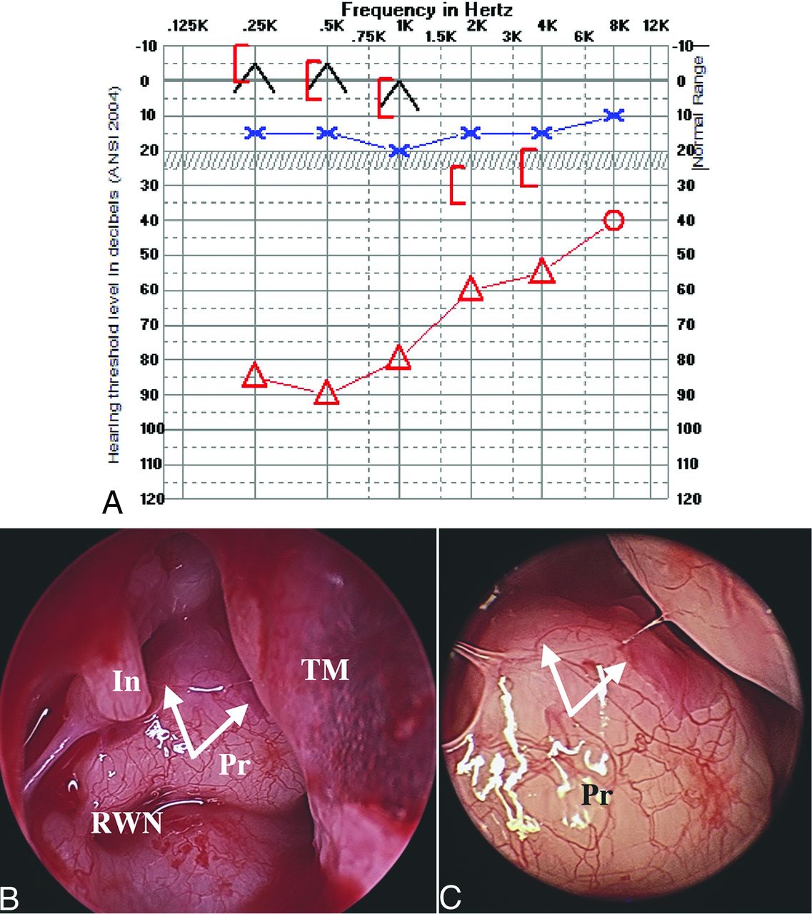
- FIG 1.
A, Preoperative audiogram demonstrates a right-sided near-maximal CHL and normal hearing thresholds in the left ear. B and C, Right middle ear intraoperative endoscopy demonstrates right PSA (white arrows) coursing over the surface of the cochlear promontory. Notably, the inferior half of the artery was encased within promontory bone; however, the superior aspect was dehiscent and easily visible during surgery. In this patient, the artery and facial nerve were pulsatile. In = incus; Pr = promontory of cochlea; RWN = round window niche; TM = tympanic membrane.
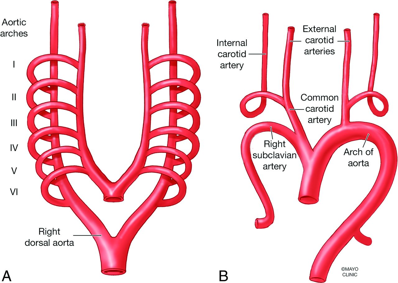
- FIG 2.
Primitive aortic arches embryology. A, Paired 6 aortic arches course from the aortic sac to the ipsilateral descending dorsal aorta (right dorsal aorta is annotated). B, First and 2nd arches give rise to the ventral pharyngeal artery, later to become ECA; the 3rd arch becomes proximal ICA; and the ventral 3rd–4th arch junction becomes the common carotid artery. Used with permission of Mayo Foundation for Medical Education and Research, all rights reserved.
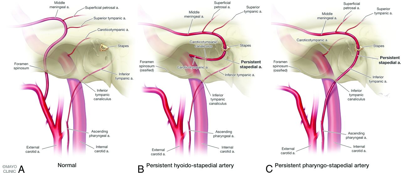
- FIG 3.
Normal carotid vascular anatomy (A) and anatomic variants of persistent stapedial artery (PSA, B and C). B, PSA is supplied by an enlarged caroticotympanic artery, a remnant of the embryonic hyoid artery stem arising from the petrous ICA via caroticotympanic canaliculus; this anatomic variant is referred to as persistent hyoidostapedial artery. C, PSA is supplied by an enlarged inferior tympanic artery, a branch of the ascending pharyngeal artery, which travels through the inferior tympanic canaliculus into the middle ear, anastomosing with the embryonic hyoid artery; this anatomic variant is referred to as persistent pharyngostapedial artery. Used with permission of Mayo Foundation for Medical Education and Research, all rights reserved.
- FIG 4.
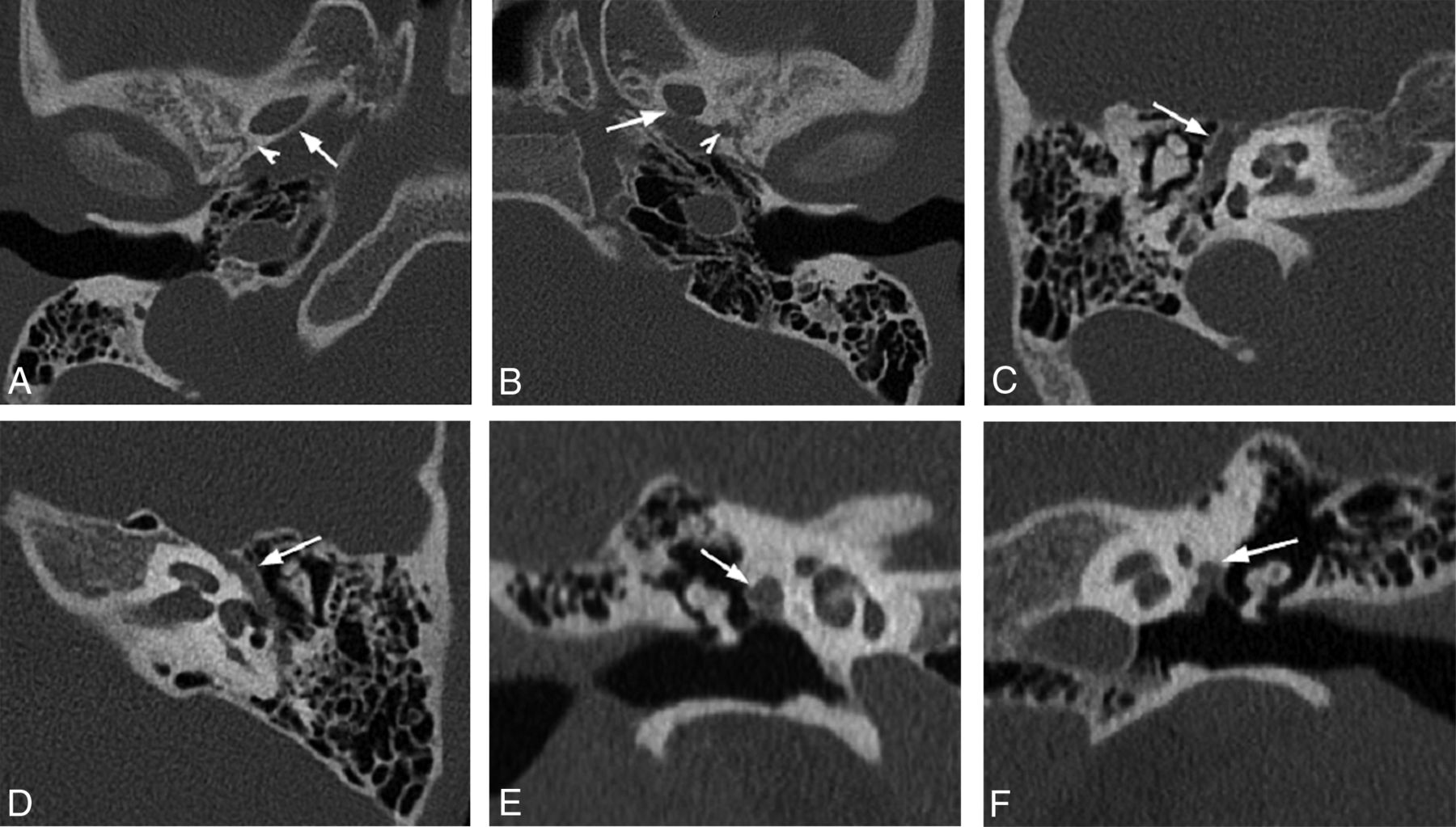
Nonspecific findings of PSA. Axial oblique CT reconstructions of right (A) and left (B) temporal bones in the same patient with the right PSA demonstrate normal foramen spinosum on the left and absent on the right at its expected location (arrowheads); note the normal foramen ovale on both sides (arrows). Axial (C) and coronal (E) oblique reconstructions of nonduplicated, enlarged tympanic segment of the right facial nerve canal (FNC). Nonduplicated FNC is enlarged to accommodate facial nerve and PSA (arrows). Compare with normal nonenlarged left FNC in similar planes, D and F (arrows).
- FIG 5.
Right ear (A) and left ear (B) coronal CT images from 2 patients demonstrate an additional lumen lateral to the tympanic facial canal (“3-eyed snail”) representing the bifurcation of the PSA as it traverses the floor of the middle cranial fossa to give rise the middle meningeal artery (white arrows). Labyrinthine (black arrows) and tympanic (white arrowheads) segments of the facial nerve represent usually seen “2 eyes of the snail.” Right ear (C) and left ear (D) axial CT images in the same patients demonstrate duplication of the anterior segment of the tympanic facial nerve canal (white arrows). The labyrinthine segment (black arrows), tympanic segment (white arrowheads), and PSA canal intersect to form an “N” on the left (D) and a reverse “N” on the right (C). Geniculate ganglion is seen anteromedially (black arrowheads). TT = tensor tympani.
- FIG 6.
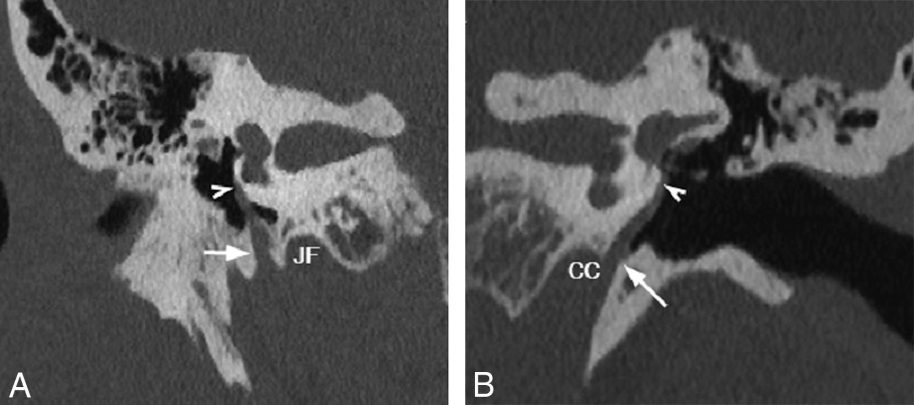
Coronal oblique CT reconstructions demonstrate PSA coursing over the lateral surface of the cochlear promontory (string sign). Right PSA (A) arises from the inferior tympanic canaliculus (arrow), and left PSA (B) takes its origin from the carotid canal via caroticotympanic canaliculus (arrow), both traversing cephalad over the cochlear promontory (arrowheads). CC = carotid canal; JF = jugular foramen.
- FIG 7.
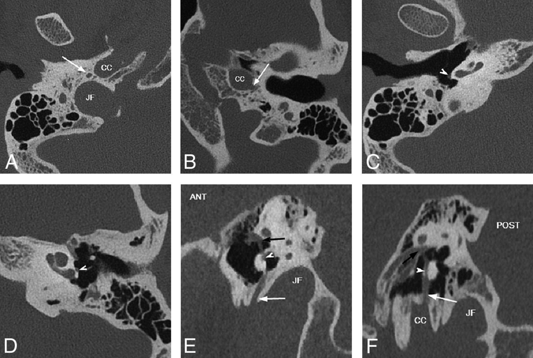
Variable origin of the PSA. Axial (A–D) and sagittal oblique (E and F) CT reconstructions. In A, C, and E, PSA arises from the ITC posterolateral to the carotid canal (CC) and anterior to the jugular foramen (JF), (white arrows). In B, D, and F, PSA arises directly from CC via CTC (white arrows). Note the posterior position of the PSA on the cochlear promontory with ITC origin versus the anterior position with CTC origin (white arrowheads), best seen on sagittal reconstructions, E and F, PSA traverses oval window niche to enter tympanic segment of the facial nerve canal (black arrows). ANT = anterior; POST = posterior.
- FIG 8.
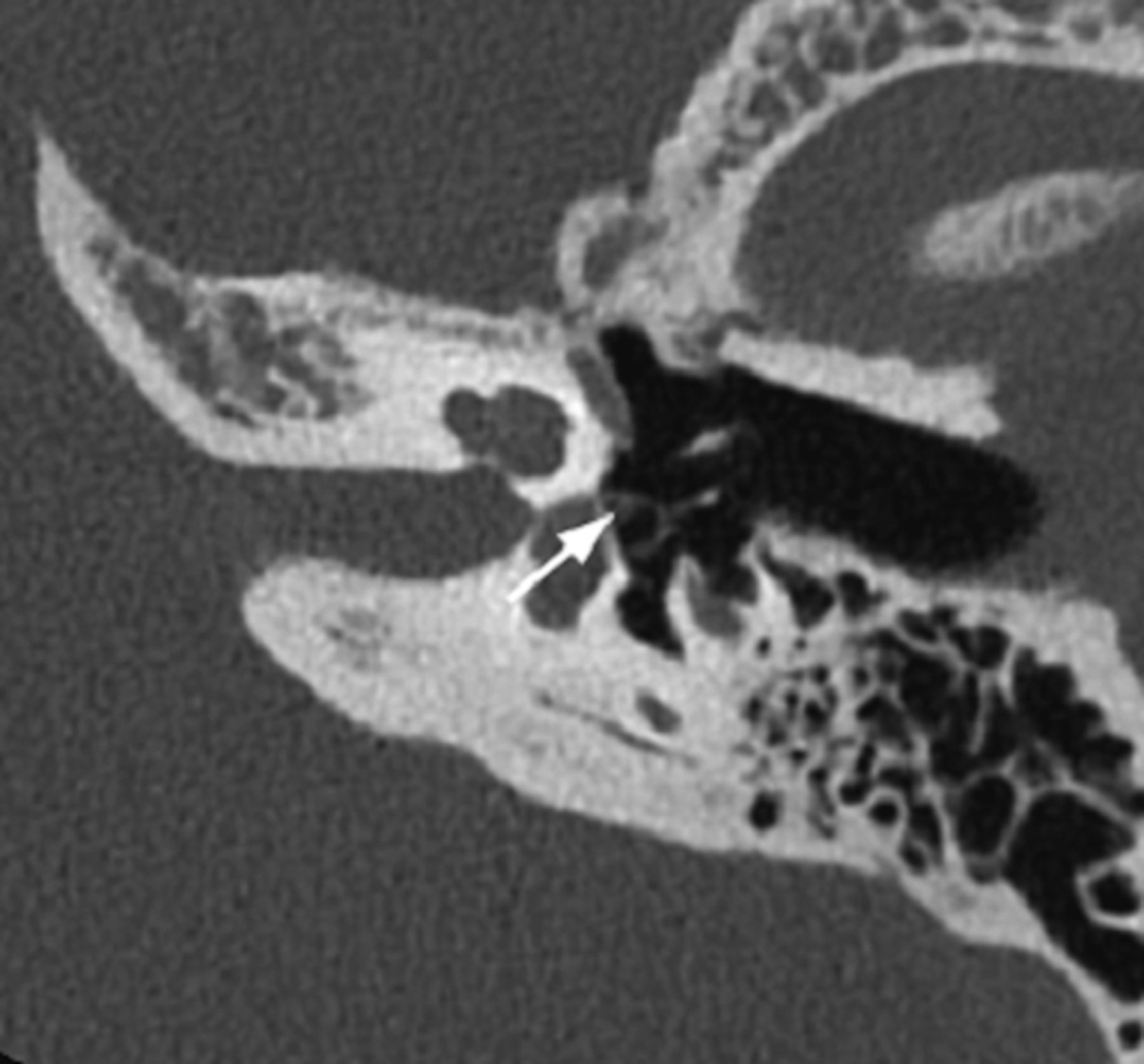
PSA courses through the obturator foramen of the stapes. Oblique axial CT reconstruction demonstrates cross-section of the artery along the inner margin of the anterior crus of the stapes (arrow) resembling a horseshoe on a stake (ringer sign).


Tables
Patient characteristics and prevalence of associated persistent stapedial artery findings
Case Side Associated Anomalies CHL Duplicated FNC Fr Spn Coch Prom Vessel Large CTC Large ITC Stapes Obturator Artery Surgical Confirmation 1 L None + + − + + − + − 2 L Ipsilateral congenital cholesteatoma and dysplastic anterior stapedial crus + − − + + − + + 3 L None N/A + − + − + + − 4 L None − − − + − + + − 5 R Ipsilateral congenital aerodigestive venolymphatic malformation and dysplastic stapes + − − + + − −c + 6 R Chromosome 3q duplication or trisomy N/A + − + + − + − 7 R None − + − + − + + − 8 L None − − − + + − + − 9a R None + + − + + − −d + 10a L None +b + − + + − + − ↵a #9 and #10: bilateral PSA; same patient.
↵b #10 remote middle ear surgery, not otherwise specified.
↵c #5 PSA anterior to the dysplastic crus.
↵d #9 granulation tissue obscured artery.
Note:—Findings are marked as either present (+) or absent (−) in each patient. Coch Prom indicates cochlear promontory; FNC = facial nerve canal; Fr Spn = foramen spinosum; N/A = not applicable.


Jump to section
Related Articles
Cited By...
- No citing articles found.