Advertisement

Main menu

  • Home
  • Content
    • Current Issue
    • Accepted Manuscripts
    • Article Preview
    • Past Issue Archive
    • AJNR Case Collection
    • Case of the Week Archive
    • Classic Case Archive
    • Case of the Month Archive
  • Special Collections
    • Spinal CSF Leak Articles (Jan 2020-June 2024)
    • 2024 AJNR Journal Awards
    • Most Impactful AJNR Articles
  • Multimedia
    • AJNR Podcast
    • AJNR Scantastics
    • Video Articles
  • For Authors
    • Submit a Manuscript
    • Author Policies
    • Fast publishing of Accepted Manuscripts
    • Graphical Abstract Preparation
    • Manuscript Submission Guidelines
    • Imaging Protocol Submission
    • Submit a Case for the Case Collection
  • About Us
    • About AJNR
    • Editorial Board
  • More
    • Become a Reviewer/Academy of Reviewers
    • Subscribers
    • Permissions
    • Alerts
    • Feedback
    • Advertisers
    • ASNR Home
  • Other Publications
    • ajnr

User menu

  • Alerts
  • Log in

Search

  • Advanced search
American Journal of Neuroradiology

American Journal of Neuroradiology

ASHNR American Society of Functional Neuroradiology ASHNR American Society of Pediatric Neuroradiology ASSR
  • Alerts
  • Log in

Advanced Search

  • Home
  • Content
    • Current Issue
    • Accepted Manuscripts
    • Article Preview
    • Past Issue Archive
    • AJNR Case Collection
    • Case of the Week Archive
    • Classic Case Archive
    • Case of the Month Archive
  • Special Collections
    • Spinal CSF Leak Articles (Jan 2020-June 2024)
    • 2024 AJNR Journal Awards
    • Most Impactful AJNR Articles
  • Multimedia
    • AJNR Podcast
    • AJNR Scantastics
    • Video Articles
  • For Authors
    • Submit a Manuscript
    • Author Policies
    • Fast publishing of Accepted Manuscripts
    • Graphical Abstract Preparation
    • Manuscript Submission Guidelines
    • Imaging Protocol Submission
    • Submit a Case for the Case Collection
  • About Us
    • About AJNR
    • Editorial Board
  • More
    • Become a Reviewer/Academy of Reviewers
    • Subscribers
    • Permissions
    • Alerts
    • Feedback
    • Advertisers
    • ASNR Home
  • Follow AJNR on Twitter
  • Visit AJNR on Facebook
  • Follow AJNR on Instagram
  • Join AJNR on LinkedIn
  • RSS Feeds

Welcome to the new AJNR, Updated Hall of Fame, and more. Read the full announcements.


AJNR is seeking candidates for the position of Associate Section Editor, AJNR Case Collection. Read the full announcement.


Getting new auth cookie, if you see this message a lot, tell someone!
Research ArticleAdult Brain
Open Access

Brain MR Spectroscopic Findings in 3 Consecutive Patients with COVID-19: Preliminary Observations

O. Rapalino, A. Weerasekera, S.J. Moum, K. Eikermann-Haerter, B.L. Edlow, D. Fischer, A. Torrado-Carvajal, M.L. Loggia, S.S. Mukerji, P.W. Schaefer, R.G. Gonzalez, M.H. Lev and E.-M. Ratai
American Journal of Neuroradiology January 2021, 42 (1) 37-41; DOI: https://doi.org/10.3174/ajnr.A6877
O. Rapalino
aFrom the Departments of Radiology (O.R., A.W., K.E.-H., M.L.L., P.W.S., R.G.G., M.H.L., E.R.)
  • Find this author on Google Scholar
  • Find this author on PubMed
  • Search for this author on this site
  • ORCID record for O. Rapalino
A. Weerasekera
aFrom the Departments of Radiology (O.R., A.W., K.E.-H., M.L.L., P.W.S., R.G.G., M.H.L., E.R.)
cA.A. Martinos Center for Biomedical Imaging (A.W., A.T.-C., M.L.L., E.R.), Charlestown, Massachusetts
  • Find this author on Google Scholar
  • Find this author on PubMed
  • Search for this author on this site
  • ORCID record for A. Weerasekera
S.J. Moum
dAnn & Robert H. Lurie Children’s Hospital of Chicago and Northwestern University Feinberg School of Medicine (S.J.M.)
  • Find this author on Google Scholar
  • Find this author on PubMed
  • Search for this author on this site
  • ORCID record for S.J. Moum
K. Eikermann-Haerter
aFrom the Departments of Radiology (O.R., A.W., K.E.-H., M.L.L., P.W.S., R.G.G., M.H.L., E.R.)
  • Find this author on Google Scholar
  • Find this author on PubMed
  • Search for this author on this site
  • ORCID record for K. Eikermann-Haerter
B.L. Edlow
bNeurology (B.L.E., D.F., S.S.M.), Massachusetts General Hospital and Harvard Medical School, Boston, Massachusetts
  • Find this author on Google Scholar
  • Find this author on PubMed
  • Search for this author on this site
  • ORCID record for B.L. Edlow
D. Fischer
bNeurology (B.L.E., D.F., S.S.M.), Massachusetts General Hospital and Harvard Medical School, Boston, Massachusetts
  • Find this author on Google Scholar
  • Find this author on PubMed
  • Search for this author on this site
  • ORCID record for D. Fischer
A. Torrado-Carvajal
cA.A. Martinos Center for Biomedical Imaging (A.W., A.T.-C., M.L.L., E.R.), Charlestown, Massachusetts
eMedical Image Analysis and Biometry Laboratory (A.T.-C.), Universidad Rey Juan Carlos, Madrid, Spain.
  • Find this author on Google Scholar
  • Find this author on PubMed
  • Search for this author on this site
  • ORCID record for A. Torrado-Carvajal
M.L. Loggia
aFrom the Departments of Radiology (O.R., A.W., K.E.-H., M.L.L., P.W.S., R.G.G., M.H.L., E.R.)
cA.A. Martinos Center for Biomedical Imaging (A.W., A.T.-C., M.L.L., E.R.), Charlestown, Massachusetts
  • Find this author on Google Scholar
  • Find this author on PubMed
  • Search for this author on this site
  • ORCID record for M.L. Loggia
S.S. Mukerji
bNeurology (B.L.E., D.F., S.S.M.), Massachusetts General Hospital and Harvard Medical School, Boston, Massachusetts
  • Find this author on Google Scholar
  • Find this author on PubMed
  • Search for this author on this site
  • ORCID record for S.S. Mukerji
P.W. Schaefer
aFrom the Departments of Radiology (O.R., A.W., K.E.-H., M.L.L., P.W.S., R.G.G., M.H.L., E.R.)
  • Find this author on Google Scholar
  • Find this author on PubMed
  • Search for this author on this site
  • ORCID record for P.W. Schaefer
R.G. Gonzalez
aFrom the Departments of Radiology (O.R., A.W., K.E.-H., M.L.L., P.W.S., R.G.G., M.H.L., E.R.)
  • Find this author on Google Scholar
  • Find this author on PubMed
  • Search for this author on this site
  • ORCID record for R.G. Gonzalez
M.H. Lev
aFrom the Departments of Radiology (O.R., A.W., K.E.-H., M.L.L., P.W.S., R.G.G., M.H.L., E.R.)
  • Find this author on Google Scholar
  • Find this author on PubMed
  • Search for this author on this site
  • ORCID record for M.H. Lev
E.-M. Ratai
aFrom the Departments of Radiology (O.R., A.W., K.E.-H., M.L.L., P.W.S., R.G.G., M.H.L., E.R.)
cA.A. Martinos Center for Biomedical Imaging (A.W., A.T.-C., M.L.L., E.R.), Charlestown, Massachusetts
  • Find this author on Google Scholar
  • Find this author on PubMed
  • Search for this author on this site
  • ORCID record for E.-M. Ratai
  • Article
  • Figures & Data
  • Info & Metrics
  • Responses
  • References
  • PDF
Loading

Abstract

SUMMARY: Brain multivoxel MR spectroscopic imaging was performed in 3 consecutive patients with coronavirus disease 2019 (COVID-19). These included 1 patient with COVID-19-associated necrotizing leukoencephalopathy, another patient who had a recent pulseless electrical activity cardiac arrest with subtle white matter changes, and a patient without frank encephalopathy or a recent severe hypoxic episode. The MR spectroscopic imaging findings were compared with those of 2 patients with white matter pathology not related to Severe Acute Respiratory Syndrome coronavirus 2 infection and a healthy control subject. The NAA reduction, choline elevation, and glutamate/glutamine elevation found in the patient with COVID-19-associated necrotizing leukoencephalopathy and, to a lesser degree, the patient with COVID-19 postcardiac arrest, follow a similar pattern as seen with the patient with delayed posthypoxic leukoencephalopathy. Lactate elevation was most pronounced in the patient with COVID-19 necrotizing leukoencephalopathy.

ABBREVIATIONS:

COVID-19
coronavirus disease 2019
DPL
delayed posthypoxic leukoencephalopathy
Lac
lactate
PEA
pulseless electrical activity
SAE
sepsis-associated encephalopathy
SARS-CoV-2
Severe Acute Respiratory Syndrome coronavirus 2

Coronavirus disease 2019 (COVID-19), caused by Severe Acute Respiratory Syndrome coronavirus 2 (SARS-CoV-2), was declared a pandemic by the World Health Organization on March 11, 2020.1 COVID-19 primarily affects the lower respiratory tract, but many organs can be involved, including the central nervous system.2,3 Neurologic manifestations of SARS-CoV-2 infection are increasingly reported and include cerebrovascular complications, leukoencephalopathy, and other CNS disorders.4 Although COVID-19-associated white matter abnormalities resemble other forms of diffuse white matter injury (such as posthypoxic leukoencephalopathy or sepsis-associated leukoencephalopathy), there are also notable differences, such as the neuroanatomic distribution of white matter lesions. The pathogenesis of COVID-19 white matter abnormalities remains unknown, though “silent hypoxia” has been hypothesized to have a central role in their development.5,6

MR spectroscopy represents a noninvasive in vivo diagnostic tool for evaluating white matter injury and can provide valuable information regarding the underlying pathogenesis of white matter pathologies. In the setting of diffuse white matter injury, MR spectroscopy offers a complementary evaluation to structural MR brain imaging by providing a sensitive measurement of various in vivo metabolites. Most important, MR spectroscopy can identify neurochemical abnormalities even in the absence of corresponding findings on structural MR brain imaging.7,8

The metabolic profile of COVID-19-associated leukoencephalopathy using MRS has not been well established in the literature to date. We present examples of MR spectra in patients with COVID-19 and compare them with both other patients with leukoencephalopathy and a control.

Case Series

Six 3D multivoxel MRS datasets from 3 patients with COVID-19 (Fig 1 and Table 1), 2 control patients with leukoencephalopathy (posthypoxic and sepsis-related leukoencephalopathy), and 1 healthy age-matched control were acquired using 3T MR imaging scanners (Siemens, Erlangen, Germany). This 3D-MRS sequence was acquired using a Localization by Adiabatic SElective Refocusing (LASER) pulse sequence with a fast spiral k-space acquisition.9 Acquisition parameters included the following: TE = 30 ms and 288 ms (except for the control and patient with delayed posthypoxic leukoencephalopathy [DPL]), TR = 1500 ms, Number of Acquisitions = 5, isotropic resolution 1 cc. Excitation VOIs were placed using FLAIR or T2-weighted images to include the white matter abnormalities.

FIG 1.
  • Download figure
  • Open in new tab
  • Download powerpoint
FIG 1.

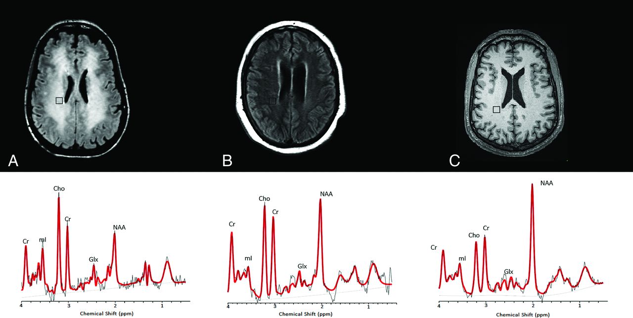
1H-MR spectra of 3 consecutive patients with COVID-19. Upper row: Axial FLAIR images at the corona radiata level show representative MRS voxels (black squares) from sampled periventricular regions. Lower row: Corresponding spectrum (black) and LCModel fit (red) from each patient acquired at TE = 30 ms (upper row) and TE = 288 ms (lower row). A, A patient with COVID-19-associated multifocal necrotizing leukoencephalopathy shows diffuse patchy WM lesions with markedly increased Cho and decreased NAA, as well as elevated Lac. B, A patient with COVID-19 after recent PEA cardiac arrest with subtle FLAIR hyperintense white matter changes also shows elevated Cho/Cr and decreased NAA/Cr ratios. However, these derangements are less severe than in the patient in A. There is no clear elevation of Lac. C, A patient with COVID-19 without encephalopathy or recent severe hypoxia has normal Cho/Cr, with mildly decreased NAA/Cr and no lactate elevation. Cho, Choline; NAA, N-Acetyl-Aspartate; mI, Myo-Inositol; Lac, Lactate; Glx, Glutamate + Glutamine.

View this table:
  • View inline
  • View popup
Table 1:

Demographics and clinical information

Raw data files were processed using the LCModel software package, Version 6.3 (http://www.lcmodel.com/) with the appropriate basis set.10 For the data analysis, 6–8 white matter MR spectroscopy voxels of the section with the most prominent white matter abnormalities were chosen, and their metabolite ratios were averaged.

Of the patients with COVID-19, 1 patient had MR imaging findings compatible with a necrotizing leukoencephalopathy with abnormal reduced diffusivity and cavitation within the white matter lesions (COVID-A) (Fig 1A). The second patient had pulseless electrical activity (PEA) arrest while in the intensive care unit and subsequently developed altered mental status. His MR imaging showed cerebellar cortical and hippocampal signal abnormalities suggestive of prior hypoxic-ischemic injury and subtle bilateral cerebellar and supratentorial white matter T2/FLAIR hyperintensities (COVID-B) (Fig 1B). The third patient with COVID-19 without an encephalopathic syndrome had a history of parkinsonism and developed catatonia of unclear etiology (COVID-C) (Fig 1C). This patient’s MR imaging showed mild nonspecific periventricular and deep white matter changes suggestive of small-vessel disease (considering their neuroanatomic distribution). None of these patients were found to have other neurologic comorbidities (eg, other metabolic encephalopathies, hydrocephalus, and so forth).

Data from these 3 patients were contrasted to multivoxel MRS data obtained before the COVID-19 pandemic from 2 patients with other leukoencephalopathies. One patient had DPL and developed severe encephalopathy, rigidity, and mutism, likely related to a prior toxic exposure (opiates) or a hypoxic episode (Fig 2A). The other patient without COVID-19 had sepsis-associated encephalopathy (SAE), with mild diffuse white matter abnormalities on brain MR imaging studies (Fig 2B).

FIG 2.
  • Download figure
  • Open in new tab
  • Download powerpoint
FIG 2.

1H-MR spectra from 2 non-COVID leukoencephalopathy patients, compared with a spectrum from a healthy control patient. Upper row: Axial noncontrast FLAIR (A and B) and T1-weighted (C) images at the level of the corona radiata show representative MRS voxels (black squares) from sampled periventricular regions. Lower row: Corresponding spectrum (black) and LCModel fit (red) from each patient acquired at TE = 30 ms. A, A patient with delayed posthypoxic/toxic leukoencephalopathy shows an increase of Cho/Cr and decreased NAA/Cr ratios. B, A patient with sepsis-associated encephalopathy shows milder degrees of Cho/Cr elevation and NAA/Cr reduction. C, A healthy control subject with a normal MR spectrum. Cho, Choline; NAA, N-Acetyl-Aspartate; mI, Myo-Inositol; Lac, Lactate; Glx, Glutamate + Glutamine.

Last, data of all 5 patients with leukoencephalopathy were compared with a dataset acquired using the same protocol from a healthy control subject, a 65-year-old man who volunteered for a research study (Fig 2C).

MRS datasets with short TEs (TE = 30 ms) were analyzed to quantify ratios of NAA, Cho, mI, and Glx relative to Cr. Lactate (Lac)/Cr levels were quantified using MRS datasets with long TEs (TE = 288 ms). Table 2 summarizes the means, SDs, and percentage differences of metabolite levels compared with the control.

View this table:
  • View inline
  • View popup
Table 2:

Mean, SD, and percentage difference relative to controls of brain MRS metabolite ratios in 3 patients with and 3 patients without COVID-19a

Comparative analysis showed decreased NAA/Cr levels within the white matter in 2 of the patients positive for COVID-19 compared with the control. The patient with necrotizing leukoencephalopathy (COVID-A) had the most diminished NAA levels, which were completely absent in several MR spectroscopy voxels, followed by the patient with recent cardiac arrest and mild diffuse white matter changes without necrosis (COVID-B). The patient with COVID-C without recent cardiac arrest or overt leukoencephalopathy (Fig 1) had slightly decreased NAA/Cr levels compared with the age-matched control. The metabolic profiles of the 3 patients with COVID-19 were contrasted to those of 2 patients with other leukoencephalopathies without COVID-19 (described above) (Fig 2). The NAA/Cr levels of these patients without COVID leukoencephalopathy were also decreased but still higher than those in the patient with COVID-A (Table 2).

Compared with the control, 2 of the 3 patients with COVID-19 had elevated Cho/Cr levels (Table 2). The third patient with COVID-19 without leukoencephalopathy (COVID-C) showed Cho/Cr levels indistinguishable from those of the control. The patient without COVID-19 with DPL and extensive white matter abnormalities showed increased Cho/Cr ratios, while the patient with SAE had a relatively normal range of Cho/Cr ratios.

All 3 patients with COVID-19 and the patient without COVID-19 with DPL showed elevated mI/Cr levels (Table 2). Most interesting, our patient with sepsis-associated encephalopathy had decreased mI/Cr ratios. Glx/Cr was markedly increased in the 2 patients with COVID-19 with necrotizing leukoencephalopathy and after cardiac arrest as well as in the patient without COVID-19 with DPL. Lac/Cr ratios were increased in the patient with COVID-19 with necrotizing leukoencephalopathy on long-TE spectra. No elevation of Lac/Cr ratios was seen in the patient with SAE. The patient with DPL and control did not have long-TE spectra. Therefore, we cannot confirm or exclude the presence of lactate in these subjects (Fig 2A).

DISCUSSION

Multiple recent reports in the medical literature have confirmed the development of leukoencephalopathy as a potential CNS complication in SARS-CoV-2 infection,11,12 though no study has reported the MR spectroscopic findings in these patients. We evaluated the metabolic differences among 3 patients with COVID-19 (1 with necrotizing leukoencephalopathy, another after cardiac arrest, and the third with mild nonspecific white matter changes without clinical encephalopathy) and other control groups with multivoxel MR spectroscopy to better understand the underlying pathophysiology of this disease. Markedly increased Cho/Cr, decreased NAA/Cr, and increased Lac/Cr ratios were observed in the patient with COVID-19 with multifocal necrotizing leukoencephalopathy. Less pronounced changes in Cho/Cr and NAA/Cr ratios were noted in the patient with COVID-19 with prior PEA arrest and subtle nonspecific white matter signal abnormalities. Notably, the magnitude of the Cho and NAA abnormalities in the patient with COVID-19 associated necrotizing leukoencephalopathy was more pronounced compared with the patients with delayed post-hypoxic leukoencephalopathy and SAE.

In our series, similar patterns of metabolic changes were observed in the setting of COVID-19-associated leukoencephalopathy and DPL, namely elevated Cho, elevated Lac, decreased NAA, increased mI, and increased Glx ratios in relation to Cr. This spectral pattern has been previously reported in the setting of DPL following carbon monoxide poisoning or medication overdose.13⇓-15 Prolonged impaired oxygenation to the subcortical white matter is thought to promote anaerobic metabolism and leads to elevated tissue Lactate.14,15

The observed increase of Cho/Cr ratios in the patients with leukoencephalopathy, particularly in the case of COVID-associated necrotizing leukoencephalopathy, likely reflects demyelination within these white matter lesions as recently described in an initial neuropathologic study of a patient with COVID-19.16 Cho elevation could also be related to cell death and/or immune cellular infiltration within areas of demyelination.17 Axonal damage was also reported on pathology16 and likely contributes to the decreased NAA/Cr ratios we observed.

Increased levels of Myo-Inositol (mI) may reflect neuroinflammation, which when coupled with choline elevations in demyelinating pathologies may reflect glial proliferation.18 The presence of elevated Glx levels has been reported in cases of acute excitotoxic leukoencephalopathy19 and viral-associated acute leukoencephalopathy with restricted diffusion.20 Both conditions are thought to be mediated by excitotoxic injury to the cerebral white matter and exhibit prominent reduced diffusivity within white matter lesions.

Notably, the metabolic derangements seen in the setting of COVID-19-associated multifocal necrotizing leukoencephalopathy are similar to those observed with DPL. However, more evidence is required to validate this conclusion. Some of the previously published MR spectroscopy studies of necrotizing encephalopathies from other etiologies21 also showed increased Lac along with decreased NAA and increased Cho,22 while others showed Glx and lipid elevations predominantly.23 Certainly, additional research is warranted in this new field of COVID-19-related metabolic derangement.

The small patient cohort of our study limits our ability to generalize our observations. MRS was only performed in patients with SARS-CoV-2 infection when requested by referring providers for specific clinical indications. Furthermore, the controls without COVID-19 are slightly younger, especially the patient with DPL. Another limitation of our study is that only a single time point was assessed; thus, we cannot exclude other baseline conditions before the SARS-CoV-2 infection, such as chronic small-vessel disease, that may affect our results. Finally, some of these subjects (the patient with DPL and the healthy control) did not have a long-TE MRS acquisition, limiting our ability to unequivocally determine the presence or absence of Lac.

In conclusion, the reported spectroscopic abnormalities within the white matter lesions of COVID-19-associated leukoencephalopathy may reflect several pathophysiologic processes, including but not limited to the following: 1) an anaerobic metabolic environment produced by the well-described “silent hypoxia” seen in these patients, resulting in elevation of Lac levels; 2) neuronal dysfunction and axonal injury with decreased NAA/Cr ratios; and 3) increased membrane destruction or turnover with elevated Cho/Cr ratios. Continued data collection in a larger cohort is required to validate these observations and better elucidate their significance in the pathophysiology of SARS-CoV-2 infection.

Footnotes

  • O. Rapalino and A. Weerasekera contributed equally to this article.

  • M.L. Loggia was funded by the National Institutes of Health grant 1R01NS095937-01A1.

  • Disclosures: Otto Rapalino—UNRELATED: Travel and hotel expenses as a lecturer at an event were sponsored by GE Healthcare in August 2019. This lecture did not have any commercial content and did not have any relationship to the current work. Brian L. Edlow—RELATED: Grant: James S. McDonnell Foundation, Comments: McDonnell Foundation COVID-19 Recovery of Consciousness Consortium.* David Fischer—RELATED: Grant: R25NS06574309. Shibani Mukerji—UNRELATED: Grants/Grants Pending: National Institute of Mental Health, Comments: K23MH115812.* Michael H. Lev—UNRELATED: Consultancy: Takeda Pharmaceutical Company, GE Healthcare, Comments: Consultant; Grants/Grants Pending: National Institutes of Health, Comments: Coinvestigator on 2 technical development grants; Patents (Planned, Pending or Issued): Electrical Impedance Spectroscopy, Artificial Intelligence-related. Eva-Maria Ratai—UNRELATED: Employment: Massachusetts General Hospital; Grants/Grants Pending: National Institutes of Health, National Science Foundation, Comments: R01NS080816, R01HD065762, R01HD085813, U01NS104326, R01DA047088, P01-AT009965, R01CA190901, R01NS112694.* Marco L. Loggia—RELATED: Grant: National Institute of Neurological Disorders and Stroke, Comments: 1R01NS095937-01A1*; UNRELATED: Grants/Grants Pending: National Institutes of Health. Angel Torrado-Carvajal—UNRELATED: Employment: Universidad Rey Juan Carlos, Assistant Professor (September 2019 until now); Massachusetts General Hospital, Research Fellow (August 2017 to August 2019); Grants/Grants Pending: Comunidad de Madrid, MIMC3 PET/MR Project.* *Money paid to the institution.

Indicates open access to non-subscribers at www.ajnr.org

References

  1. 1.↵
    1. Cucinotta D,
    2. Vanelli M
    . WHO declares COVID-19 a pandemic. Acta Biomed 2020;91:157–60 doi:10.23750/abm.v91i1.9397 pmid:32191675
    CrossRefPubMed
  2. 2.↵
    1. Berlin DA,
    2. Gulick RM,
    3. Martinez FJ
    . Severe Covid-19. N Engl J Med 2020 May 15. [Epub ahead of print] doi:10.1056/NEJMcp2009575 pmid:32412710
    CrossRefPubMed
  3. 3.↵
    1. Romero-Sanchez CM,
    2. Diaz-Maroto I,
    3. Fernandez-Diaz E, et al
    . Neurologic manifestations in hospitalized patients with COVID-19: the ALBACOVID registry. Neurology 2020;95:e1060–70 doi:10.1212/WNL.0000000000009937 pmid:32482845
    Abstract/FREE Full Text
  4. 4.↵
    1. Helms J,
    2. Kremer S,
    3. Merdji H, et al
    . Neurologic features in severe SARS-CoV-2 infection. N Engl J Med 2020;382:2268–70 doi:10.1056/NEJMc2008597 pmid:32294339
    CrossRefPubMed
  5. 5.↵
    1. Couzin-Frankel J
    . The mystery of the pandemic’s ‘happy hypoxia.’ Science 2020;368:455–56 doi:10.1126/science.368.6490.455 pmid:32355007
    Abstract/FREE Full Text
  6. 6.↵
    1. Solomon IH,
    2. Normandin E,
    3. Bhattacharyya S, et al
    . Neuropathological features of Covid-19. N Engl J Med 2020;383:989–92 doi:10.1056/NEJMc2019373 pmid:32530583
    CrossRefPubMed
  7. 7.↵
    1. Sun J,
    2. Song H,
    3. Yang Y, et al
    . Metabolic changes in normal appearing white matter in multiple sclerosis patients using multivoxel magnetic resonance spectroscopy imaging. Medicine (Baltimore) 2017;96:e6534 doi:10.1097/MD.0000000000006534 pmid:28383419
    CrossRefPubMed
  8. 8.↵
    1. Bizzi A,
    2. Castelli G,
    3. Bugiani M, et al
    . Classification of childhood white matter disorders using proton MR spectroscopic imaging. AJNR Am J Neuroradiol 2008;29:1270–75 doi:10.3174/ajnr.A1106 pmid:18483189
    Abstract/FREE Full Text
  9. 9.↵
    1. Andronesi OC,
    2. Gagoski BA,
    3. Sorensen AG
    . Neurologic 3D MR spectroscopic imaging with low-power adiabatic pulses and fast spiral acquisition. Radiology 2012;262:647–61 doi:10.1148/radiol.11110277 pmid:22187628
    CrossRefPubMed
  10. 10.↵
    1. Provencher SW
    . Automatic quantitation of localized in vivo 1H spectra with LCModel. NMR Biomed 2001;14:260–64 doi:10.1002/nbm.698 pmid:11410943
    CrossRefPubMed
  11. 11.↵
    1. Radmanesh A,
    2. Derman A,
    3. Lui YW, et al
    . COVID-19-associated diffuse leukoencephalopathy and microhemorrhages. Radiology 2020 May 21. [Epub ahead of print] doi:10.1148/radiol.2020202040 pmid:32437314
    CrossRefPubMed
  12. 12.↵
    1. Sachs JR,
    2. Gibbs KW,
    3. Swor DE, et al
    . COVID-19-associated leukoencephalopathy. Radiology 2020;296:E184–85 doi:10.1148/radiol.2020201753 pmid:32407258
    CrossRefPubMed
  13. 13.↵
    1. Beeskow AB,
    2. Oberstadt M,
    3. Saur D, et al
    . Delayed post-hypoxic leukoencephalopathy (DPHL): an uncommon variant of hypoxic brain damage in adults. Front Neurol 2018;9:708 doi:10.3389/fneur.2018.00708 pmid:30210433
    CrossRefPubMed
  14. 14.↵
    1. Shprecher DR,
    2. Flanigan KM,
    3. Smith AG, et al
    . Clinical and diagnostic features of delayed hypoxic leukoencephalopathy. J Neuropsychiatry Clin Neurosci 2008;20:473–77 doi:10.1176/jnp.2008.20.4.473 pmid:19196933
    CrossRefPubMed
  15. 15.↵
    1. Terajima K,
    2. Igarashi H,
    3. Hirose M, et al
    . Serial assessments of delayed encephalopathy after carbon monoxide poisoning using magnetic resonance spectroscopy and diffusion tensor imaging on 3.0T system. Eur Neurol 2008;59:55–61 doi:10.1159/000109262 pmid:17917459
    CrossRefPubMed
  16. 16.↵
    1. Reichard RR,
    2. Kashani KB,
    3. Boire NA, et al
    . Neuropathology of COVID-19: a spectrum of vascular and acute disseminated encephalomyelitis (ADEM)-like pathology. Acta Neuropathol 2020;140:1–6 doi:10.1007/s00401-020-02166-2 pmid:32449057
    CrossRefPubMed
  17. 17.↵
    1. Brenner RE,
    2. Munro PM,
    3. Williams SC, et al
    . The proton NMR spectrum in acute EAE: the significance of the change in the Cho:Cr ratio. Magn Reson Med 1993;29:737–45 doi:10.1002/mrm.1910290605 pmid:8350716
    CrossRefPubMed
  18. 18.↵
    1. Bitsch A,
    2. Bruhn H,
    3. Vougioukas V, et al
    . Inflammatory CNS demyelination: histopathologic correlation with in vivo quantitative proton MR spectroscopy. AJNR Am J Neuroradiol 1999;20:1619–27 pmid:10543631
    Abstract/FREE Full Text
  19. 19.↵
    1. Takanashi JI,
    2. Murofushi Y,
    3. Hirai N, et al
    . Prognostic value of MR spectroscopy in patients with acute excitotoxic encephalopathy. J Neurol Sci 2020;408:116636 doi:10.1016/j.jns.2019.116636 pmid:31879119
    CrossRefPubMed
  20. 20.↵
    1. Kamate M
    . Acute leukoencephalopathy with restricted diffusion. Indian J Crit Care Med 2018;22:519–23 doi:10.4103/ijccm.IJCCM_139_18 pmid:30111927
    CrossRefPubMed
  21. 21.↵
    1. Premji S,
    2. Kang L,
    3. Rojiani MV, et al
    . Multifocal necrotizing leukoencephalopathy: expanding the clinicopathologic spectrum. J Neuropathol Exp Neurol 2019;78:340–47 doi:10.1093/jnen/nlz003 pmid:30753632
    CrossRefPubMed
  22. 22.↵
    1. Raghavendra S,
    2. Nair MD,
    3. Chemmanam T, et al
    . Disseminated necrotizing leukoencephalopathy following low-dose oral methotrexate. Eur J Neurol 2007;14:309–14 doi:10.1111/j.1468-1331.2006.01659.x pmid:17355553
    CrossRefPubMed
  23. 23.↵
    1. Aydin H,
    2. Ozgul E,
    3. Agildere AM
    . Acute necrotizing encephalopathy secondary to diphtheria, tetanus toxoid and whole-cell pertussis vaccination: diffusion-weighted imaging and proton MR spectroscopy findings. Pediatr Radiol 2010;40:1281–84 doi:10.1007/s00247-009-1498-9 pmid:20119724
    CrossRefPubMed
  • Received June 14, 2020.
  • Accepted after revision August 13, 2020.
  • © 2021 by American Journal of Neuroradiology
PreviousNext
Back to top

In this issue

American Journal of Neuroradiology: 42 (1)
American Journal of Neuroradiology
Vol. 42, Issue 1
1 Jan 2021
  • Table of Contents
  • Index by author
  • Complete Issue (PDF)
Advertisement
Print
Download PDF
Email Article

Thank you for your interest in spreading the word on American Journal of Neuroradiology.

NOTE: We only request your email address so that the person you are recommending the page to knows that you wanted them to see it, and that it is not junk mail. We do not capture any email address.

Enter multiple addresses on separate lines or separate them with commas.
Brain MR Spectroscopic Findings in 3 Consecutive Patients with COVID-19: Preliminary Observations
(Your Name) has sent you a message from American Journal of Neuroradiology
(Your Name) thought you would like to see the American Journal of Neuroradiology web site.
CAPTCHA
This question is for testing whether or not you are a human visitor and to prevent automated spam submissions.
Cite this article
O. Rapalino, A. Weerasekera, S.J. Moum, K. Eikermann-Haerter, B.L. Edlow, D. Fischer, A. Torrado-Carvajal, M.L. Loggia, S.S. Mukerji, P.W. Schaefer, R.G. Gonzalez, M.H. Lev, E.-M. Ratai
Brain MR Spectroscopic Findings in 3 Consecutive Patients with COVID-19: Preliminary Observations
American Journal of Neuroradiology Jan 2021, 42 (1) 37-41; DOI: 10.3174/ajnr.A6877

Citation Manager Formats

  • BibTeX
  • Bookends
  • EasyBib
  • EndNote (tagged)
  • EndNote 8 (xml)
  • Medlars
  • Mendeley
  • Papers
  • RefWorks Tagged
  • Ref Manager
  • RIS
  • Zotero
0 Responses
Respond to this article
Share
Bookmark this article
Brain MR Spectroscopic Findings in 3 Consecutive Patients with COVID-19: Preliminary Observations
O. Rapalino, A. Weerasekera, S.J. Moum, K. Eikermann-Haerter, B.L. Edlow, D. Fischer, A. Torrado-Carvajal, M.L. Loggia, S.S. Mukerji, P.W. Schaefer, R.G. Gonzalez, M.H. Lev, E.-M. Ratai
American Journal of Neuroradiology Jan 2021, 42 (1) 37-41; DOI: 10.3174/ajnr.A6877
del.icio.us logo Twitter logo Facebook logo Mendeley logo
  • Tweet Widget
  • Facebook Like
  • Google Plus One
Purchase

Jump to section

  • Article
    • Abstract
    • ABBREVIATIONS:
    • DISCUSSION
    • Footnotes
    • References
  • Figures & Data
  • Info & Metrics
  • Responses
  • References
  • PDF

Related Articles

  • No related articles found.
  • PubMed
  • Google Scholar

Cited By...

  • No citing articles found.
  • Crossref (17)
  • Google Scholar

This article has been cited by the following articles in journals that are participating in Crossref Cited-by Linking.

  • Susceptibility-weighted imaging reveals cerebral microvascular injury in severe COVID-19
    John Conklin, Matthew P. Frosch, Shibani S. Mukerji, Otto Rapalino, Mary D. Maher, Pamela W. Schaefer, Michael H. Lev, R.G. Gonzalez, Sudeshna Das, Samantha N. Champion, Colin Magdamo, Pritha Sen, G. Kyle Harrold, Haitham Alabsi, Erica Normandin, Bennett Shaw, Jacob E. Lemieux, Pardis C. Sabeti, John A. Branda, Emery N. Brown, M. Brandon Westover, Susie Y. Huang, Brian L. Edlow
    Journal of the Neurological Sciences 2021 421
  • Effect of Hypoxia on the Function of the Human Serotonin1A Receptor
    Aritri Dutta, Parijat Sarkar, Sandeep Shrivastava, Amitabha Chattopadhyay
    ACS Chemical Neuroscience 2022 13 9
  • Neuroradiologic Imaging of Neurologic and Neuro-Ophthalmic Complications of Coronavirus-19 Infection
    Marcela Marsiglia, Bart K. Chwalisz, Mary Maher
    Journal of Neuro-Ophthalmology 2021 41 4
  • Resistance exercise effects on hippocampus subfield volumes and biomarkers of neuroplasticity and neuroinflammation in older adults with low and high risk of mild cognitive impairment: a randomized controlled trial
    Wouter A. J. Vints, Julija Šeikinaitė, Evrim Gökçe, Simona Kušleikienė, Milda Šarkinaite, Kristina Valatkeviciene, Vida J. Česnaitienė, Jeanine Verbunt, Oron Levin, Nerijus Masiulis
    GeroScience 2024 46 4
  • Cortical GABA Levels Are Reduced in Post-Acute COVID-19 Syndrome
    Ksenija Marinkovic, David R. White, Austin Alderson Myers, Katie S. Parker, Donatello Arienzo, Graeme F. Mason
    Brain Sciences 2023 13 12
  • Brain Imaging in COVID-19
    Avantika Samkaria, Pravat Kumar Mandal
    ACS Chemical Neuroscience 2021 12 16
  • Brain Stress Mapping in COVID-19 Survivors Using MR Spectroscopy: New Avenue of Mental Health Status Monitoring$
    Avantika Samkaria, Khushboo Punjabi, Shallu Sharma, Shallu Joon, Kanika Sandal, Tirthankar Dasgupta, Pooja Sharma, Pravat K. Mandal
    Journal of Alzheimer's Disease 2021 83 2
  • COVID-19’s effects on the Egyptian population’s brain: Could MRI and specialized MR spectroscopic analysis be beneficial?
    Alaa Mohamed Reda, Ahmed Elsharkawy, Tamer Mahmoud Dawoud
    Egyptian Journal of Radiology and Nuclear Medicine 2022 53 1
  • Demographic Features, Physical Examination Findings, and Medication Use in Hospitalized, Delirious Patients With and Without COVID-19 Infection: A Retrospective Study
    Noor Beckwith, Julia Probert, Blake L. Rosenbaum, Ashika Bains, Victoria C. Angelucci, Alejandra E. Morfin Rodriguez, Stephanie London, Joshua W. Zollman, Andrea Soto Ordoñez, Nicholas Kontos, Felicia A. Smith, Christopher M. Celano, Scott R. Beach
    Journal of the Academy of Consultation-Liaison Psychiatry 2023 64 1
  • Neuropsychological and psychological dysfunctions associated with coronavirus disease 2019: a case report
    Maria Damianova
    Journal of Medical Case Reports 2021 15 1

More in this TOC Section

Adult Brain

  • Diagnostic Neuroradiology of Monoclonal Antibodies
  • Segmentation of Brain Metastases with BLAST
  • NCCT vs. MRI for Brain Atrophy in Acute Stroke
Show more Adult Brain

Functional

  • Glutaric Aciduria Type 1: DK vs. Conventional MRI
  • Kurtosis and Epileptogenic Tubers: A Pilot Study
  • Brain Iron in Niemann-Pick Type C: 7T Study
Show more Functional

Similar Articles

Advertisement

Indexed Content

  • Current Issue
  • Accepted Manuscripts
  • Article Preview
  • Past Issues
  • Editorials
  • Editors Choice
  • Fellow Journal Club
  • Letters to the Editor

Cases

  • Case Collection
  • Archive - Case of the Week
  • Archive - Case of the Month
  • Archive - Classic Case

Special Collections

  • Special Collections

Resources

  • News and Updates
  • Turn around Times
  • Submit a Manuscript
  • Author Policies
  • Manuscript Submission Guidelines
  • Evidence-Based Medicine Level Guide
  • Publishing Checklists
  • Graphical Abstract Preparation
  • Imaging Protocol Submission
  • Submit a Case
  • Become a Reviewer/Academy of Reviewers
  • Get Peer Review Credit from Publons

Multimedia

  • AJNR Podcast
  • AJNR SCANtastic
  • Video Articles

About Us

  • About AJNR
  • Editorial Board
  • Not an AJNR Subscriber? Join Now
  • Alerts
  • Feedback
  • Advertise with us
  • Librarian Resources
  • Permissions
  • Terms and Conditions

American Society of Neuroradiology

  • Not an ASNR Member? Join Now

© 2025 by the American Society of Neuroradiology All rights, including for text and data mining, AI training, and similar technologies, are reserved.
Print ISSN: 0195-6108 Online ISSN: 1936-959X

Powered by HighWire