Advertisement

Main menu

  • Home
  • Content
    • Current Issue
    • Accepted Manuscripts
    • Article Preview
    • Past Issue Archive
    • Video Articles
    • AJNR Case Collection
    • Case of the Week Archive
    • Case of the Month Archive
    • Classic Case Archive
  • Special Collections
    • AJNR Awards
    • Low-Field MRI
    • Alzheimer Disease
    • ASNR Foundation Special Collection
    • Photon-Counting CT
    • View All
  • Multimedia
    • AJNR Podcasts
    • AJNR SCANtastic
    • Trainee Corner
    • MRI Safety Corner
    • Imaging Protocols
  • For Authors
    • Submit a Manuscript
    • Submit a Video Article
    • Submit an eLetter to the Editor/Response
    • Manuscript Submission Guidelines
    • Statistical Tips
    • Fast Publishing of Accepted Manuscripts
    • Graphical Abstract Preparation
    • Imaging Protocol Submission
    • Author Policies
  • About Us
    • About AJNR
    • Editorial Board
    • Editorial Board Alumni
  • More
    • Become a Reviewer/Academy of Reviewers
    • Subscribers
    • Permissions
    • Alerts
    • Feedback
    • Advertisers
    • ASNR Home

User menu

  • Alerts
  • Log in

Search

  • Advanced search
American Journal of Neuroradiology

American Journal of Neuroradiology

ASHNR American Society of Functional Neuroradiology ASHNR American Society of Pediatric Neuroradiology ASSR
  • Alerts
  • Log in

Advanced Search

  • Home
  • Content
    • Current Issue
    • Accepted Manuscripts
    • Article Preview
    • Past Issue Archive
    • Video Articles
    • AJNR Case Collection
    • Case of the Week Archive
    • Case of the Month Archive
    • Classic Case Archive
  • Special Collections
    • AJNR Awards
    • Low-Field MRI
    • Alzheimer Disease
    • ASNR Foundation Special Collection
    • Photon-Counting CT
    • View All
  • Multimedia
    • AJNR Podcasts
    • AJNR SCANtastic
    • Trainee Corner
    • MRI Safety Corner
    • Imaging Protocols
  • For Authors
    • Submit a Manuscript
    • Submit a Video Article
    • Submit an eLetter to the Editor/Response
    • Manuscript Submission Guidelines
    • Statistical Tips
    • Fast Publishing of Accepted Manuscripts
    • Graphical Abstract Preparation
    • Imaging Protocol Submission
    • Author Policies
  • About Us
    • About AJNR
    • Editorial Board
    • Editorial Board Alumni
  • More
    • Become a Reviewer/Academy of Reviewers
    • Subscribers
    • Permissions
    • Alerts
    • Feedback
    • Advertisers
    • ASNR Home
  • Follow AJNR on Twitter
  • Visit AJNR on Facebook
  • Follow AJNR on Instagram
  • Join AJNR on LinkedIn
  • RSS Feeds

AJNR Awards, New Junior Editors, and more. Read the latest AJNR updates

Research ArticleAdult Brain

Artificial Intelligence–Based 3D Angiography for Visualization of Complex Cerebrovascular Pathologies

S. Lang, P. Hoelter, M. Schmidt, C. Strother, C. Kaethner, M. Kowarschik and A. Doerfler
American Journal of Neuroradiology October 2021, 42 (10) 1762-1768; DOI: https://doi.org/10.3174/ajnr.A7252
S. Lang
aFrom the Department of Neuroradiology (S.L., P.H., M.S., A.D.), University of Erlangen-Nuremberg, Erlangen, Germany
  • Find this author on Google Scholar
  • Find this author on PubMed
  • Search for this author on this site
  • ORCID record for S. Lang
P. Hoelter
aFrom the Department of Neuroradiology (S.L., P.H., M.S., A.D.), University of Erlangen-Nuremberg, Erlangen, Germany
  • Find this author on Google Scholar
  • Find this author on PubMed
  • Search for this author on this site
  • ORCID record for P. Hoelter
M. Schmidt
aFrom the Department of Neuroradiology (S.L., P.H., M.S., A.D.), University of Erlangen-Nuremberg, Erlangen, Germany
  • Find this author on Google Scholar
  • Find this author on PubMed
  • Search for this author on this site
  • ORCID record for M. Schmidt
C. Strother
bDepartment of Radiology (C.S.), University of Wisconsin School of Medicine and Public Health, E3/366 Clinical Sciences Center, Madison, Wisconsin
  • Find this author on Google Scholar
  • Find this author on PubMed
  • Search for this author on this site
  • ORCID record for C. Strother
C. Kaethner
cAdvanced Therapies (C.K., M.K.), Siemens Healthcare GmbH, Forchheim, Germany
  • Find this author on Google Scholar
  • Find this author on PubMed
  • Search for this author on this site
  • ORCID record for C. Kaethner
M. Kowarschik
cAdvanced Therapies (C.K., M.K.), Siemens Healthcare GmbH, Forchheim, Germany
  • Find this author on Google Scholar
  • Find this author on PubMed
  • Search for this author on this site
  • ORCID record for M. Kowarschik
A. Doerfler
aFrom the Department of Neuroradiology (S.L., P.H., M.S., A.D.), University of Erlangen-Nuremberg, Erlangen, Germany
  • Find this author on Google Scholar
  • Find this author on PubMed
  • Search for this author on this site
  • ORCID record for A. Doerfler
  • Article
  • Figures & Data
  • Info & Metrics
  • Responses
  • References
  • PDF
Loading

Article Figures & Data

Figures

  • Tables
  • FIG 1.
    • Download figure
    • Open in new tab
    • Download powerpoint
    FIG 1.

    Key aspects of the chosen network architecture. In addition to the in- and output of the network, kernel sizes, dilation rates, number of filters, and activation function (ie, rectified linear unit [ReLu]) are shown. Conv3D indicates 3D convolutional neural network.

  • FIG 2.
    • Download figure
    • Open in new tab
    • Download powerpoint
    FIG 2.

    Sample measurement of the vascular dimensions of an ICA (C4 segment) in a 3D-DSA data set. The maximum vessel diameters have been assessed for both 3D-DSA and 3DA in 2 projections each (VD1 and VD2) using multiplanar reconstructions. A, Sagittal orientation of the C4 segment. B, Coronary orientation of the C4 segment. C, Finally, both vessel diameters allow the calculation of the VGI (VGI = VD1/VD2).
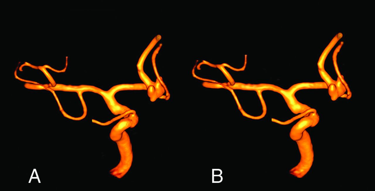
  • FIG 3.
    • Download figure
    • Open in new tab
    • Download powerpoint
    FIG 3.

    Illustrative case 1. Sample visualization of a right-sided AVM with 3D-DSA (A) and the AI-based 3DA (B) using VRT. 3DA (B) shows both feeding arteries originating from the MCA and the nidus and drainage via superficial veins, equivalent to 3D-DSA. Not only the flow-associated aneurysm of the MCA bifurcation (red arrow) but also the venous ectasia adjacent to the nidus is comparably visualized without loss of information using 3DA.

  • FIG 4.
    • Download figure
    • Open in new tab
    • Download powerpoint
    FIG 4.

    Illustrative case 2. Sample visualization of a left-sided dAVF with 3D-DSA (A) and the AI–based 3DA (B) using VRT. 3DA (B) offers comparable visualization of the influx of the fistula via the dilated middle meningeal artery and the drainage of the fistula via an ectatic cerebral vein compared with 3D-DSA (A).

  • FIG 5.
    • Download figure
    • Open in new tab
    • Download powerpoint
    FIG 5.

    Illustrative case 3. Sample 3D visualization of an irregular aneurysm of the anterior communicating artery in VRT reconstructions using 3D-DSA (A) and 3DA (B). Despite differences concerning the reconstruction algorithm, AI-based 3DA (B) provides all relevant information on the aneurysmal configuration equivalent to 3D-DSA.

Tables

  • Figures
    • View popup
    Table 1:

    Image quality

    GradeCharacteristics
    4Excellent (high contrast, no artifacts)
    3Good (high contrast; minimal artifacts, eg, due to movement or implants)
    2Compromised (eg, noticeable movement artifacts and/or reduced homogeneity of the vessel contrast)
    1Heavily compromised (low contrast and/or strong movement artifacts)
    0Not diagnostic (vasculature is not differentiable due to heavy artifacts and/or missing contrast)
    • View popup
    Table 2:

    VD and VGI

    Parameter3DA (Mean)3D-DSA (Mean)rP
    VD1AVM3.82 (SD, 0.47) mm3.81 (SD, 0.55) mm0.988<.001
    VD2AVM3.97 (SD, 0.60) mm3.94 (SD, 0.59) mm0.984<.001
    VGIAVM0.96 (SD, 0.04) mm0.97 (SD, 0.03) mm0.835.003
    VD1CA4.39 (SD, 0.84) mm4.34 (SD, 0.87) mm0.998<.001
    VD2CA4.61 (SD, 0.81) mm4.54 (SD, 0.85) mm0.996<.001
    VGICA0.95 (SD, 0.04) mm0.95 (SD, 0.03) mm0.955<.001
    VD1dAVF4.24 (SD, 0.72) mm4.22 (SD, 0.77) mm0.993<.001
    VD2dAVF4.44 (SD, 0.780) mm4.43 (SD, 0.81) mm0.992<.001
    VGIdAVF0.95 (SD, 0.02) mm0.95 (SD, 0.02) mm0.824.003
PreviousNext
Back to top

In this issue

American Journal of Neuroradiology: 42 (10)
American Journal of Neuroradiology
Vol. 42, Issue 10
1 Oct 2021
  • Table of Contents
  • Index by author
  • Complete Issue (PDF)
Advertisement
Print
Download PDF
Email Article

Thank you for your interest in spreading the word on American Journal of Neuroradiology.

NOTE: We only request your email address so that the person you are recommending the page to knows that you wanted them to see it, and that it is not junk mail. We do not capture any email address.

Enter multiple addresses on separate lines or separate them with commas.
Artificial Intelligence–Based 3D Angiography for Visualization of Complex Cerebrovascular Pathologies
(Your Name) has sent you a message from American Journal of Neuroradiology
(Your Name) thought you would like to see the American Journal of Neuroradiology web site.
CAPTCHA
This question is for testing whether or not you are a human visitor and to prevent automated spam submissions.
Cite this article
S. Lang, P. Hoelter, M. Schmidt, C. Strother, C. Kaethner, M. Kowarschik, A. Doerfler
Artificial Intelligence–Based 3D Angiography for Visualization of Complex Cerebrovascular Pathologies
American Journal of Neuroradiology Oct 2021, 42 (10) 1762-1768; DOI: 10.3174/ajnr.A7252

Citation Manager Formats

  • BibTeX
  • Bookends
  • EasyBib
  • EndNote (tagged)
  • EndNote 8 (xml)
  • Medlars
  • Mendeley
  • Papers
  • RefWorks Tagged
  • Ref Manager
  • RIS
  • Zotero
0 Responses
Respond to this article
Share
Bookmark this article
Artificial Intelligence–Based 3D Angiography for Visualization of Complex Cerebrovascular Pathologies
S. Lang, P. Hoelter, M. Schmidt, C. Strother, C. Kaethner, M. Kowarschik, A. Doerfler
American Journal of Neuroradiology Oct 2021, 42 (10) 1762-1768; DOI: 10.3174/ajnr.A7252
del.icio.us logo Twitter logo Facebook logo Mendeley logo
  • Tweet Widget
  • Facebook Like
  • Google Plus One
Purchase

Jump to section

  • Article
    • Abstract
    • ABBREVIATIONS:
    • MATERIALS AND METHODS
    • RESULTS
    • DISCUSSION
    • CONCLUSIONS
    • Footnotes
    • References
  • Figures & Data
  • Info & Metrics
  • Responses
  • References
  • PDF

Related Articles

  • No related articles found.
  • PubMed
  • Google Scholar

Cited By...

  • No citing articles found.
  • Crossref
  • Google Scholar

This article has not yet been cited by articles in journals that are participating in Crossref Cited-by Linking.

More in this TOC Section

Adult Brain

  • Diagnostic Neuroradiology of Monoclonal Antibodies
  • Segmentation of Brain Metastases with BLAST
  • Cerebral ADC Changes in Fabry Disease
Show more Adult Brain

Functional

  • Glutaric Aciduria Type 1: DK vs. Conventional MRI
  • Kurtosis and Epileptogenic Tubers: A Pilot Study
  • Multiparametric MRI in PEDS Pontine Glioma
Show more Functional

Similar Articles

Advertisement

Indexed Content

  • Current Issue
  • Accepted Manuscripts
  • Article Preview
  • Past Issues
  • Editorials
  • Editor's Choice
  • Fellows' Journal Club
  • Letters to the Editor
  • Video Articles

Cases

  • Case Collection
  • Archive - Case of the Week
  • Archive - Case of the Month
  • Archive - Classic Case

More from AJNR

  • Trainee Corner
  • Imaging Protocols
  • MRI Safety Corner
  • Book Reviews

Multimedia

  • AJNR Podcasts
  • AJNR Scantastics

Resources

  • Turnaround Time
  • Submit a Manuscript
  • Submit a Video Article
  • Submit an eLetter to the Editor/Response
  • Manuscript Submission Guidelines
  • Statistical Tips
  • Fast Publishing of Accepted Manuscripts
  • Graphical Abstract Preparation
  • Imaging Protocol Submission
  • Evidence-Based Medicine Level Guide
  • Publishing Checklists
  • Author Policies
  • Become a Reviewer/Academy of Reviewers
  • News and Updates

About Us

  • About AJNR
  • Editorial Board
  • Editorial Board Alumni
  • Alerts
  • Permissions
  • Not an AJNR Subscriber? Join Now
  • Advertise with Us
  • Librarian Resources
  • Feedback
  • Terms and Conditions
  • AJNR Editorial Board Alumni

American Society of Neuroradiology

  • Not an ASNR Member? Join Now

© 2025 by the American Society of Neuroradiology All rights, including for text and data mining, AI training, and similar technologies, are reserved.
Print ISSN: 0195-6108 Online ISSN: 1936-959X

Powered by HighWire