Advertisement

Main menu

  • Home
  • Content
    • Current Issue
    • Accepted Manuscripts
    • Article Preview
    • Past Issue Archive
    • Video Articles
    • AJNR Case Collection
    • Case of the Week Archive
    • Case of the Month Archive
    • Classic Case Archive
  • Special Collections
    • AJNR Awards
    • Low-Field MRI
    • Alzheimer Disease
    • ASNR Foundation Special Collection
    • Photon-Counting CT
    • View All
  • Multimedia
    • AJNR Podcasts
    • AJNR SCANtastic
    • Trainee Corner
    • MRI Safety Corner
    • Imaging Protocols
  • For Authors
    • Submit a Manuscript
    • Submit a Video Article
    • Submit an eLetter to the Editor/Response
    • Manuscript Submission Guidelines
    • Statistical Tips
    • Fast Publishing of Accepted Manuscripts
    • Graphical Abstract Preparation
    • Imaging Protocol Submission
    • Author Policies
  • About Us
    • About AJNR
    • Editorial Board
    • Editorial Board Alumni
  • More
    • Become a Reviewer/Academy of Reviewers
    • Subscribers
    • Permissions
    • Alerts
    • Feedback
    • Advertisers
    • ASNR Home

User menu

  • Alerts
  • Log in

Search

  • Advanced search
American Journal of Neuroradiology

American Journal of Neuroradiology

ASHNR American Society of Functional Neuroradiology ASHNR American Society of Pediatric Neuroradiology ASSR
  • Alerts
  • Log in

Advanced Search

  • Home
  • Content
    • Current Issue
    • Accepted Manuscripts
    • Article Preview
    • Past Issue Archive
    • Video Articles
    • AJNR Case Collection
    • Case of the Week Archive
    • Case of the Month Archive
    • Classic Case Archive
  • Special Collections
    • AJNR Awards
    • Low-Field MRI
    • Alzheimer Disease
    • ASNR Foundation Special Collection
    • Photon-Counting CT
    • View All
  • Multimedia
    • AJNR Podcasts
    • AJNR SCANtastic
    • Trainee Corner
    • MRI Safety Corner
    • Imaging Protocols
  • For Authors
    • Submit a Manuscript
    • Submit a Video Article
    • Submit an eLetter to the Editor/Response
    • Manuscript Submission Guidelines
    • Statistical Tips
    • Fast Publishing of Accepted Manuscripts
    • Graphical Abstract Preparation
    • Imaging Protocol Submission
    • Author Policies
  • About Us
    • About AJNR
    • Editorial Board
    • Editorial Board Alumni
  • More
    • Become a Reviewer/Academy of Reviewers
    • Subscribers
    • Permissions
    • Alerts
    • Feedback
    • Advertisers
    • ASNR Home
  • Follow AJNR on Twitter
  • Visit AJNR on Facebook
  • Follow AJNR on Instagram
  • Join AJNR on LinkedIn
  • RSS Feeds

AJNR Awards, New Junior Editors, and more. Read the latest AJNR updates

Research ArticleHead and Neck Imaging
Open Access

Intraindividual Comparison between the Contrast-Enhanced Golden-Angle Radial Sparse Parallel Sequence and the Conventional Fat-Suppressed Contrast-Enhanced T1-Weighted Spin-Echo Sequence for Head and Neck MRI

S.-J. Hur, Y. Choi, J. Yoon, J. Jang, N.-Y. Shin, K.-J. Ahn and B.-S. Kim
American Journal of Neuroradiology November 2021, 42 (11) 2009-2015; DOI: https://doi.org/10.3174/ajnr.A7285
S.-J. Hur
aFrom the Department of Radiology, Seoul St. Mary’s Hospital, College of Medicine, The Catholic University of Korea, Seoul, Republic of Korea
  • Find this author on Google Scholar
  • Find this author on PubMed
  • Search for this author on this site
  • ORCID record for S.-J. Hur
Y. Choi
aFrom the Department of Radiology, Seoul St. Mary’s Hospital, College of Medicine, The Catholic University of Korea, Seoul, Republic of Korea
  • Find this author on Google Scholar
  • Find this author on PubMed
  • Search for this author on this site
  • ORCID record for Y. Choi
J. Yoon
aFrom the Department of Radiology, Seoul St. Mary’s Hospital, College of Medicine, The Catholic University of Korea, Seoul, Republic of Korea
  • Find this author on Google Scholar
  • Find this author on PubMed
  • Search for this author on this site
  • ORCID record for J. Yoon
J. Jang
aFrom the Department of Radiology, Seoul St. Mary’s Hospital, College of Medicine, The Catholic University of Korea, Seoul, Republic of Korea
  • Find this author on Google Scholar
  • Find this author on PubMed
  • Search for this author on this site
  • ORCID record for J. Jang
N.-Y. Shin
aFrom the Department of Radiology, Seoul St. Mary’s Hospital, College of Medicine, The Catholic University of Korea, Seoul, Republic of Korea
  • Find this author on Google Scholar
  • Find this author on PubMed
  • Search for this author on this site
  • ORCID record for N.-Y. Shin
K.-J. Ahn
aFrom the Department of Radiology, Seoul St. Mary’s Hospital, College of Medicine, The Catholic University of Korea, Seoul, Republic of Korea
  • Find this author on Google Scholar
  • Find this author on PubMed
  • Search for this author on this site
  • ORCID record for K.-J. Ahn
B.-S. Kim
aFrom the Department of Radiology, Seoul St. Mary’s Hospital, College of Medicine, The Catholic University of Korea, Seoul, Republic of Korea
  • Find this author on Google Scholar
  • Find this author on PubMed
  • Search for this author on this site
  • ORCID record for B.-S. Kim
  • Article
  • Figures & Data
  • Info & Metrics
  • Responses
  • References
  • PDF
Loading

Article Figures & Data

Figures

  • Tables
  • FIG 1.
    • Download figure
    • Open in new tab
    • Download powerpoint
    FIG 1.

    Flow chart of the patient-selection process.

  • FIG 2.
    • Download figure
    • Open in new tab
    • Download powerpoint
    FIG 2.

    A 55-year-old female patient with a history of recurrent squamous cell carcinoma of the tongue (top row); the lesion is better delineated in GRASP-VIBE (A) than in T1-TSE (B). A 76-year-old male patient with a history of left hypopharyngeal cancer with transoral robotic surgery (middle row). Compared with GRASP-VIBE (C), a significant respiratory motion artifact is observed in T1-TSE (D). A 70-year-old male patient with a history of left vocal cord squamous cell carcinoma in situ and laser cordectomy (bottom row). Compared with GRASP-VIBE (E), there is a noticeable pulsation artifact from blood vessels in T1-TSE (F).

  • FIG 3.
    • Download figure
    • Open in new tab
    • Download powerpoint
    FIG 3.

    A 73-year-old male patient with a history of oropharyngeal squamous cell carcinoma of the right tonsil; the susceptibility artifacts due to dental amalgam are more prominent in GRASP-VIBE (A) than in T1-TSE (B). In right submental region of the same patient, fat suppression is weaker in GRASP-VIBE (C) than in T1-TSE (D).

  • FIG 4.
    • Download figure
    • Open in new tab
    • Download powerpoint
    FIG 4.

    Boxplots of qualitative assessments for GRASP-VIBE and T1-TSE.
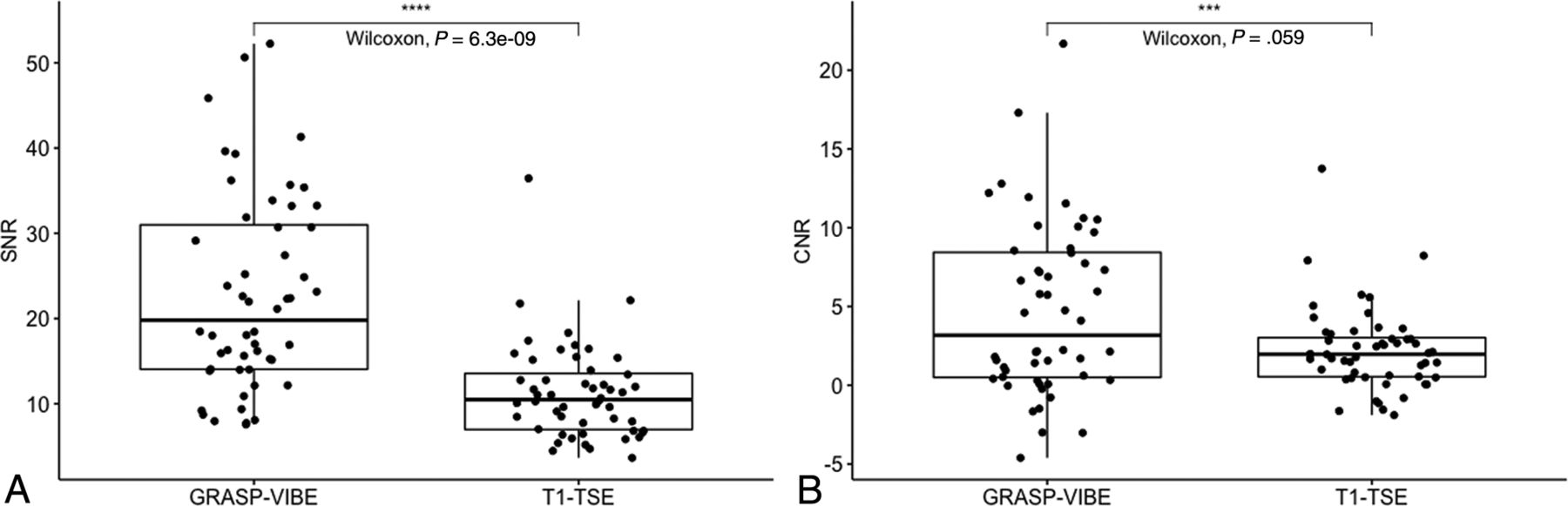
  • FIG 5.
    • Download figure
    • Open in new tab
    • Download powerpoint
    FIG 5.

    Boxplots of SNR (A) and CNR (B) of GRASP-VIBE and T1-TSE.

Tables

  • Figures
    • View popup
    Table 1:

    Detailed acquisition parameters of the 2 sequences

    SequenceGRASP-VIBET1-TSE
    FOV200 × 200200 × 200
    Matrix224 × 224384 × 230
    Section thickness (mm)44
    Gap (mm)01
    No. of slices2632
    In-plane resolution (mm)1.04 × 1.040.52 × 0.52
    Echo-train14
    Flip angle12°160°
    TR/TE (ms)4.1/1.9690/12
    Bandwidth (Hz/px)496 Hz/px310 Hz/px
    No. of excitations11
    Fat-suppression techniqueChemical shift selectiveDixon
    Scanning typeGradient-echoTSE
    Scan time (min/sec)4/46 2/6 
    No. of dynamic acquisitions (temporal resolution) (sec)95 (2.5)NA
    Acquisition type3D2D
    • Note:—NA indicates not applicable.

    • View popup
    Table 2:

    Baseline characteristics of included patientsa

    VariableN = 52
    Age (mean) (yr)60 (SD, 14)
    Sex
     Male37 (71.2)
     Female15 (28.8)
    Diagnosis
     Head and neck malignancies50 (94.3)
      Buccal1 (1.9)
      Floor of mouth1 (1.9)
      Vocal cord10 (19.2)
      Supraglottis1 (1.9)
      Hypopharynx7 (13.5)
      Salivary gland6 (11.5)
      Tongue13 (25.0)
      Tonsil10 (19.2)
     Other malignancies3 (5.7)
      Primary lesion visible
       Yes10 (19.2)
       No42 (80.8)
      Postoperative state
       Yes45 (86.5)
       No7 (13.5)
    • ↵a All values are presented as No. (%) unless otherwise specified.

    • View popup
    Table 3:

    Qualitative evaluations and κ values for overall image quality, overall artifacts, and anatomic conspicuities of GRASP-VIBE and T1-TSE

    GRASP-VIBET1-TSEP Valueb
    Scores (mean)κP ValueaScores (mean)κP Valuea
    Overall image quality
     Rater A4.74 (SD, 0.49)0.29.0033.81 (SD, 0.68)0.594<.001
     Rater B4.62 (SD, 0.53)3.51 (SD, 0.91)
     Average of two raters4.68 (SD, 0.41)3.66 (SD, 0.73)<.001
    Overall artifacts0.21.0410.542<.001
     Rater A4.26 (SD, 0.68)3.60 (SD, 0.60)
     Rater B4.68 (SD, 0.51)3.55 (SD, 0.97)
     Average of two raters4.47 (SD, 0.48)3.58 (SD, 0.71)<.001
    Oropharyngeal mucosal conspicuity0.80<.0010.5<.001
     Rater A4.87 (SD, 0.39)4.15 (SD, 0.74)
     Rater B4.83 (SD, 0.47)4.04 (SD, 1.06)
     Average of two raters4.85 (SD, 0.41)4.11 (SD, 0.79)<.001
    Hypopharyngeal mucosal conspicuity0.30.0160.422.002
     Rater A4.89 (SD, 0.32)3.58 (SD, 0.84)
     Rater B4.79 (SD, 0.49)3.57 (SD, 1.05)
     Average of two raters4.84 (SD, 0.34)3.58 (SD, 0.81)<.001
    Cervical lymph node conspicuity0.18.0720.34.001
     Rater A4.64 (SD, 0.48)4.34 (SD, 0.65)
     Rater B4.94 (SD, 0.30)3.81 (SD, 0.83)
     Average of two raters4.79 (SD, 0.32)4.08 (SD, 0.64)<.001
    • ↵a P value for κ.

    • ↵b P values for paired Wilcoxon rank sum tests comparing the scores of the 2 sequences.

PreviousNext
Back to top

In this issue

American Journal of Neuroradiology: 42 (11)
American Journal of Neuroradiology
Vol. 42, Issue 11
1 Nov 2021
  • Table of Contents
  • Index by author
  • Complete Issue (PDF)
Advertisement
Print
Download PDF
Email Article

Thank you for your interest in spreading the word on American Journal of Neuroradiology.

NOTE: We only request your email address so that the person you are recommending the page to knows that you wanted them to see it, and that it is not junk mail. We do not capture any email address.

Enter multiple addresses on separate lines or separate them with commas.
Intraindividual Comparison between the Contrast-Enhanced Golden-Angle Radial Sparse Parallel Sequence and the Conventional Fat-Suppressed Contrast-Enhanced T1-Weighted Spin-Echo Sequence for Head and Neck MRI
(Your Name) has sent you a message from American Journal of Neuroradiology
(Your Name) thought you would like to see the American Journal of Neuroradiology web site.
CAPTCHA
This question is for testing whether or not you are a human visitor and to prevent automated spam submissions.
Cite this article
S.-J. Hur, Y. Choi, J. Yoon, J. Jang, N.-Y. Shin, K.-J. Ahn, B.-S. Kim
Intraindividual Comparison between the Contrast-Enhanced Golden-Angle Radial Sparse Parallel Sequence and the Conventional Fat-Suppressed Contrast-Enhanced T1-Weighted Spin-Echo Sequence for Head and Neck MRI
American Journal of Neuroradiology Nov 2021, 42 (11) 2009-2015; DOI: 10.3174/ajnr.A7285

Citation Manager Formats

  • BibTeX
  • Bookends
  • EasyBib
  • EndNote (tagged)
  • EndNote 8 (xml)
  • Medlars
  • Mendeley
  • Papers
  • RefWorks Tagged
  • Ref Manager
  • RIS
  • Zotero
0 Responses
Respond to this article
Share
Bookmark this article
Intraindividual Comparison between the Contrast-Enhanced Golden-Angle Radial Sparse Parallel Sequence and the Conventional Fat-Suppressed Contrast-Enhanced T1-Weighted Spin-Echo Sequence for Head and Neck MRI
S.-J. Hur, Y. Choi, J. Yoon, J. Jang, N.-Y. Shin, K.-J. Ahn, B.-S. Kim
American Journal of Neuroradiology Nov 2021, 42 (11) 2009-2015; DOI: 10.3174/ajnr.A7285
del.icio.us logo Twitter logo Facebook logo Mendeley logo
  • Tweet Widget
  • Facebook Like
  • Google Plus One
Purchase

Jump to section

  • Article
    • Abstract
    • ABBREVIATIONS:
    • MATERIALS AND METHODS
    • RESULTS
    • DISCUSSION
    • CONCLUSIONS
    • Acknowledgments
    • Footnotes
    • References
  • Figures & Data
  • Info & Metrics
  • Responses
  • References
  • PDF

Related Articles

  • No related articles found.
  • PubMed
  • Google Scholar

Cited By...

  • No citing articles found.
  • Crossref
  • Google Scholar

This article has not yet been cited by articles in journals that are participating in Crossref Cited-by Linking.

More in this TOC Section

  • Hydrops Herniation into the Semicircular Canals
  • ASL Sensitivity for Head and Neck Paraganglioma
  • Post SRS Peritumoral Hyperintense Signal of VSs
Show more HEAD AND NECK IMAGING

Similar Articles

Advertisement

Indexed Content

  • Current Issue
  • Accepted Manuscripts
  • Article Preview
  • Past Issues
  • Editorials
  • Editor's Choice
  • Fellows' Journal Club
  • Letters to the Editor
  • Video Articles

Cases

  • Case Collection
  • Archive - Case of the Week
  • Archive - Case of the Month
  • Archive - Classic Case

More from AJNR

  • Trainee Corner
  • Imaging Protocols
  • MRI Safety Corner
  • Book Reviews

Multimedia

  • AJNR Podcasts
  • AJNR Scantastics

Resources

  • Turnaround Time
  • Submit a Manuscript
  • Submit a Video Article
  • Submit an eLetter to the Editor/Response
  • Manuscript Submission Guidelines
  • Statistical Tips
  • Fast Publishing of Accepted Manuscripts
  • Graphical Abstract Preparation
  • Imaging Protocol Submission
  • Evidence-Based Medicine Level Guide
  • Publishing Checklists
  • Author Policies
  • Become a Reviewer/Academy of Reviewers
  • News and Updates

About Us

  • About AJNR
  • Editorial Board
  • Editorial Board Alumni
  • Alerts
  • Permissions
  • Not an AJNR Subscriber? Join Now
  • Advertise with Us
  • Librarian Resources
  • Feedback
  • Terms and Conditions
  • AJNR Editorial Board Alumni

American Society of Neuroradiology

  • Not an ASNR Member? Join Now

© 2025 by the American Society of Neuroradiology All rights, including for text and data mining, AI training, and similar technologies, are reserved.
Print ISSN: 0195-6108 Online ISSN: 1936-959X

Powered by HighWire