
Article Figures & Data
Figures
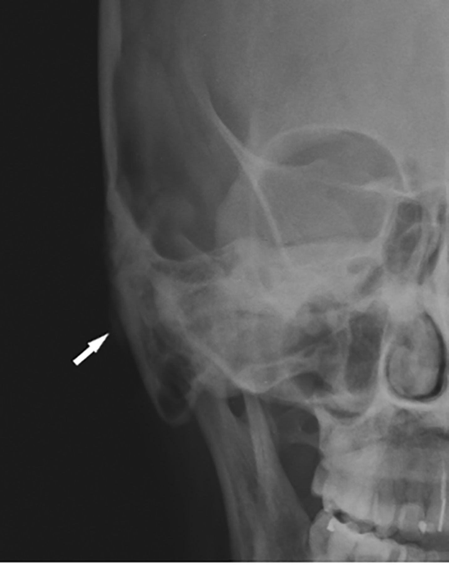
- FIG 1.
Case 1. Plain radiographs of the skull show a small, exophytic sclerotic/mineralized lesion with bony thickening of the right retroauricular cortex (white arrow).
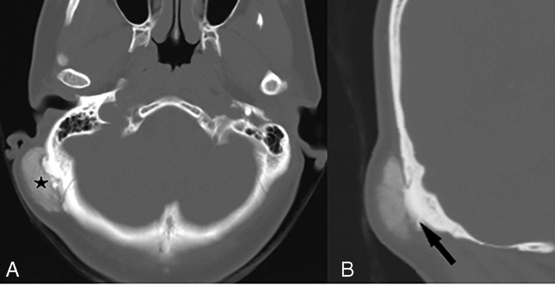
- FIG 2.
Case 1. Axial (A) and coronal (B) noncontrast CT images through the temporal bone show a well-defined, exophytic, protuberant calcified mass with ground-glass density (star) in the retroauricular region, seen emanating from the outer cortex of the right temporal bone near the occipitomastoid suture causing elevation of the overlying scalp without intramedullary or intracranial extension. A broad-based stalk-like attachment to the occipitomastoid suture is noted (arrow).
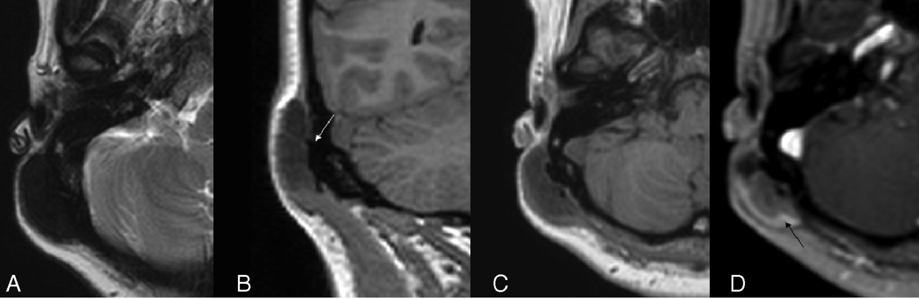
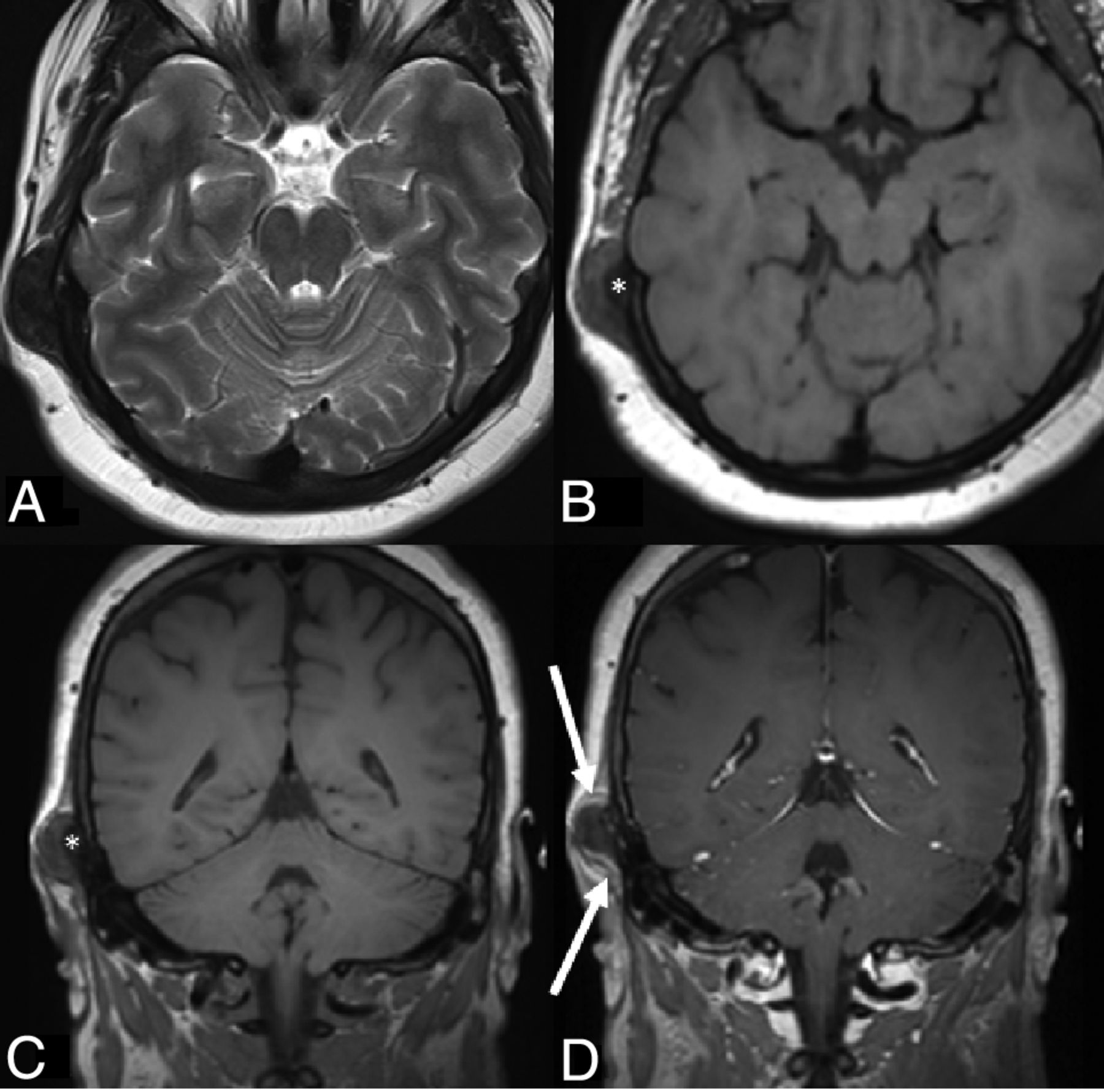
- FIG 3.
Case 1. MR images with and without contrast. Axial T2 (A), coronal and axial T1 (B and C), and postgadolinium axial (D) T1-weighted images show a T1- and T2-hypointense (to muscle), mildly enhancing broad-based, exophytic postauricular lesion emanating from the outer table of the right temporal bone (arrow in B). There is no signal change in the underlying bone and no intramedullary or intracranial extension. A thin, peripheral rim of enhancement is seen (arrow in D).
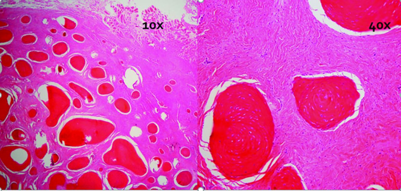
- FIG 4.
Case 1. Histology under low-power (10×) and high-power (40×) fields. Note numerous rounded ossified bodies composed of either woven or lamellar bone within a bland fibrous stroma. No atypia, mitotic activity or osteoblastic rimming are identified.
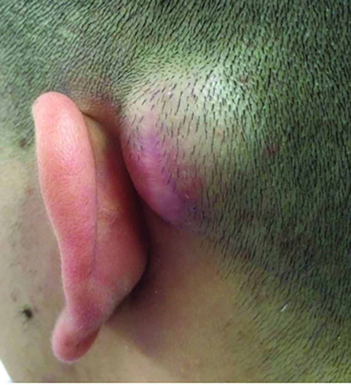
- FIG 5.
Case 2. On clinical examination, a 3 × 2 cm left retroauricular bony protuberance over the mastoid area is noted.
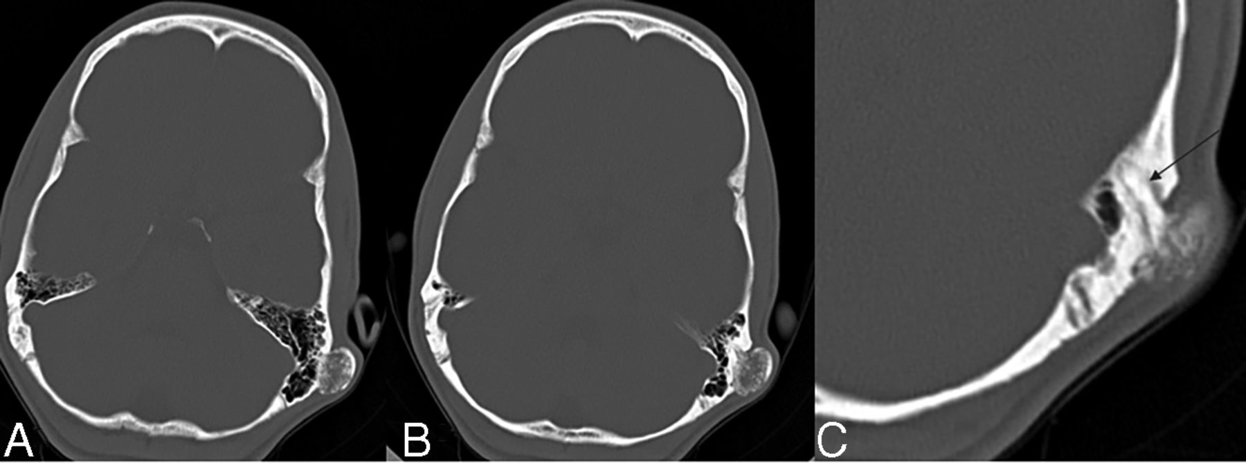
- FIG 6.
Case 2. Axial noncontrast CT images through the temporal bones (A and B) show a well-marginated exophytic mass based along the lateral mastoid cortex with a densely calcified rim and a central heterogeneously calcified component. Magnified axial CT image (C) shows a densely calcified stalk (black arrow) that extends into the occipitomastoid suture.
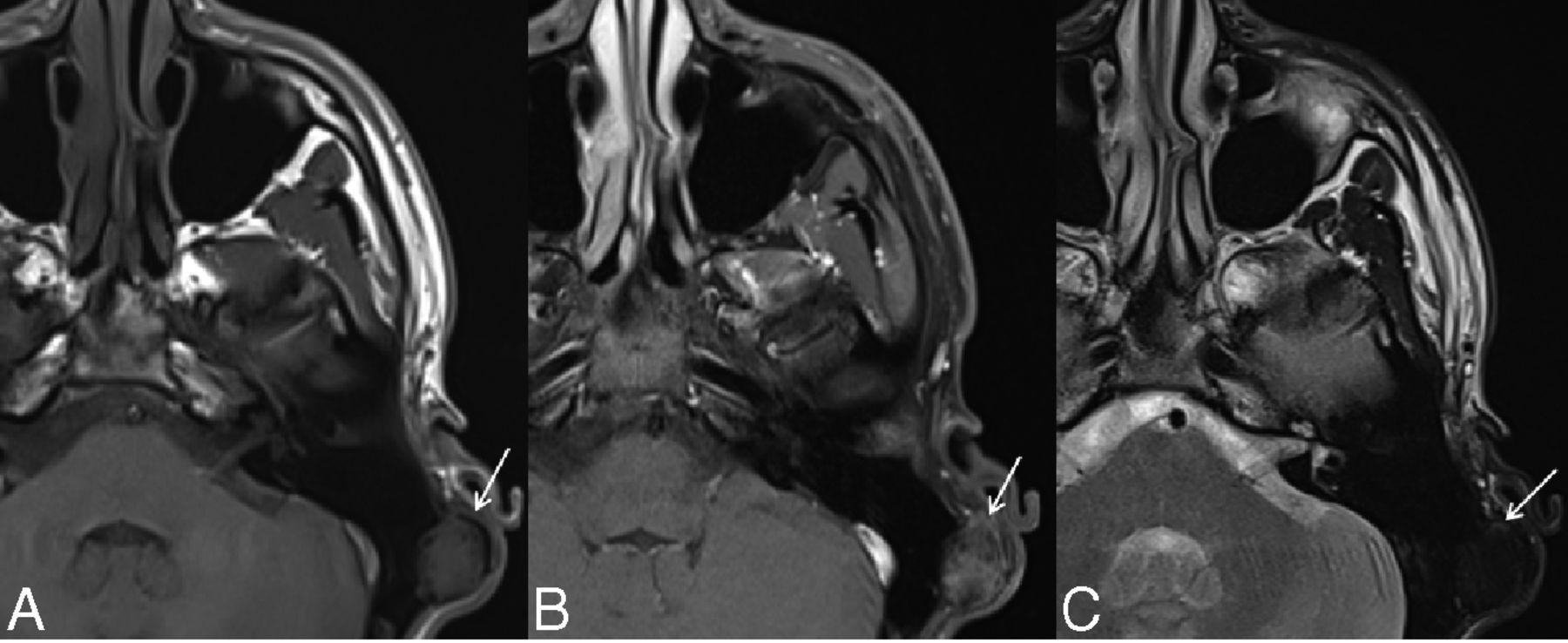
- FIG 7.
Case 2. MR images with and without contrast. Axial T1 (A), axial T1 postgadolinium fat-saturated (B), and axial T2-weighted (C) images show a thin peripheral signal void (arrow in A) and central intermediate signal intensity on noncontrast T1WI with mild heterogeneous enhancement (arrow in B) and diffusely low T2 signal intensity (arrow in C).
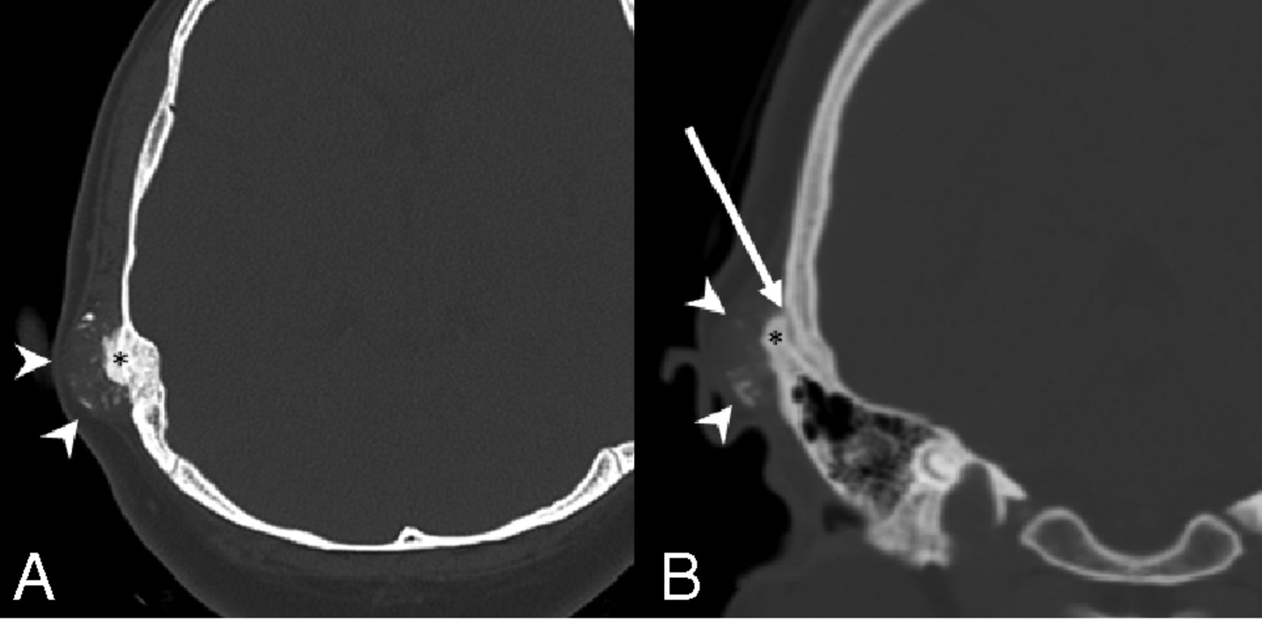
- FIG 8.
Case 3. Axial and coronal noncontrast CT images (A and B) demonstrate an irregularly calcified mass (white arrowheads) centered over the right temporal outer cortex at the level of the occipitomastoid suture (white arrow). Note the solidly calcified bony stalk (black asterisk). No gross destruction of the outer table cortex is seen, and there is no intraosseous or intracranial extension. The lesion uplifts the overlying scalp.
- FIG 9.
Case 3. Axial noncontrast CT images across time demonstrating the slow, interval growth of the retroauricular mass along the outer cortex of the right temporal bone. Also note that the lesion was initially a solidly calcified bony stalk, imaging similar to a peripheral ivory osteoma (white arrow in A). As the lesion grew, the ossification was less solid at the periphery and more irregular (white arrowheads in C) with persistence of the initial bony stalk (arrow in C).
- FIG 10.
Case 3. MR images with and without contrast. Axial T2-weighted image (A) demonstrates the homogeneous T2-hypointense signal of the lesion. The lesion is predominantly T1-hypointense on the axial and coronal noncontrast T1-weighted images (B and C), particularly centrally, corresponding to the more densely calcified bony stalk (white asterisk). A largely peripheral cap of enhancement (white arrows) is noted on the coronal, postcontrast T1-weighted image.
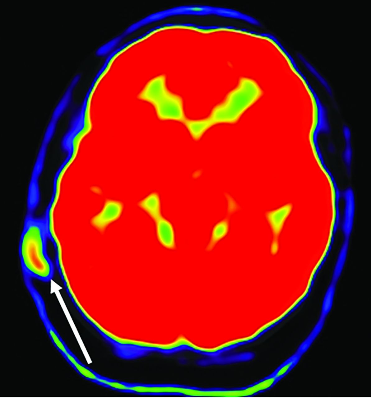
- FIG 11.
Case 3. FDG-PET image demonstrates FDG-avidity of the lesion (white arrow), consistent with the occasional mitotic features at histology.
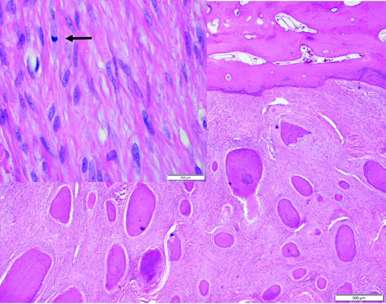
- FIG 12.
Case 3. Histology under a low-power field demonstrates numerous round ossified bodies within a proliferation of bland spindle cells. The fibrous stroma shows occasional mitotic figures (arrow in inset); however, no atypia is identified.


Jump to section
Related Articles
Cited By...
- No citing articles found.