Advertisement

Main menu

  • Home
  • Content
    • Current Issue
    • Accepted Manuscripts
    • Article Preview
    • Past Issue Archive
    • AJNR Case Collection
    • Case of the Week Archive
    • Classic Case Archive
    • Case of the Month Archive
  • Special Collections
    • Spinal CSF Leak Articles (Jan 2020-June 2024)
    • 2024 AJNR Journal Awards
    • Most Impactful AJNR Articles
  • Multimedia
    • AJNR Podcast
    • AJNR Scantastics
    • Video Articles
  • For Authors
    • Submit a Manuscript
    • Author Policies
    • Fast publishing of Accepted Manuscripts
    • Graphical Abstract Preparation
    • Manuscript Submission Guidelines
    • Imaging Protocol Submission
    • Submit a Case for the Case Collection
  • About Us
    • About AJNR
    • Editorial Board
  • More
    • Become a Reviewer/Academy of Reviewers
    • Subscribers
    • Permissions
    • Alerts
    • Feedback
    • Advertisers
    • ASNR Home
  • Other Publications
    • ajnr

User menu

  • Alerts
  • Log in

Search

  • Advanced search
American Journal of Neuroradiology

American Journal of Neuroradiology

ASHNR American Society of Functional Neuroradiology ASHNR American Society of Pediatric Neuroradiology ASSR
  • Alerts
  • Log in

Advanced Search

  • Home
  • Content
    • Current Issue
    • Accepted Manuscripts
    • Article Preview
    • Past Issue Archive
    • AJNR Case Collection
    • Case of the Week Archive
    • Classic Case Archive
    • Case of the Month Archive
  • Special Collections
    • Spinal CSF Leak Articles (Jan 2020-June 2024)
    • 2024 AJNR Journal Awards
    • Most Impactful AJNR Articles
  • Multimedia
    • AJNR Podcast
    • AJNR Scantastics
    • Video Articles
  • For Authors
    • Submit a Manuscript
    • Author Policies
    • Fast publishing of Accepted Manuscripts
    • Graphical Abstract Preparation
    • Manuscript Submission Guidelines
    • Imaging Protocol Submission
    • Submit a Case for the Case Collection
  • About Us
    • About AJNR
    • Editorial Board
  • More
    • Become a Reviewer/Academy of Reviewers
    • Subscribers
    • Permissions
    • Alerts
    • Feedback
    • Advertisers
    • ASNR Home
  • Follow AJNR on Twitter
  • Visit AJNR on Facebook
  • Follow AJNR on Instagram
  • Join AJNR on LinkedIn
  • RSS Feeds

Welcome to the new AJNR, Updated Hall of Fame, and more. Read the full announcements.


AJNR is seeking candidates for the position of Associate Section Editor, AJNR Case Collection. Read the full announcement.


Getting new auth cookie, if you see this message a lot, tell someone!
Research ArticleSpine

Spinal CSF-Venous Fistulas in Morbidly and Super Obese Patients with Spontaneous Intracranial Hypotension

W.I. Schievink, M. Maya, R.S. Prasad, V.S. Wadhwa, R.B. Cruz and F.G. Moser
American Journal of Neuroradiology February 2021, 42 (2) 397-401; DOI: https://doi.org/10.3174/ajnr.A6895
W.I. Schievink
aFrom the Departments of Neurosurgery (W.I.S., R.B.C.)
  • Find this author on Google Scholar
  • Find this author on PubMed
  • Search for this author on this site
  • ORCID record for W.I. Schievink
M. Maya
bImaging (M.M., R.S.P., V.S.W., F.G.M.), Cedars-Sinai Medical Center, Los Angeles, California
  • Find this author on Google Scholar
  • Find this author on PubMed
  • Search for this author on this site
  • ORCID record for M. Maya
R.S. Prasad
bImaging (M.M., R.S.P., V.S.W., F.G.M.), Cedars-Sinai Medical Center, Los Angeles, California
  • Find this author on Google Scholar
  • Find this author on PubMed
  • Search for this author on this site
  • ORCID record for R.S. Prasad
V.S. Wadhwa
bImaging (M.M., R.S.P., V.S.W., F.G.M.), Cedars-Sinai Medical Center, Los Angeles, California
  • Find this author on Google Scholar
  • Find this author on PubMed
  • Search for this author on this site
  • ORCID record for V.S. Wadhwa
R.B. Cruz
aFrom the Departments of Neurosurgery (W.I.S., R.B.C.)
  • Find this author on Google Scholar
  • Find this author on PubMed
  • Search for this author on this site
  • ORCID record for R.B. Cruz
F.G. Moser
bImaging (M.M., R.S.P., V.S.W., F.G.M.), Cedars-Sinai Medical Center, Los Angeles, California
  • Find this author on Google Scholar
  • Find this author on PubMed
  • Search for this author on this site
  • ORCID record for F.G. Moser
  • Article
  • Figures & Data
  • Info & Metrics
  • Responses
  • References
  • PDF
Loading

Abstract

BACKGROUND AND PURPOSE: Spinal CSF-venous fistulas are increasingly recognized as the cause of spontaneous intracranial hypotension. Here, we describe the challenges in the care of patients with CSF-venous fistulas who are morbidly or super obese.

MATERIALS AND METHODS: A review was undertaken of all patients with spontaneous intracranial hypotension and a body mass index of >40 who underwent digital subtraction myelography in the lateral decubitus position to look for CSF-venous fistulas.

RESULTS: Eight patients with spontaneous intracranial hypotension with a body mass index  of >40 underwent lateral decubitus digital subtraction myelography. The mean age of these 5 women and 3 men was 53 years (range, 45 to 68 years). Six patients were morbidly obese (body mass indexes = 40.2, 40.6, 41, 41.8, 45.4, and 46.9), and 2 were super obese (body mass indexes = 53.7 and 56.3). Lumbar puncture showed an elevated opening pressure in 5 patients (26.5–47 cm H2O). The combination of an elevated opening pressure and normal conventional spine imaging findings resulted in a misdiagnosis (midbrain glioma and demyelinating disease, respectively) in 2 patients. Prior treatment included surgical nerve root ligation for suspected CSF-venous fistula in 3 patients. Digital subtraction myelography demonstrated a CSF-venous fistula in 6 patients (75%). Rebound high-pressure headache occurred in all 6 patients following surgical ligation of the fistula, and papilledema developed in 3.

CONCLUSIONS: In our series, opening pressure was generally elevated in patients with morbid or super obesity. The yield of identifying CSF-venous fistulas with digital subtraction myelography in this patient population can approach that of the nonobese patient population. These patients may be at higher risk of developing rebound high-pressure headaches and papilledema.

ABBREVIATIONS:

BMI
body mass index
DSM
digital subtraction myelography
SIH
spontaneous intracranial hypotension

Spontaneous intracranial hypotension (SIH) is an increasingly recognized cause of secondary headache and has also been associated with a wide variety of other neurologic disorders.1-4 The cause of SIH is a spinal CSF leak; at least 3 distinct types have been identified.5 A direct communication between the subarachnoid space and a spinal epidural vein, a so-called CSF-venous fistula, is the most recently discovered type of spinal CSF leak and may be detected in up to one-fourth of patients with SIH.6 Since the first description of spontaneous spinal CSF-venous fistulas in 2014,7 several groups have confirmed their importance, but much remains unknown about these fistulas.6⇓⇓⇓⇓⇓⇓⇓⇓-15 The main reason that this type of CSF leak remained undiscovered for so long is that it is not associated with an extradural CSF collection and its detection requires sophisticated imaging, eg, digital subtraction myelography (DSM) or dynamic CT myelography.6⇓⇓⇓⇓⇓⇓⇓⇓-15

Obesity has emerged as a major global and national health problem.16 Obesity is defined as a body mass index (BMI) of ≥30 and is associated with various diseases, such as diabetes, metabolic syndrome, and cardiovascular disease. The prevalence of obesity is increasing, and in the United States, it is among the highest in the world. According to the National Health and Nutrition Examination Survey 2015 and 2016 dataset, 39.8% of US adults live with obesity.17 Morbid obesity (BMI ≥ 40) and super obesity (BMI ≥ 50) have shown the most rapid rise in prevalence, and with data from the Behavioral Risk Factor Surveillance System, it was estimated that 6.6% of Americans had a BMI of >40 in 2010.18 Despite the importance of obesity, there is very limited information available on obesity in patients with spontaneous spinal CSF-venous fistulas. Actually, no information on BMI is available in any of the prior reports, including our own, on spontaneous spinal CSF-venous fistulas.6⇓⇓⇓⇓⇓⇓⇓⇓-15 We have noted that particularly morbid and super obesity can result in unique challenges in the diagnosis, detection, and treatment of patients with SIH with spontaneous spinal CSF-venous fistulas; therefore, we reviewed our experience with this patient population.

MATERIALS AND METHODS

This study was approved by the Cedars-Sinai Medical Center institutional review board.

Using a prospectively maintained registry, we identified all patients with SIH and a BMI of ≥40 who underwent DSM in the lateral decubitus position to look for CSF-venous fistulas. The diagnosis of SIH was based on the criteria of the International Classification of Headache Disorders, third edition,19 with minor modifications. These criteria require objective evidence of SIH, consisting of brain MR imaging showing stigmata of SIH (ie, pachymeningeal enhancement, brain sagging, or subdural fluid collections), spinal imaging showing a CSF leak (ie, the presence of extradural CSF), or low CSF opening pressure (ie, <6.0 cm H2O). The modification consists of also including patients who do not have headaches but whose symptoms are best explained by SIH.

All patients underwent brain MR imaging and MR myelography (heavily T2-weighted MR imaging without intrathecal contrast). For DSM, the technique as described by Hoxworth et al20 was used with some minor modifications.6,8 Briefly, DSM is performed with the patient under general endotracheal anesthesia with deep paralysis and suspended respiration for maximal detail and temporal resolution. Patients are positioned in the lateral decubitus position in a biplane angiography suite, with tilt-table capability. Pillows or foam padding are placed to optimize cervicothoracic alignment. A fluoroscopically guided lumbar puncture is performed at the L2–3 level with a 22-ga needle. An opening pressure is obtained at this time using standard manometry. Then, accurate needle position is confirmed with an injection of 0.5 mL of iohexol (Omnipaque; GE Healthcare). Patients are then further positioned on the basis of the area of interest, and the table is tilted to achieve contrast flow to the cervicothoracic spine. Great care is taken, especially in these patients with obesity, to maximize contrast opacification of the lateral dural sac by adjusting the degree of tilting to patient-specific spinal curvature and anatomy. Finally, contrast is injected manually at 1 mL per second with suspended respiration for 40–100 seconds while acquiring biplane subtraction images at 2 frames per second. If the first DSM fails to identify a CSF-venous fistula, then the study is repeated on another day with the patient in the lateral decubitus position on the contralateral side.

Radiation exposure during DSM was calculated for these patients and for 2 age- and sex-matched controls, each with a BMI of <40.

RESULTS

Eight patients with SIH with a BMI of ≥40 underwent lateral decubitus DSM (Fig 1). The mean age of these 5 women and 3 men was 53 years (range, 45–68 years). Six patients were morbidly obese (BMI = 40.2, 40.6, 41, 41.8, 45.4, and 46.9), and 2 were super obese (BMI = 53.7 and 56.3). None of the patients had undergone bariatric surgery. Four patients presented with isolated orthostatic headaches; 3, with orthostatic headaches with Valsalva-induced worsening; and 1, with isolated Valsalva-induced headache. Fundoscopic examination findings were normal in all 8 patients. Brain MR imaging showed brain sagging in 7 patients and meningeal enhancement in 5 patients.

FIG 1.
  • Download figure
  • Open in new tab
  • Download powerpoint
FIG 1.

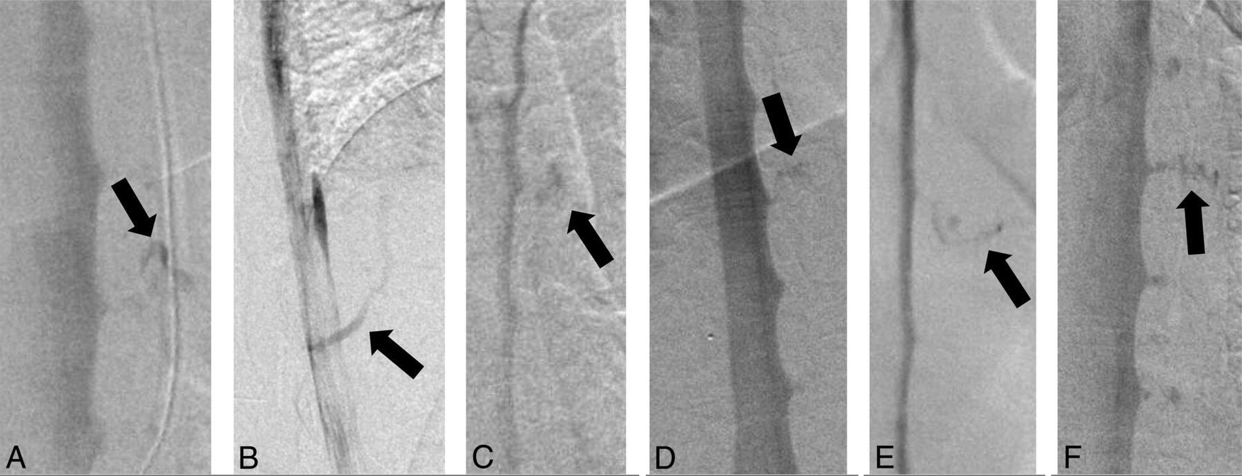
A, Anterior-posterior DSM demonstrates a CSF-venous fistula (BMI = 46.9 kg/m2, weight 149 kg, height = 178 cm). B, Lateral DSM demonstrates a CSF-venous fistula (BMI = 40.6 kg/m2, weight = 94.5 kg, height = 152 cm). C, Anterior-posterior DSM demonstrates a CSF-venous fistula (BMI = 40.2 kg/m2, weight = 113 kg, height = 167 cm). D, Anterior-posterior DSM demonstrates a CSF-venous fistula (BMI = 53.7 kg/m2, weight = 151 kg, height = 167 cm). E, Anterior-posterior DSM demonstrates a CSF-venous fistula (BMI = 41 kg/m2, weight = 137 kg, height = 183 cm). F, Anterior-posterior DSM demonstrates a CSF-venous fistula (BMI = 45.4 kg/m2, weight = 161 kg, height = 188 cm). In 2 other patients (BMI = 56.3 kg/m2, weight = 149 kg, height = 163 cm and BMI = 41.8 kg/m2, weight = 140 kg, height = 182 cm, respectively), DSM failed to demonstrate a CSF-venous fistula. Arrows indicates CSF-venous fistulas.

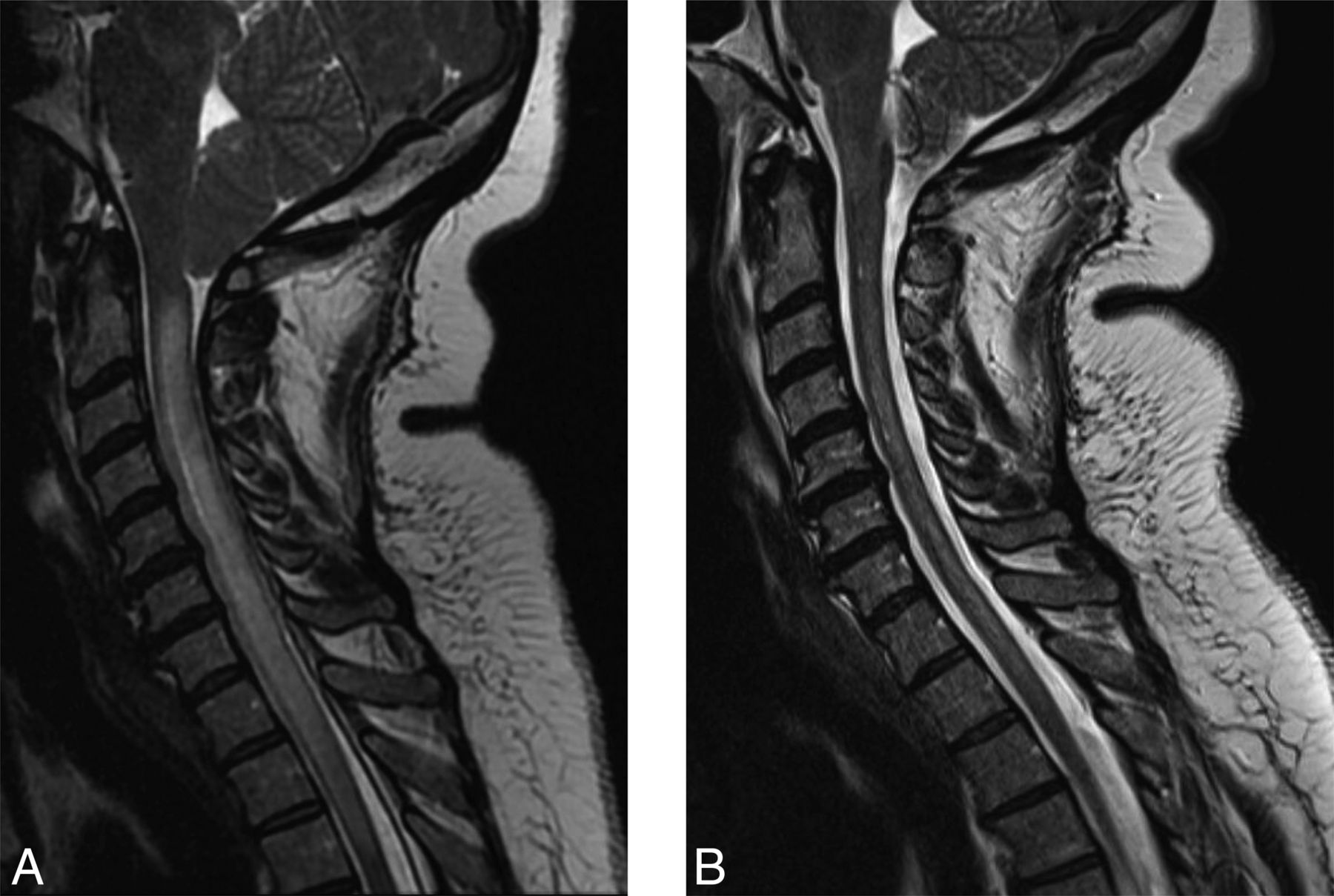
Before referral to our medical center, a lumbar puncture in the lateral decubitus position had been performed in 6 patients, showing a low opening pressure (<6 cm H2O) in 1 patient and an elevated opening pressure (26.5–36 cm H2O) in 5 patients. A lumbar puncture performed with the patient in the prone position yielded a normal opening pressure (11.5 cm H2O) in the remaining patient. All patients had undergone a spine MR imaging, 5 had undergone a conventional CT myelogram, 3 had undergone dynamic CT myelography, and 2 had undergone DSMs in the lateral decubitus position (using general or local anesthesia). None of these investigations had shown extradural CSF collections or a clear evidence of a CSF-venous fistula. The combination of an elevated opening pressure and normal spine imaging findings resulted in the diagnosis of SIH being discarded in 2 patients who were subsequently misdiagnosed with demyelinating disease (T2 signal abnormality of the spinal cord in the setting of severe brain sagging, Fig 2) and a midbrain glioma (T2 signal abnormality in the setting of severe brain sagging), respectively. Three patients had been treated with multiple epidural blood patches or fibrin glue injections as well as surgical ligation of thoracic nerve roots for the suspicion of a CSF-venous fistula. One patient had undergone an exploratory operation at the site of a disc herniation.

FIG 2.
  • Download figure
  • Open in new tab
  • Download powerpoint
FIG 2.

Sagittal T2-weighted MRIs showing brain sagging and T2 signal change within the cervical spinal cord (presyrinx) (A) and resolution of the presyrinx after surgical ligation of the thoracic CSF-venous fistula due to resolution of brain sagging and cerebellar tonsillar herniation (B).

Using DSM with the patient in the lateral decubitus position, we were able to detect a CSF-venous fistula in 6 of the 8 patients (75%). All fistulas were located in the thoracic spine, 3 on the right side and 3 on the left side. All were associated with a spinal meningeal diverticulum. A normal opening pressure was found in 3 patients (13, 17, and 20 cm H2O), a borderline elevated opening pressure was found in 2 patients (23 and 24 cm H2O), and an elevated opening pressure was found in 3 patients (28, 29, and 47 cm H2O). These opening pressure readings were obtained with the patient in the lateral decubitus position under general anesthesia. Radiation exposure was higher in these patients (mean, 1753 mGy; range, 1271–2314 mGy) than in controls (mean, 488 mGy; range, 167–979 mGy).

All 6 patients with a spinal CSF-venous fistula underwent an uneventful thoracic foraminotomy, with ligation of the fistula resulting in symptom resolution and normalization of the MR imaging findings. However, rebound high-pressure headache requiring treatment with acetazolamide developed in all 6 patients, and this was complicated by visual blurring and papilledema in 3 patients. A lumbar puncture was performed with the patient in the lateral decubitus position in 2 of these patients with papilledema, and this showed an increase of opening pressure from 24 to 30 cm H2O in one patient (spontaneous respirations) and from 47 to 51 cm H2O in the other (at the time of repeat DSM with general anesthesia). Vision symptoms resolved with the acetazolamide treatment.

DISCUSSION

In this study of morbidly and super obese patients with spontaneous spinal CSF-venous fistulas, we found specific challenges in the diagnosis, imaging, and treatment.

Although it has been well-documented since the 1990s that patients with SIH often do not have abnormally low CSF pressure,21⇓-23 the current study showed that opening pressure can be markedly elevated in these patients. This finding shows that just like the presence of a normal opening pressure, an abnormally high opening pressure does not preclude the diagnosis of SIH. Prior studies had already concluded that some patients with SIH may have borderline elevated CSF pressure,22,23 and a patient with SIH and an opening pressure of 31 cm H2O was reported by Kranz et al.23 The combination of an elevated opening pressure in addition to normal results of conventional spinal imaging, ie, the absence of any extradural CSF collection, caused the diagnosis of SIH to be discarded in some of the presently reported patients, despite the presence of the typical brain MR imaging findings. This step resulted in an erroneous diagnosis of demyelinating disease in one patient and of a midbrain glioma in another. The presence of elevated CSF pressure also has possible pathophysiologic implications for the development of spinal CSF-venous fistulas. Similar to spontaneous skull base CSF leaks in patients with idiopathic intracranial hypertension,24⇓⇓–27 chronically elevated CSF pressure could also predispose to spontaneous spinal CSF leaks. This possibility also had been reported in patients with pre-existing idiopathic intracranial hypertension who developed SIH.2 The presently reported patients had normal fundoscopic examination findings on presentation and did not have idiopathic intracranial hypertension. Of note, none of the patients in the present study had undergone bariatric surgery, a risk factor for developing SIH.28

The detection of CSF-venous fistulas requires sophisticated imaging, and we have been using lateral decubitus DSM with the patient under general anesthesia for this purpose with excellent results.6 Although the body habitus of morbidly and super obese patients may be intimidating, it should not result in a defeatist attitude. In the present study, we detected CSF-venous fistulas in three-fourths of morbidly and super obese patients with SIH and no extradural CSF collections on spinal imaging, thus demonstrating that the yield of identifying such fistulas in this patient population can approach that of the nonobese patient population.6 In a prior study of 23 consecutive patients with SIH and the stigmata of SIH on brain MR imaging but no extradural CSF collections on spinal imaging who underwent lateral decubitus DSM, we found CSF-venous fistulas in 17 patients (74%).6

Postoperatively, rebound high-pressure headaches occurred in all 6 patients who underwent surgical ligation of the CSF-venous fistula, and this was complicated by papilledema and visual symptoms in 3 patients. The development of papilledema was associated with only a modest increase in opening pressure. This compares with a previous study in which about one-fourth of 113 patients developed rebound high-pressure headache requiring administration of acetazolamide following treatment of SIH, and papilledema was noted in only 2 patients.29 Similarly, Wang et al30 reported rebound high-pressure headaches requiring treatment with acetazolamide in 5 (25%) of 20 patients with SIH following surgical ligation of a spinal CSF-venous fistula. Although the number of patients in the current study is small, this suggests that morbidly and super obese patients with CSF-venous fistulas are at increased risk of developing rebound high-pressure headaches and papilledema postoperatively and that consideration should be given to pharmacologic prophylaxis with, for example acetazolamide, before treating the CSF-venous fistula and that careful ophthalmologic evaluation postoperatively is indicated.

CONCLUSIONS

In patients with SIH due to a spinal CSF-venous fistula who are morbidly or super obese, CSF pressure often is elevated and the risk of posttreatment rebound high-pressure headache and papilledema is increased. The yield of finding a CSF-venous fistula in this patient population using DSM is similar to that in patients who are not obese.

Footnotes

  • Disclosures: Wouter I. Schievink—UNRELATED: Board Membership: I am on the medical advisory board of the Spinal CSF Leak Foundation; this is a nonpaying position, and I have not received any funds toward this project. Marcel Maya—UNRELATED: Board Membership: I am on the medical advisory board of the Spinal CSF Leak Foundation; this is a nonpaying position, and I have not received any funds toward this project.

References

  1. 1.↵
    1. Schievink WI
    . Spontaneous spinal cerebrospinal fluid leaks and intracranial hypotension. JAMA 2006;295:2286–96 doi:10.1001/jama.295.19.2286 pmid:16705110
    CrossRefPubMed
  2. 2.↵
    1. Mokri B
    . Spontaneous intracranial hypotension. Continuum (Minneap Minn) 2015;21:1086–1108 doi:10.1212/CON.0000000000000193 pmid:26252593
    CrossRefPubMed
  3. 3.
    1. Kranz PG,
    2. Malinzak MD,
    3. Amrhein TJ, et al
    . Update on the diagnosis and treatment of spontaneous intracranial hypotension. Curr Pain Headache Rep 2017;21:37 doi:10.1007/s11916-017-0639-3 pmid:28755201
    CrossRefPubMed
  4. 4.↵
    1. Robblee J,
    2. Secora KA,
    3. Alhilali LM, et al
    . Spontaneous intracranial hypotension. Pract Neurol 2020. https://practicalneurology.com/articles/2020-may/spontaneous-intracranial-hypotension-1. Accessed August 1, 2020
  5. 5.↵
    1. Schievink WI,
    2. Maya MM,
    3. Jean-Pierre S, et al
    . A classification system of spontaneous spinal CSF leaks. Neurology 2016;87:673–79 doi:10.1212/WNL.0000000000002986 pmid:27440149
    Abstract/FREE Full Text
  6. 6.↵
    1. Schievink WI,
    2. Maya MM,
    3. Moser FG, et al
    . Lateral decubitus digital subtraction myelography to identify spinal CSF-venous fistulas in spontaneous intracranial hypotension. J Neurosurg Spine 2019 Sept 13. [Epub ahead of print] doi:10.3171/2019.6.SPINE19487 pmid:31518974
    CrossRefPubMed
  7. 7.↵
    1. Schievink WI,
    2. Moser FG,
    3. Maya MM
    . CSF-venous fistula in spontaneous intracranial hypotension. Neurology 2014;83:472–73 doi:10.1212/WNL.0000000000000639 pmid:24951475
    Abstract/FREE Full Text
  8. 8.↵
    1. Schievink WI,
    2. Moser FG,
    3. Maya MM, et al
    . Digital subtraction myelography for the identification of spontaneous spinal CSF-venous fistulas. J Neurosurg Spine 2016;24:960–64 doi:10.3171/2015.10.SPINE15855 pmid:26849709
    CrossRefPubMed
  9. 9.↵
    1. Kumar N,
    2. Diehn FE,
    3. Carr CM, et al
    . Spinal CSF venous fistula: a treatable etiology for CSF leaks in craniospinal hypovolemia. Neurology 2016;86:2310–12 doi:10.1212/WNL.0000000000002776 pmid:27178701
    Abstract/FREE Full Text
  10. 10.↵
    1. Kranz PG,
    2. Amrhein TJ,
    3. Schievink WI, et al
    . The “hyperdense paraspinal vein” sign: a marker of CSF-venous fistula. AJNR Am J Neuroradiol 2016;37:1379–81 doi:10.3174/ajnr.A4682 pmid:26869470
    Abstract/FREE Full Text
  11. 11.↵
    1. Kranz PG,
    2. Amrhein TJ,
    3. Gray L
    . CSF venous fistulas in spontaneous intracranial hypotension: imaging characteristics on dynamic and CT myelography. AJR Am J Roentgenol 2017;209:1360–66 doi:10.2214/AJR.17.18351 pmid:29023155
    CrossRefPubMed
  12. 12.↵
    1. Farb RI,
    2. Nicholson PJ,
    3. Peng PW, et al
    . Spontaneous intracranial hypotension: a systematic imaging approach for CSF leak localization and management based on MRI and digital subtraction myelography. AJNR Am J Neuroradiol 2019;40:745–53 doi:10.3174/ajnr.A6016 pmid:30923083
    Abstract/FREE Full Text
  13. 13.↵
    1. Schievink WI,
    2. Maya MM,
    3. Moser FG, et al
    . Spontaneous spinal CSF-venous fistulas associated with venous/venolymphatic vascular malformations: report of 3 cases. J Neurosurg Spine 2019;32:305–10 doi:10.3171/2019.8.SPINE19716 pmid:31675703
    CrossRefPubMed
  14. 14.↵
    1. Duvall JR,
    2. Robertson CE,
    3. Cutsforth-Gregory JK, et al
    . Headache due to spontaneous spinal cerebrospinal fluid leak secondary to cerebrospinal fluid-venous fistula: case series. Cephalalgia 2019;39:1847–54 doi:10.1177/0333102419881673 pmid:31597463
    CrossRefPubMed
  15. 15.↵
    1. Chazen JL,
    2. Robbins MS,
    3. Strauss SB, et al
    . MR myelography for the detection of CSF-venous fistulas. AJNR Am J Neuroradiol 2020;41:938–40 doi:10.3174/ajnr.A6521 pmid:32354709
    Abstract/FREE Full Text
  16. 16.↵
    NCD Risk Factor Collaboration (NCD-RisC). Trends in adult body-mass index in 200 countries from 1975 to 2014: a pooled analysis of 1698 population-based measurement studies with 19.2 million participants. Lancet 2016;387:1377–96 doi:10.1016/S0140-6736(16)30054-X pmid:27115820
    CrossRefPubMed
  17. 17.↵
    Health, United States, 2018–Data Finder. https://www.cdc.gov/nchs/hus/contents2018.htm#Table_021. Accessed June 9, 2020
  18. 18.↵
    1. Sturm R,
    2. Hattori A
    . Morbid obesity rates continue to rise rapidly in the United States. Int J Obes (Lond) 2013;37:889–91 doi:10.1038/ijo.2012.159 pmid:22986681
    CrossRefPubMed
  19. 19.↵
    Headache Classification Committee of the International Headache Society: The International Classification of Headache Disorders, 3rd edition. Cephalalgia 2018 38:1–211 doi:10.1177/0333102417738202 pmid:29368949
    CrossRefPubMed
  20. 20.↵
    1. Hoxworth JM,
    2. Patel AC,
    3. Bosch EP, et al
    . Localization of a rapid CSF leak with digital subtraction myelography. AJNR Am J Neuroradiol 2009;30:516–19 doi:10.3174/ajnr.A1294 pmid:18842766
    Abstract/FREE Full Text
  21. 21.↵
    1. Mokri B,
    2. Hunter SF,
    3. Atkinson JL, et al
    . Orthostatic headaches caused by CSF leak but with normal CSF pressures. Neurology 1998;51:786–90 doi:10.1212/wnl.51.3.786 pmid:9748027
    Abstract/FREE Full Text
  22. 22.↵
    1. Luetmer PH,
    2. Schwartz KM,
    3. Eckel LJ, et al
    . When should I do dynamic CT myelography? Predicting fast spinal CSF leaks in patients with spontaneous intracranial hypotension. AJNR Am J Neuroradiol 2012;33:690–94 doi:10.3174/ajnr.A2849 pmid:22194380
    Abstract/FREE Full Text
  23. 23.↵
    1. Kranz PG,
    2. Tanpitukpongse TP,
    3. Choudhury KR, et al
    . How common is normal cerebrospinal fluid pressure in spontaneous intracranial hypotension? Cephalalgia 2016;36:1209–17 doi:10.1177/0333102415623071 pmid:26682575
    CrossRefPubMed
  24. 24.↵
    1. Brisman R,
    2. Hughes JE,
    3. Mount LA
    . Cerebrospinal fluid rhinorrhea. Arch Neurol 1970;22:245–52 doi:10.1001/archneur.1970.00480210055007 pmid:5411680
    CrossRefPubMed
  25. 25.↵
    1. Aaron G,
    2. Doyle J,
    3. Vaphiades MS, et al
    . Increased intracranial pressure in spontaneous CSF leak patients not associated with papilledema. Otolaryngol Head Neck Surg 2014;151:1061–66 doi:10.1177/0194599814551122 pmid:25248848
    CrossRefPubMed
  26. 26.↵
    1. Bidot S,
    2. Levy JM,
    3. Saindane AM, et al
    . Do most patients with a spontaneous cerebrospinal fluid leak have idiopathic intracranial hypertension? J Neuroophthalmol 2019;39:487–95 doi:10.1097/WNO.0000000000000761 pmid:30747786
    CrossRefPubMed
  27. 27.↵
    1. Tam EK,
    2. Gilbert AL
    . Spontaneous cerebrospinal fluid leak and idiopathic intracranial hypertension. Curr Opin Ophthalmol 2019;30:467–71 doi:10.1097/ICU.0000000000000603 pmid:31449087
    CrossRefPubMed
  28. 28.↵
    1. Schievink WI,
    2. Goseland A,
    3. Cunneen S
    . Bariatric surgery as a possible risk factor for spontaneous intracranial hypotension. Neurology 2014;83:1819–22 doi:10.1212/WNL.0000000000000985 pmid:25339216
    Abstract/FREE Full Text
  29. 29.↵
    1. Schievink WI,
    2. Maya MM,
    3. Jean-Pierre S, et al
    . Rebound high-pressure headache after treatment of spontaneous intracranial hypotension: MRV study. Neurol Clin Pract 2019;9:93–100 doi:10.1212/CPJ.0000000000000550 pmid:31041122
    Abstract/FREE Full Text
  30. 30.↵
    1. Wang TY,
    2. Karikari IO,
    3. Amrhein TJ, et al
    . Clinical outcomes following surgical ligation of cerebrospinal fluid–venous fistula in patients with spontaneous intracranial hypotension: a prospective case series. Oper Neurosurg (Hagerstown) 2020;18:239–45 doi:10.1093/ons/opz134 pmid:31134267
    CrossRefPubMed
  • Received August 23, 2020.
  • Accepted after revision September 15, 2020.
  • © 2021 by American Journal of Neuroradiology
PreviousNext
Back to top

In this issue

American Journal of Neuroradiology: 42 (2)
American Journal of Neuroradiology
Vol. 42, Issue 2
1 Feb 2021
  • Table of Contents
  • Index by author
  • Complete Issue (PDF)
Advertisement
Print
Download PDF
Email Article

Thank you for your interest in spreading the word on American Journal of Neuroradiology.

NOTE: We only request your email address so that the person you are recommending the page to knows that you wanted them to see it, and that it is not junk mail. We do not capture any email address.

Enter multiple addresses on separate lines or separate them with commas.
Spinal CSF-Venous Fistulas in Morbidly and Super Obese Patients with Spontaneous Intracranial Hypotension
(Your Name) has sent you a message from American Journal of Neuroradiology
(Your Name) thought you would like to see the American Journal of Neuroradiology web site.
CAPTCHA
This question is for testing whether or not you are a human visitor and to prevent automated spam submissions.
Cite this article
W.I. Schievink, M. Maya, R.S. Prasad, V.S. Wadhwa, R.B. Cruz, F.G. Moser
Spinal CSF-Venous Fistulas in Morbidly and Super Obese Patients with Spontaneous Intracranial Hypotension
American Journal of Neuroradiology Feb 2021, 42 (2) 397-401; DOI: 10.3174/ajnr.A6895

Citation Manager Formats

  • BibTeX
  • Bookends
  • EasyBib
  • EndNote (tagged)
  • EndNote 8 (xml)
  • Medlars
  • Mendeley
  • Papers
  • RefWorks Tagged
  • Ref Manager
  • RIS
  • Zotero
0 Responses
Respond to this article
Share
Bookmark this article
Spinal CSF-Venous Fistulas in Morbidly and Super Obese Patients with Spontaneous Intracranial Hypotension
W.I. Schievink, M. Maya, R.S. Prasad, V.S. Wadhwa, R.B. Cruz, F.G. Moser
American Journal of Neuroradiology Feb 2021, 42 (2) 397-401; DOI: 10.3174/ajnr.A6895
del.icio.us logo Twitter logo Facebook logo Mendeley logo
  • Tweet Widget
  • Facebook Like
  • Google Plus One
Purchase

Jump to section

  • Article
    • Abstract
    • ABBREVIATIONS:
    • MATERIALS AND METHODS
    • RESULTS
    • DISCUSSION
    • CONCLUSIONS
    • Footnotes
    • References
  • Figures & Data
  • Info & Metrics
  • Responses
  • References
  • PDF

Related Articles

  • No related articles found.
  • PubMed
  • Google Scholar

Cited By...

  • Spinal CSF Leaks: The Neuroradiologist Transforming Care
  • The Spatial Relationship between Spinal Osteoarthritis and CSF Venous Fistulas in Patients with Spontaneous Intracranial Hypotension
  • Perspectives from the Inaugural "Spinal CSF Leak: Bridging the Gap" Conference: A Convergence of Clinical and Patient Expertise
  • Factors Predictive of Treatment Success in CT-Guided Fibrin Occlusion of CSF-Venous Fistulas: A Multicenter Retrospective Cross-Sectional Study
  • Resisted Inspiration Improves Visualization of CSF-Venous Fistulas in Spontaneous Intracranial Hypotension
  • Spontaneous Spinal CSF Leaks Stratified by Age, Body Mass Index, and Spinal Level
  • Crossref (9)
  • Google Scholar

This article has been cited by the following articles in journals that are participating in Crossref Cited-by Linking.

  • Spontaneous intracranial hypotension: diagnostic and therapeutic workup
    Niklas Luetzen, Philippe Dovi-Akue, Christian Fung, Juergen Beck, Horst Urbach
    Neuroradiology 2021 63 11
  • Factors Predictive of Treatment Success in CT-Guided Fibrin Occlusion of CSF-Venous Fistulas: A Multicenter Retrospective Cross-Sectional Study
    Andrew L. Callen, Lalani Carlton Jones, Vincent M. Timpone, Jack Pattee, Daniel J. Scoffings, David Butteriss, Thien Huynh, Peter Y. Shen, Mark D. Mamlouk
    American Journal of Neuroradiology 2023 44 11
  • Spontaneous Spinal CSF Leaks Stratified by Age, Body Mass Index, and Spinal Level
    M.D. Mamlouk, P.Y. Shen, P. Jun, M.F. Sedrak
    American Journal of Neuroradiology 2022 43 7
  • Resisted Inspiration Improves Visualization of CSF-Venous Fistulas in Spontaneous Intracranial Hypotension
    P.G. Kranz, M.D. Malinzak, L. Gray, J. Willhite, T.J. Amrhein
    American Journal of Neuroradiology 2023 44 8
  • The reversible impairment of behavioral variant frontotemporal brain sagging syndrome: Challenges and opportunities
    Wouter I. Schievink, Marcel Maya, Zachary Barnard, Rachelle B. Taché, Ravi S. Prasad, Vikram S. Wadhwa, Franklin G. Moser, Miriam Nuño
    Alzheimer's & Dementia: Translational Research & Clinical Interventions 2022 8 1
  • Rebound Intracranial Hypertension
    Simy K. Parikh
    Current Pain and Headache Reports 2024 28 5
  • Spinal CSF Leaks: The Neuroradiologist Transforming Care
    Mark D. Mamlouk, Andrew L. Callen, Ajay A. Madhavan, Niklas Lützen, Lalani Carlton Jones, Ian T. Mark, Waleed Brinjikji, John C. Benson, Jared T. Verdoorn, D.K. Kim, Timothy J. Amrhein, Linda Gray, William P. Dillon, Marcel M. Maya, Thien J. Huynh, Vinil N. Shah, Tomas Dobrocky, Eike I. Piechowiak, Joseph Levi Chazen, Michael D. Malinzak, Jessica L. Houk, Peter G. Kranz
    American Journal of Neuroradiology 2024 45 11
  • The Spatial Relationship between Spinal Osteoarthritis and CSF Venous Fistulas in Patients with Spontaneous Intracranial Hypotension
    Emma M.Z. Sechrist, Samantha L. Pisani Petrucci, Nadya Andonov, Peter Lennarson, Andrew L. Callen
    American Journal of Neuroradiology 2024 45 7
  • Perspectives from the Inaugural “Spinal CSF Leak: Bridging the Gap” Conference: A Convergence of Clinical and Patient Expertise
    Andrew L. Callen, Samantha L. Pisani Petrucci, Peter Lennarson, Marius Birlea, Jennifer MacKenzie, Andrea J. Buchanan
    American Journal of Neuroradiology 2024 45 7

More in this TOC Section

  • Bern Score Validity for SIH
  • MP2RAGE 7T in MS Lesions of the Cervical Spine
  • Resisted Inspiration for CSF-Venous Fistula
Show more Spine

Similar Articles

Advertisement

Indexed Content

  • Current Issue
  • Accepted Manuscripts
  • Article Preview
  • Past Issues
  • Editorials
  • Editors Choice
  • Fellow Journal Club
  • Letters to the Editor

Cases

  • Case Collection
  • Archive - Case of the Week
  • Archive - Case of the Month
  • Archive - Classic Case

Special Collections

  • Special Collections

Resources

  • News and Updates
  • Turn around Times
  • Submit a Manuscript
  • Author Policies
  • Manuscript Submission Guidelines
  • Evidence-Based Medicine Level Guide
  • Publishing Checklists
  • Graphical Abstract Preparation
  • Imaging Protocol Submission
  • Submit a Case
  • Become a Reviewer/Academy of Reviewers
  • Get Peer Review Credit from Publons

Multimedia

  • AJNR Podcast
  • AJNR SCANtastic
  • Video Articles

About Us

  • About AJNR
  • Editorial Board
  • Not an AJNR Subscriber? Join Now
  • Alerts
  • Feedback
  • Advertise with us
  • Librarian Resources
  • Permissions
  • Terms and Conditions

American Society of Neuroradiology

  • Not an ASNR Member? Join Now

© 2025 by the American Society of Neuroradiology All rights, including for text and data mining, AI training, and similar technologies, are reserved.
Print ISSN: 0195-6108 Online ISSN: 1936-959X

Powered by HighWire