Advertisement

Main menu

  • Home
  • Content
    • Current Issue
    • Accepted Manuscripts
    • Article Preview
    • Past Issue Archive
    • AJNR Case Collection
    • Case of the Week Archive
    • Classic Case Archive
    • Case of the Month Archive
  • Special Collections
    • Spinal CSF Leak Articles (Jan 2020-June 2024)
    • 2024 AJNR Journal Awards
    • Most Impactful AJNR Articles
  • Multimedia
    • AJNR Podcast
    • AJNR Scantastics
    • Video Articles
  • For Authors
    • Submit a Manuscript
    • Author Policies
    • Fast publishing of Accepted Manuscripts
    • Graphical Abstract Preparation
    • Manuscript Submission Guidelines
    • Imaging Protocol Submission
    • Submit a Case for the Case Collection
  • About Us
    • About AJNR
    • Editorial Board
  • More
    • Become a Reviewer/Academy of Reviewers
    • Subscribers
    • Permissions
    • Alerts
    • Feedback
    • Advertisers
    • ASNR Home
  • Other Publications
    • ajnr

User menu

  • Alerts
  • Log in

Search

  • Advanced search
American Journal of Neuroradiology

American Journal of Neuroradiology

ASHNR American Society of Functional Neuroradiology ASHNR American Society of Pediatric Neuroradiology ASSR
  • Alerts
  • Log in

Advanced Search

  • Home
  • Content
    • Current Issue
    • Accepted Manuscripts
    • Article Preview
    • Past Issue Archive
    • AJNR Case Collection
    • Case of the Week Archive
    • Classic Case Archive
    • Case of the Month Archive
  • Special Collections
    • Spinal CSF Leak Articles (Jan 2020-June 2024)
    • 2024 AJNR Journal Awards
    • Most Impactful AJNR Articles
  • Multimedia
    • AJNR Podcast
    • AJNR Scantastics
    • Video Articles
  • For Authors
    • Submit a Manuscript
    • Author Policies
    • Fast publishing of Accepted Manuscripts
    • Graphical Abstract Preparation
    • Manuscript Submission Guidelines
    • Imaging Protocol Submission
    • Submit a Case for the Case Collection
  • About Us
    • About AJNR
    • Editorial Board
  • More
    • Become a Reviewer/Academy of Reviewers
    • Subscribers
    • Permissions
    • Alerts
    • Feedback
    • Advertisers
    • ASNR Home
  • Follow AJNR on Twitter
  • Visit AJNR on Facebook
  • Follow AJNR on Instagram
  • Join AJNR on LinkedIn
  • RSS Feeds

Welcome to the new AJNR, Updated Hall of Fame, and more. Read the full announcements.


AJNR is seeking candidates for the position of Associate Section Editor, AJNR Case Collection. Read the full announcement.


ReplyLETTER

Reply:

M.F.V.V. Aragão, M.C. Leal, T.M. Fonseca, O.Q. Cartaxo Filho and M.M Valença
American Journal of Neuroradiology February 2021, 42 (2) E2-E3; DOI: https://doi.org/10.3174/ajnr.A6943
M.F.V.V. Aragão
aUniversidade Federal de PernambucoRecife, Brazil
bCentro Diagnostico MultimagemRecife, Brazil
  • Find this author on Google Scholar
  • Find this author on PubMed
  • Search for this author on this site
  • ORCID record for M.F.V.V. Aragão
M.C. Leal
cUniversidade Federal de PernambucoRecife, Brazil
dReal Hospital Português de Beneficência em PernambucoRecife, Brazil
  • Find this author on Google Scholar
  • Find this author on PubMed
  • Search for this author on this site
  • ORCID record for M.C. Leal
T.M. Fonseca
eReal Hospital Português de Beneficência em PernambucoRecife, Brazil
  • Find this author on Google Scholar
  • Find this author on PubMed
  • Search for this author on this site
  • ORCID record for T.M. Fonseca
O.Q. Cartaxo Filho
eReal Hospital Português de Beneficência em PernambucoRecife, Brazil
  • Find this author on Google Scholar
  • Find this author on PubMed
  • Search for this author on this site
  • ORCID record for O.Q. Cartaxo Filho
M.M Valença
fUniversidade Federal de PernambucoRecife, Brazil
  • Find this author on Google Scholar
  • Find this author on PubMed
  • Search for this author on this site
  • ORCID record for M.M Valença
  • Article
  • Figures & Data
  • Info & Metrics
  • Responses
  • References
  • PDF
Loading

We are not always absolutely certain that what we see on MRI truly represents tissue injury, but there are consistent changes in the images of the olfactory bulbs in patients with coronavirus disease 2019 (COVID-19).

We understand the reason for Dr Alex Mamourian’s questions and confess that initially, we also had the same doubts that he expressed in his letter, when for the first time, we were faced with the images of olfactory bulb injuries in these patients with COVID-19. The question arises about whether these are artifacts or do they truly represent an abnormality in the olfactory bulbs? To make sure that the findings we were identifying in the olfactory bulbs of patients with COVID-19 were real and abnormal, we reviewed healthy olfactory pathway of subjects from our data base with pre- and/or postcontrast fat-suppression T1WI and STIR of the orbit MRIs performed before the COVID-19 pandemic. We compared them with our data base because we did not find any article in the literature describing the normal aspect of the olfactory bulbs in pre- and postcontrast fat-suppression T1WI or on STIR.

We would like to reply to Dr Alex Mamourian, pointing out that all considerations regarding the difficulty of analyzing the olfactory bulbs were noted in our article in the last paragraph, which discusses the limitations1 of our retrospective study during this terrible pandemic, which is still threatening humanity.

This region is difficult to analyze with fat-suppression T1WI because of the susceptibility artifacts in the interface with the air, and we spoke about these in the last paragraph of the Discussion in our article.1 These artifacts are generally well-recognized by an experienced radiologist. In fact, if one reviews the image in Fig 1 of our article (case 1)1 (referred to by Dr Mamourian as an artifact), it seems that it does not correspond to an artifact. It is unilateral, located in the left bulb, with well-defined margins (the susceptibility artifacts usually have ill-defined margins that vanish, and they are often bilateral). Another important argument against the possibility of an artifact is that besides being seen in pre- and postcontrast fat-suppression T1WI (where susceptibility artifacts often appear), it is also clearly identified on the STIR image; thus, it would seem that it corresponds to a real image. STIR is not a sequence that identifies this kind of artifact.2 Moreover, if you read the legend, you will see that we stated “probable”1 methemoglobin.

In the other 4 cases (Fig 2 of our article), the bulbs seemed to enhance on the postcontrast series, but because there were no precontrast series, the differential diagnosis would be made with the presence of methemoglobin (this is also very clear in the Materials and Methods).1

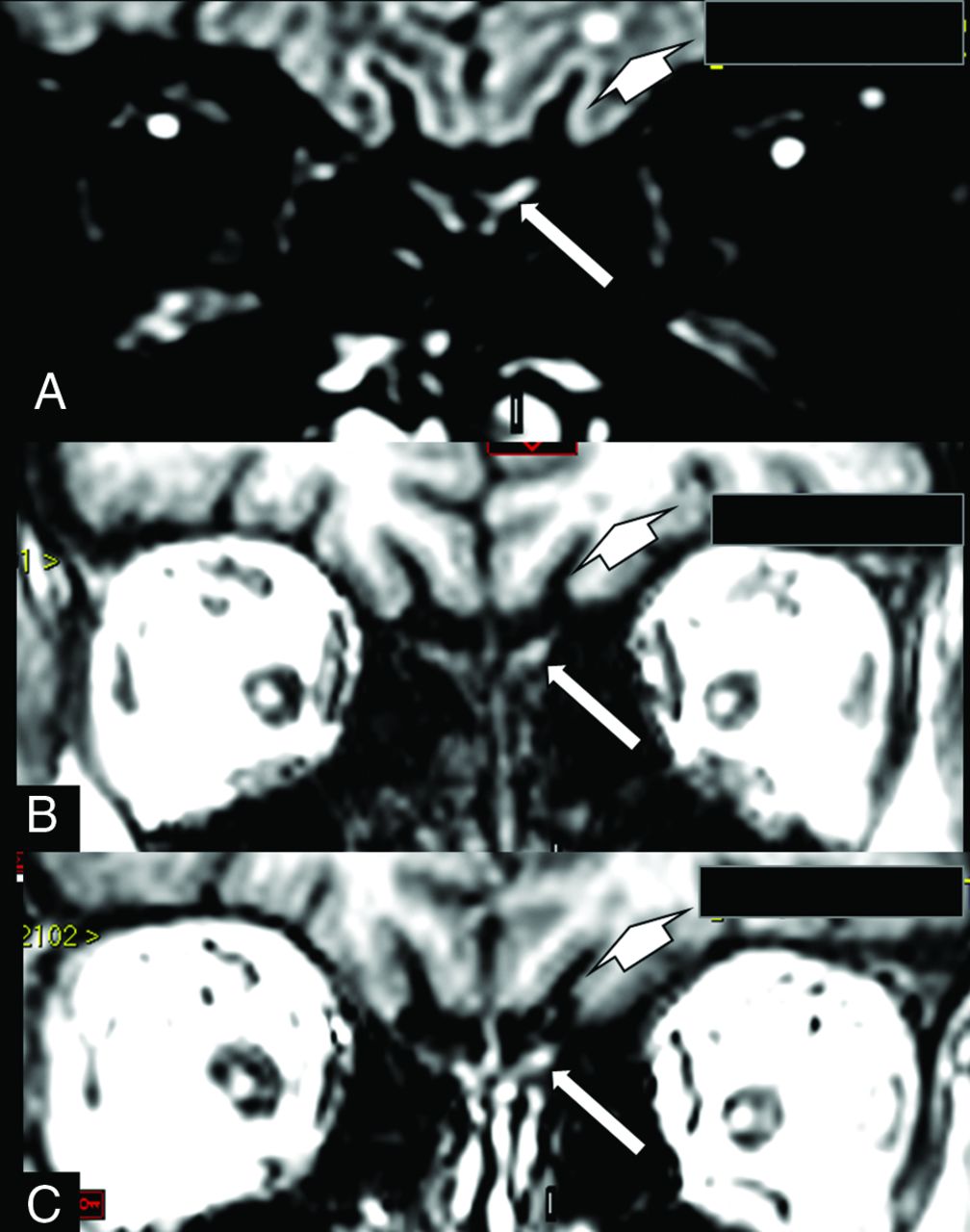
The cases in our retrospective study did not have any coronal FLAIR, FIESTA, or CISS images because the olfactory bulbs and anosmia were not the reason for the investigation by brain MR imaging.1 Thus, our study cannot be compared with the other studies about presence of edema or an increase of volume, and we also spoke about this in the introduction.1 However, one of our new cases was also investigated using a 3D-FLAIR sequence, allowing clear evidence of damage to the olfactory bulb, corresponding to the alterations observed in the pre- and postcontrast spoiled gradient recalled (SPGR) T1WI, definitively ruling out the possibility of an artifact, at least in this particular case (Figure).

FIGURE.
  • Download figure
  • Open in new tab
  • Download powerpoint
FIGURE.

A woman in her 60s with confirmed COVID-19 for which MR imaging shows a hyperintense lesion on a coronal reconstruction of 3D-FLAIR (A, arrow) and also on pre- (B, arrow) and postcontrast SPGR T1WI (C, arrow). This is suggestive of a component of probably methemoglobin in this left olfactory bulb lesion, which seems to be a little larger and asymmetric compared with the apparently normal right olfactory bulb. This asymmetry is better seen on FLAIR. On FLAIR (A, short arrow), there is also a small round hyperintense lesion in the subcortical white matter in the left frontal lobe, which is hypointense on T1WI (B, short arrow) and does not enhance on postcontrast T1WI (C, short arrow), being no specific lesion area probably corresponding to gliosis. This patient also had some areas of brain parenchymal bleeding and microbleeding, and had around 50% of the bilateral pulmonary parenchyma compromised with typical COVID-19 lesions on chest CT, not shown in this figure.

Regarding another comment, the only case reported in our publication (case 5) that did not mention anosmia presenting on the MR imaging had suggestive enhancement only in the left olfactory bulb, which is also described in the Results.1 We also held a discussion about this and concluded (fourth paragraph), “The patient hardly perceives unilateral anosmia.”1 She probably had unilateral damage to the olfactory bulb that was not sufficient to clinically result in anosmia. The human olfactory pathway projects ipsilaterally to the brain, and the central processing of its information is initially also restricted to the same hemisphere. Later processing includes bilateral cortical activation.3 The clinical consequence of this is that unilateral olfactory deficits can remain asymptomatic and thus cannot be noticed. We did not use the olfactory test evaluation, only the patient's report, because our study was retrospective. However, even using smell tests measuring olfactory function mostly reflects the best side when testing bilaterally.

Unlike the pathologist who deals directly with the injured tissue, the radiologist tries to interpret on the image what may be happening in the tissue, evaluating a possible anatomofunctional correlation. Thus, it was not only in our study1 that a relationship between anosmia and apparent alteration of the MR image was observed in patients with COVID-19 even if other MR imaging sequences have been performed and analyzed.4⇓–6 Our obligation is to present to the scientific community what we believe as an interpretation of a possible lesion demonstrated by the imaging. Obviously, we agree that there is always the risk of seeing what we want to see and not seeing what we do not want to see. Moreover, we have to always remember that in science, there are false-positives and false-negatives beyond the artifacts.

We think all would agree that anatomopathologic studies are necessary to better define neuroradiologic interpretation.

References

  1. 1.↵
    1. Aragão MF,
    2. Leal MC,
    3. Cartaxo Filho OQ, et al
    . Anosmia in COVID-19 associated with injury to the olfactory bulbs evident on MRI. AJNR Am J Neuroradiol 2020;41:1703–06 doi:10.3174/ajnr.A6675 pmid:32586960
    Abstract/FREE Full Text
  2. 2.↵
    1. Hong JH,
    2. Lee HY,
    3. Kang YH, et al
    . Improvement of fat suppression and artifact reduction using IDEAL technique in head and neck MRI at 3T. Investig Magn Reson Imaging 2016;20:44–52 doi:10.13104/imri.2016.20.1.44
    CrossRef
  3. 3.↵
    1. Lascano AM,
    2. Hummel T,
    3. Lacroix JS, et al
    . Spatio-temporal dynamics of olfactory processing in the human brain: an event-related source imaging study. Neuroscience 2010;167:700–08 doi:10.1016/j.neuroscience.2010.02.013 pmid:20153813
    CrossRefPubMed
  4. 4.↵
    1. Laurendon T,
    2. Radulesco T,
    3. Mugnier J, et al
    . Bilateral transient olfactory bulb edema during COVID-19-related anosmia. Neurology 2020;95:224–225 doi:10.1212/WNL.0000000000009850 pmid:32444492
    Abstract/FREE Full Text
  5. 5.↵
    1. Chetrit A,
    2. Lechien JR,
    3. Ammar A, et al
    . Magnetic resonance imaging of COVID-19 anosmic patients reveals abnormalities of the olfactory bulb: preliminary prospective study. The Journal of Infection 2020. doi:10.1016/j.jinf.2020.07.028 pmid:32739489
    CrossRefPubMed
  6. 6.↵
    1. Strauss SB,
    2. Lantos JE,
    3. Heier LA, et al
    . Olfactory bulb signal abnormality in patients with COVID-19 who present with neurologic symptoms. American Journal of Neuroradiology 2020;41:1882–87 doi:10.3174/ajnr.A6751
    Abstract/FREE Full Text
  • © 2021 by American Journal of Neuroradiology
PreviousNext
Back to top

In this issue

American Journal of Neuroradiology: 42 (2)
American Journal of Neuroradiology
Vol. 42, Issue 2
1 Feb 2021
  • Table of Contents
  • Index by author
  • Complete Issue (PDF)
Advertisement
Print
Download PDF
Email Article

Thank you for your interest in spreading the word on American Journal of Neuroradiology.

NOTE: We only request your email address so that the person you are recommending the page to knows that you wanted them to see it, and that it is not junk mail. We do not capture any email address.

Enter multiple addresses on separate lines or separate them with commas.
Reply:
(Your Name) has sent you a message from American Journal of Neuroradiology
(Your Name) thought you would like to see the American Journal of Neuroradiology web site.
CAPTCHA
This question is for testing whether or not you are a human visitor and to prevent automated spam submissions.
Cite this article
M.F.V.V. Aragão, M.C. Leal, T.M. Fonseca, O.Q. Cartaxo Filho, M.M Valença
Reply:
American Journal of Neuroradiology Feb 2021, 42 (2) E2-E3; DOI: 10.3174/ajnr.A6943

Citation Manager Formats

  • BibTeX
  • Bookends
  • EasyBib
  • EndNote (tagged)
  • EndNote 8 (xml)
  • Medlars
  • Mendeley
  • Papers
  • RefWorks Tagged
  • Ref Manager
  • RIS
  • Zotero
0 Responses
Respond to this article
Share
Bookmark this article
Reply:
M.F.V.V. Aragão, M.C. Leal, T.M. Fonseca, O.Q. Cartaxo Filho, M.M Valença
American Journal of Neuroradiology Feb 2021, 42 (2) E2-E3; DOI: 10.3174/ajnr.A6943
del.icio.us logo Twitter logo Facebook logo Mendeley logo
  • Tweet Widget
  • Facebook Like
  • Google Plus One
Purchase

Jump to section

  • Article
    • References
  • Figures & Data
  • Info & Metrics
  • Responses
  • References
  • PDF

Related Articles

  • No related articles found.
  • PubMed
  • Google Scholar

Cited By...

  • Virtual Biopsy: A Reality Thanks to Advances in Radiology
  • Comparative Study - The Impact and Profile of COVID-19 Patients Who Are Indicated for Neuroimaging: Vascular Phenomena Are Been Found in the Brain and Olfactory Bulbs
  • Crossref (2)
  • Google Scholar

This article has been cited by the following articles in journals that are participating in Crossref Cited-by Linking.

  • Virtual Biopsy: A Reality Thanks to Advances in Radiology
    M.F.V.V. Aragao, A.D.P. Oliveira, A.R.M.C. Lima, M.C Leal, M.M. Valenca
    American Journal of Neuroradiology 2021 42 7
  • Reply:
    M.F.V.V. Aragao, P.H.P. deAndrade, V.S. da Nobrega Segundo, M.M. Marques, H.J.G. de Azevedo, R.J.V. de Mello, M. de Carvalho Leal, M.L. Soares, M.M. Valenca
    American Journal of Neuroradiology 2021 42 9

More in this TOC Section

  • Brain AVM’s Nidus: What if We Hadn’t Understood Anything?
  • Letter to the Editor regarding “Automated Volumetric Software in Dementia: Help or Hindrance to the Neuroradiologist?”
  • Reply:
Show more Letters

Similar Articles

Advertisement

Indexed Content

  • Current Issue
  • Accepted Manuscripts
  • Article Preview
  • Past Issues
  • Editorials
  • Editors Choice
  • Fellow Journal Club
  • Letters to the Editor

Cases

  • Case Collection
  • Archive - Case of the Week
  • Archive - Case of the Month
  • Archive - Classic Case

Special Collections

  • Special Collections

Resources

  • News and Updates
  • Turn around Times
  • Submit a Manuscript
  • Author Policies
  • Manuscript Submission Guidelines
  • Evidence-Based Medicine Level Guide
  • Publishing Checklists
  • Graphical Abstract Preparation
  • Imaging Protocol Submission
  • Submit a Case
  • Become a Reviewer/Academy of Reviewers
  • Get Peer Review Credit from Publons

Multimedia

  • AJNR Podcast
  • AJNR SCANtastic
  • Video Articles

About Us

  • About AJNR
  • Editorial Board
  • Not an AJNR Subscriber? Join Now
  • Alerts
  • Feedback
  • Advertise with us
  • Librarian Resources
  • Permissions
  • Terms and Conditions

American Society of Neuroradiology

  • Not an ASNR Member? Join Now

© 2025 by the American Society of Neuroradiology All rights, including for text and data mining, AI training, and similar technologies, are reserved.
Print ISSN: 0195-6108 Online ISSN: 1936-959X

Powered by HighWire