Advertisement

Main menu

  • Home
  • Content
    • Current Issue
    • Accepted Manuscripts
    • Article Preview
    • Past Issue Archive
    • Video Articles
    • AJNR Case Collection
    • Case of the Week Archive
    • Case of the Month Archive
    • Classic Case Archive
  • Special Collections
    • AJNR Awards
    • Low-Field MRI
    • Alzheimer Disease
    • ASNR Foundation Special Collection
    • Photon-Counting CT
    • View All
  • Multimedia
    • AJNR Podcasts
    • AJNR SCANtastic
    • Trainee Corner
    • MRI Safety Corner
    • Imaging Protocols
  • For Authors
    • Submit a Manuscript
    • Submit a Video Article
    • Submit an eLetter to the Editor/Response
    • Manuscript Submission Guidelines
    • Statistical Tips
    • Fast Publishing of Accepted Manuscripts
    • Graphical Abstract Preparation
    • Imaging Protocol Submission
    • Author Policies
  • About Us
    • About AJNR
    • Editorial Board
    • Editorial Board Alumni
  • More
    • Become a Reviewer/Academy of Reviewers
    • Subscribers
    • Permissions
    • Alerts
    • Feedback
    • Advertisers
    • ASNR Home

User menu

  • Alerts
  • Log in

Search

  • Advanced search
American Journal of Neuroradiology

American Journal of Neuroradiology

ASHNR American Society of Functional Neuroradiology ASHNR American Society of Pediatric Neuroradiology ASSR
  • Alerts
  • Log in

Advanced Search

  • Home
  • Content
    • Current Issue
    • Accepted Manuscripts
    • Article Preview
    • Past Issue Archive
    • Video Articles
    • AJNR Case Collection
    • Case of the Week Archive
    • Case of the Month Archive
    • Classic Case Archive
  • Special Collections
    • AJNR Awards
    • Low-Field MRI
    • Alzheimer Disease
    • ASNR Foundation Special Collection
    • Photon-Counting CT
    • View All
  • Multimedia
    • AJNR Podcasts
    • AJNR SCANtastic
    • Trainee Corner
    • MRI Safety Corner
    • Imaging Protocols
  • For Authors
    • Submit a Manuscript
    • Submit a Video Article
    • Submit an eLetter to the Editor/Response
    • Manuscript Submission Guidelines
    • Statistical Tips
    • Fast Publishing of Accepted Manuscripts
    • Graphical Abstract Preparation
    • Imaging Protocol Submission
    • Author Policies
  • About Us
    • About AJNR
    • Editorial Board
    • Editorial Board Alumni
  • More
    • Become a Reviewer/Academy of Reviewers
    • Subscribers
    • Permissions
    • Alerts
    • Feedback
    • Advertisers
    • ASNR Home
  • Follow AJNR on Twitter
  • Visit AJNR on Facebook
  • Follow AJNR on Instagram
  • Join AJNR on LinkedIn
  • RSS Feeds

AJNR Awards, New Junior Editors, and more. Read the latest AJNR updates

Research ArticleHead and Neck Imaging

MRI Posttreatment Surveillance for Head and Neck Squamous Cell Carcinoma: Proposed MR NI-RADS Criteria

M.M. Ashour, E.A.F. Darwish, R.M. Fahiem and T.T. Abdelaziz
American Journal of Neuroradiology June 2021, 42 (6) 1123-1129; DOI: https://doi.org/10.3174/ajnr.A7058
M.M. Ashour
aFrom the Department of Diagnostic Radiology, Faculty of Medicine, Ain Shams University, Cairo, Egypt
  • Find this author on Google Scholar
  • Find this author on PubMed
  • Search for this author on this site
  • ORCID record for M.M. Ashour
E.A.F. Darwish
aFrom the Department of Diagnostic Radiology, Faculty of Medicine, Ain Shams University, Cairo, Egypt
  • Find this author on Google Scholar
  • Find this author on PubMed
  • Search for this author on this site
  • ORCID record for E.A.F. Darwish
R.M. Fahiem
aFrom the Department of Diagnostic Radiology, Faculty of Medicine, Ain Shams University, Cairo, Egypt
  • Find this author on Google Scholar
  • Find this author on PubMed
  • Search for this author on this site
  • ORCID record for R.M. Fahiem
T.T. Abdelaziz
aFrom the Department of Diagnostic Radiology, Faculty of Medicine, Ain Shams University, Cairo, Egypt
  • Find this author on Google Scholar
  • Find this author on PubMed
  • Search for this author on this site
  • ORCID record for T.T. Abdelaziz
  • Article
  • Figures & Data
  • Info & Metrics
  • Responses
  • References
  • PDF
Loading

Article Figures & Data

Figures

  • Tables
  • FIG 1.
    • Download figure
    • Open in new tab
    • Download powerpoint
    FIG 1.

    Concordance between NI-RADS category, T2 SI, and DWIs is demonstrated in this posttreatment MR imaging surveillance scan done after surgery and RTH for sinonasal squamous cell carcinoma. Coronal T2WI (A) and coronal contrast-enhanced T1WI (B) show a surgical bed discrete nodule (black arrow) that displays intermediate T2 SI (A) and postcontrast enhancement (B), fulfilling NI-RADS 3 and confirmed to be squamous cell carcinoma by histopathology. C. Axial ADC map of the surgical bed shows a corresponding low-ADC signal (black arrow). White arrows point to circumferential soft tissue thickening with sheetlike enhancement (B), which shows low T2 SI (A), absent diffusion restriction (D), and was confirmed to be posttreatment fibrous tissue.
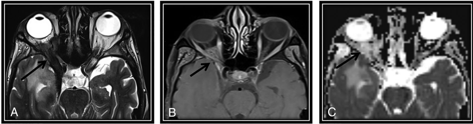
  • FIG 2.
    • Download figure
    • Open in new tab
    • Download powerpoint
    FIG 2.

    The first post-CRTH MR imaging follow-up for a known case of nasopharyngeal carcinoma with perineural tumor spread (PNTS) showing discordant findings between NI-RADS category, T2 SI, and DWI. There is a clear primary tumor site at the nasopharynx (not demonstrated here), yet a regional non-nodal target was noted. A, Axial T2WI shows orbital apex dark T2 SI tissue keeping with fibrotic scarring (arrow). B, This lesion show discrete postcontrast enhancement (arrow) (secondary to PNTS along the ophthalmic nerve) categorized as NI-RADS 3. C, Axial ADC map of the surgical bed shows a corresponding facilitated diffusion (arrow). According to our proposed modifying rules, downgrading to category 2 was done. Follow-up by PET/CT showed no FDG uptake (not demonstrated here) that was confirmed to be post-RTH fibrotic scarring by further follow-up. Note the right temporal lobe after RTH injury.

Tables

  • Figures
    • View popup
    Table 1:

    Tumor characteristics and scan order of the included population

    Patients (n)Patients (%)
    SubsiteLarynx1217.4
    Oral cavity and oropharynx2536.2
    Hypopharynx22.9
    Sinonasal1014.5
    Skull base11.4
    Nasopharynx1318.8
    Salivary68.7
    Pathologic gradeLow1420.3
    Moderate3753.6
    High1826.1
    Tumor stageaTis11.4
    T1710.14
    T22434.8
    T32231.9
    T41521.7
    Scan orderFirst follow-up5478.3
    Second follow-up810.1
    Third follow-up811.6
    • Note:—Tis indicates carcinoma in situ.

    • ↵a American Joint Committee on Cancer.20

    • View popup
    Table 2:

    Results of image analysis by T2 SI, DWI (qualitative and quantitative), NI-RADS, and NI-RADS rescoring for the primary targets

    Standard of Reference
    Negative n (%)Positive n (%)
    NI-RADSNI-RADS 120 (46.51)1 (3.85)
    NI-RADS 215 (34.88)3 (11.54)
    NI-RADS 38 (18.6)22 (84.62)
    T2 SIIsointense to surrounding tissue4 (9.3)1 (3.85)
    Dark15 (34.88)1 (3.85)
    Intermediate5 (11.63)24 (92.31)
    High19 (44.19)0 (0)
    DWIFacilitated39 (90.7)2 (7.69)
    Restricted4 (9.3)24 (92.31)
    NI-RADS combined with T2 and DWINI-RADS 130 (69.77)1 (3.85)
    NI-RADS 210 (23.26)2 (7.69)
    NI-RADS 33 (6.98)23 (88.46)
    • View popup
    Table 3:

    Diagnostic performance of T2 SI, DWI (qualitative and quantitative), NI-RADS, and NI-RADS rescoring for the detection of LTR for the primary targets

    T2 SIDWI with ADC ≤1.3NI-RADSNI-RADS-DWI-T2 SI
    TP (n)24242223
    TN (n)38393540
    FP (n)5483
    FN (n)2243
    Sensitivity (%) (95% CI)92.31 (74.87–99.05)92.3 (74.87–99.05)84.62 (65.13–95.64)88.46 (69.85–97.55)
    Specificity (%) (95% CI)88.37 (74.92–96.11)90.7 (77.86–97.41)81.4 (66.60–91.61)93.02 (80.94–98.54)
    PPV (%) (95% CI)82.76 (67.64–91.68)85.71 (70.10–93.89)73.33 (59.03–84.00)88.46 (71.84–95.84)
    NPV (%) (95% CI)95.00 (83.32–98.64)95.12 (83.69–98.67)89.74 (77.84–95.61)93.02 (82.10–97.49)
    Accuracy (%) (95% CI)89.86 (80.21–95.82)91.30 (82.03–96.74)82.61 (71.59–90.68)91.30 (82.03–96.74)
    • Note:—TP indicates true-positive; TN, true-negative.

    • View popup
    Table 4:

    Agreement between DWI (qualitative and quantitative), T2 SI, NI-RADS, and NI-RADS combined with DWI and T2 SI for the primary targets and the standard of reference

    Standard of ReferenceAgreement
    Negative n (%)Positive n (%)Kappa95% CIP Value
    DWIRestricted4 (9.3)24 (92.31)0.8180.68–0.96<.001
    Facilitated39 (90.7)2 (7.69)
    T2 SIIntermediate5 (11.63)24 (92.3)0.7890.64–0.94<.001
    No abnormality; low or high SI38 (88.37)2 (7.69)
    NI-RADS combined with T2 and DWICategory 33 (6.98)23 (88.46)0.8150.67–0.96<.001
    Categories 1 and 240 (93.02)3 (11.54)
    NI-RADSCategory 38 (18.6)22 (84.62)0.6419.46–0.82<.001
    Categories 1 and 235 (81.4)4 (15.38)
PreviousNext
Back to top

In this issue

American Journal of Neuroradiology: 42 (6)
American Journal of Neuroradiology
Vol. 42, Issue 6
1 Jun 2021
  • Table of Contents
  • Index by author
  • Complete Issue (PDF)
Advertisement
Print
Download PDF
Email Article

Thank you for your interest in spreading the word on American Journal of Neuroradiology.

NOTE: We only request your email address so that the person you are recommending the page to knows that you wanted them to see it, and that it is not junk mail. We do not capture any email address.

Enter multiple addresses on separate lines or separate them with commas.
MRI Posttreatment Surveillance for Head and Neck Squamous Cell Carcinoma: Proposed MR NI-RADS Criteria
(Your Name) has sent you a message from American Journal of Neuroradiology
(Your Name) thought you would like to see the American Journal of Neuroradiology web site.
CAPTCHA
This question is for testing whether or not you are a human visitor and to prevent automated spam submissions.
Cite this article
M.M. Ashour, E.A.F. Darwish, R.M. Fahiem, T.T. Abdelaziz
MRI Posttreatment Surveillance for Head and Neck Squamous Cell Carcinoma: Proposed MR NI-RADS Criteria
American Journal of Neuroradiology Jun 2021, 42 (6) 1123-1129; DOI: 10.3174/ajnr.A7058

Citation Manager Formats

  • BibTeX
  • Bookends
  • EasyBib
  • EndNote (tagged)
  • EndNote 8 (xml)
  • Medlars
  • Mendeley
  • Papers
  • RefWorks Tagged
  • Ref Manager
  • RIS
  • Zotero
0 Responses
Respond to this article
Share
Bookmark this article
MRI Posttreatment Surveillance for Head and Neck Squamous Cell Carcinoma: Proposed MR NI-RADS Criteria
M.M. Ashour, E.A.F. Darwish, R.M. Fahiem, T.T. Abdelaziz
American Journal of Neuroradiology Jun 2021, 42 (6) 1123-1129; DOI: 10.3174/ajnr.A7058
del.icio.us logo Twitter logo Facebook logo Mendeley logo
  • Tweet Widget
  • Facebook Like
  • Google Plus One
Purchase

Jump to section

  • Article
    • Abstract
    • ABBREVIATIONS:
    • MATERIALS AND METHODS
    • RESULTS
    • DISCUSSION
    • CONCLUSIONs
    • References
  • Figures & Data
  • Info & Metrics
  • Responses
  • References
  • PDF

Related Articles

  • No related articles found.
  • PubMed
  • Google Scholar

Cited By...

  • Performance of Neck Imaging Reporting and Data System (NI-RADS) for Diagnosis of Recurrence of Head and Neck Squamous Cell Carcinoma: A Systematic Review and Meta-analysis
  • Adding MR Diffusion Imaging and T2 Signal Intensity to Neck Imaging Reporting and Data System Categories 2 and 3 in Primary Sites of Postsurgical Oral Cavity Carcinoma Provides Incremental Diagnostic Value
  • ADC for Differentiation between Posttreatment Changes and Recurrence in Head and Neck Cancer: A Systematic Review and Meta-analysis
  • PET/MR Imaging in Evaluating Treatment Failure of Head and Neck Malignancies: A Neck Imaging Reporting and Data System-Based Study
  • Crossref
  • Google Scholar

This article has not yet been cited by articles in journals that are participating in Crossref Cited-by Linking.

More in this TOC Section

  • Hydrops Herniation into the Semicircular Canals
  • ASL Sensitivity for Head and Neck Paraganglioma
  • Post SRS Peritumoral Hyperintense Signal of VSs
Show more HEAD AND NECK IMAGING

Similar Articles

Advertisement

Indexed Content

  • Current Issue
  • Accepted Manuscripts
  • Article Preview
  • Past Issues
  • Editorials
  • Editor's Choice
  • Fellows' Journal Club
  • Letters to the Editor
  • Video Articles

Cases

  • Case Collection
  • Archive - Case of the Week
  • Archive - Case of the Month
  • Archive - Classic Case

More from AJNR

  • Trainee Corner
  • Imaging Protocols
  • MRI Safety Corner
  • Book Reviews

Multimedia

  • AJNR Podcasts
  • AJNR Scantastics

Resources

  • Turnaround Time
  • Submit a Manuscript
  • Submit a Video Article
  • Submit an eLetter to the Editor/Response
  • Manuscript Submission Guidelines
  • Statistical Tips
  • Fast Publishing of Accepted Manuscripts
  • Graphical Abstract Preparation
  • Imaging Protocol Submission
  • Evidence-Based Medicine Level Guide
  • Publishing Checklists
  • Author Policies
  • Become a Reviewer/Academy of Reviewers
  • News and Updates

About Us

  • About AJNR
  • Editorial Board
  • Editorial Board Alumni
  • Alerts
  • Permissions
  • Not an AJNR Subscriber? Join Now
  • Advertise with Us
  • Librarian Resources
  • Feedback
  • Terms and Conditions
  • AJNR Editorial Board Alumni

American Society of Neuroradiology

  • Not an ASNR Member? Join Now

© 2025 by the American Society of Neuroradiology All rights, including for text and data mining, AI training, and similar technologies, are reserved.
Print ISSN: 0195-6108 Online ISSN: 1936-959X

Powered by HighWire