
Article Figures & Data
Figures
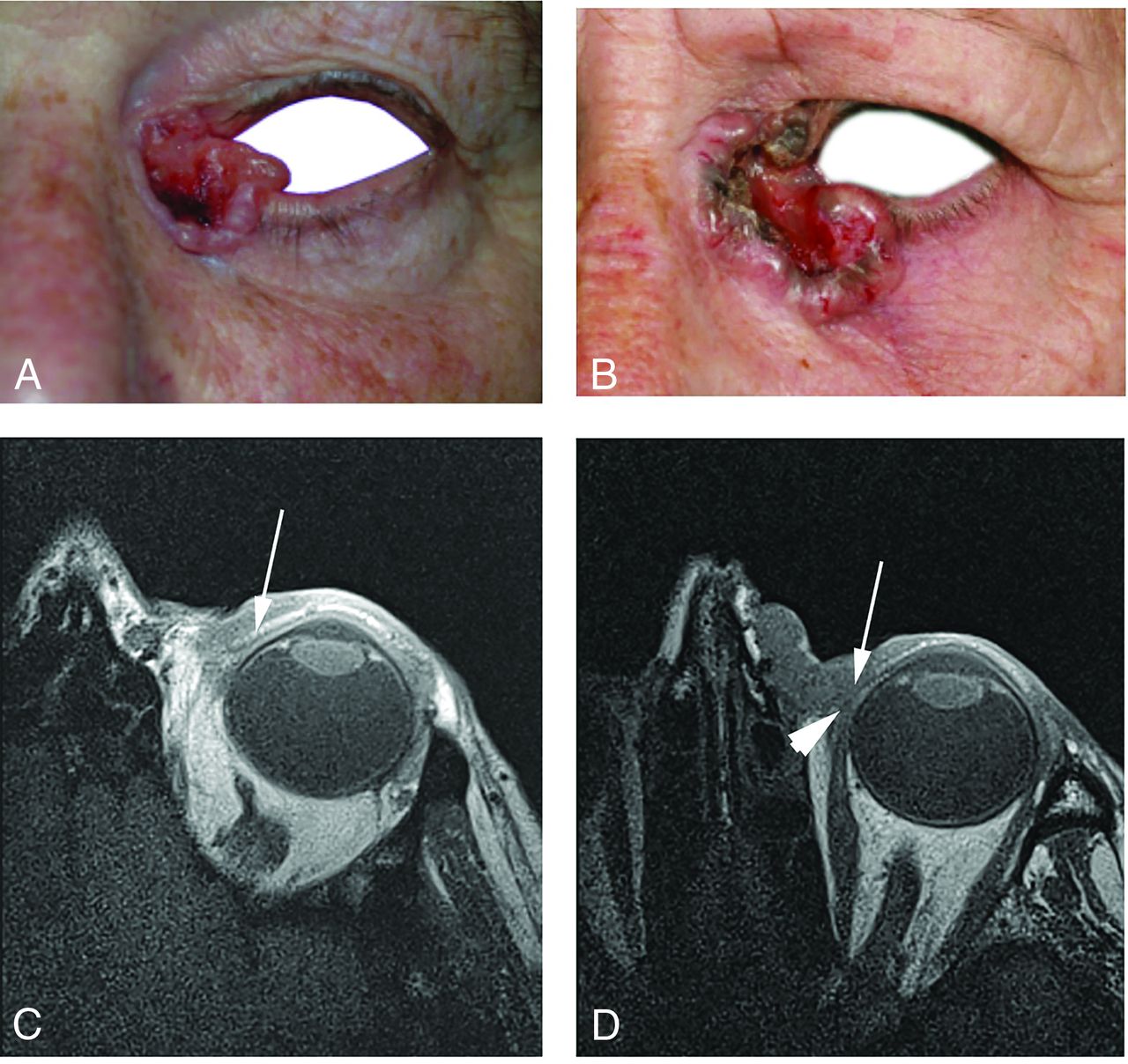
- FIG 1.
BCC of the left medial canthus in 2 elderly women. A, Clinical photograph of case 1. B, Clinical photograph of case 2, which appears superficially similar to the first case. C, T1-weighted axial MC-MR imaging of case 1 demonstrates that the superior tarsal plate is intact (arrow) and not invaded by tumor. D, T1-weighted axial MC-MR imaging of case 2 demonstrates full-thickness tumor invasion of the superior tarsal plate (arrow), requiring complex oculoplastic reconstruction surgery to optimize eyelid function after resection of the tumor. The tumor also abuts the tendinous insertion of the medial rectus muscle (arrowhead), but there was no invasion of this found during the operation.
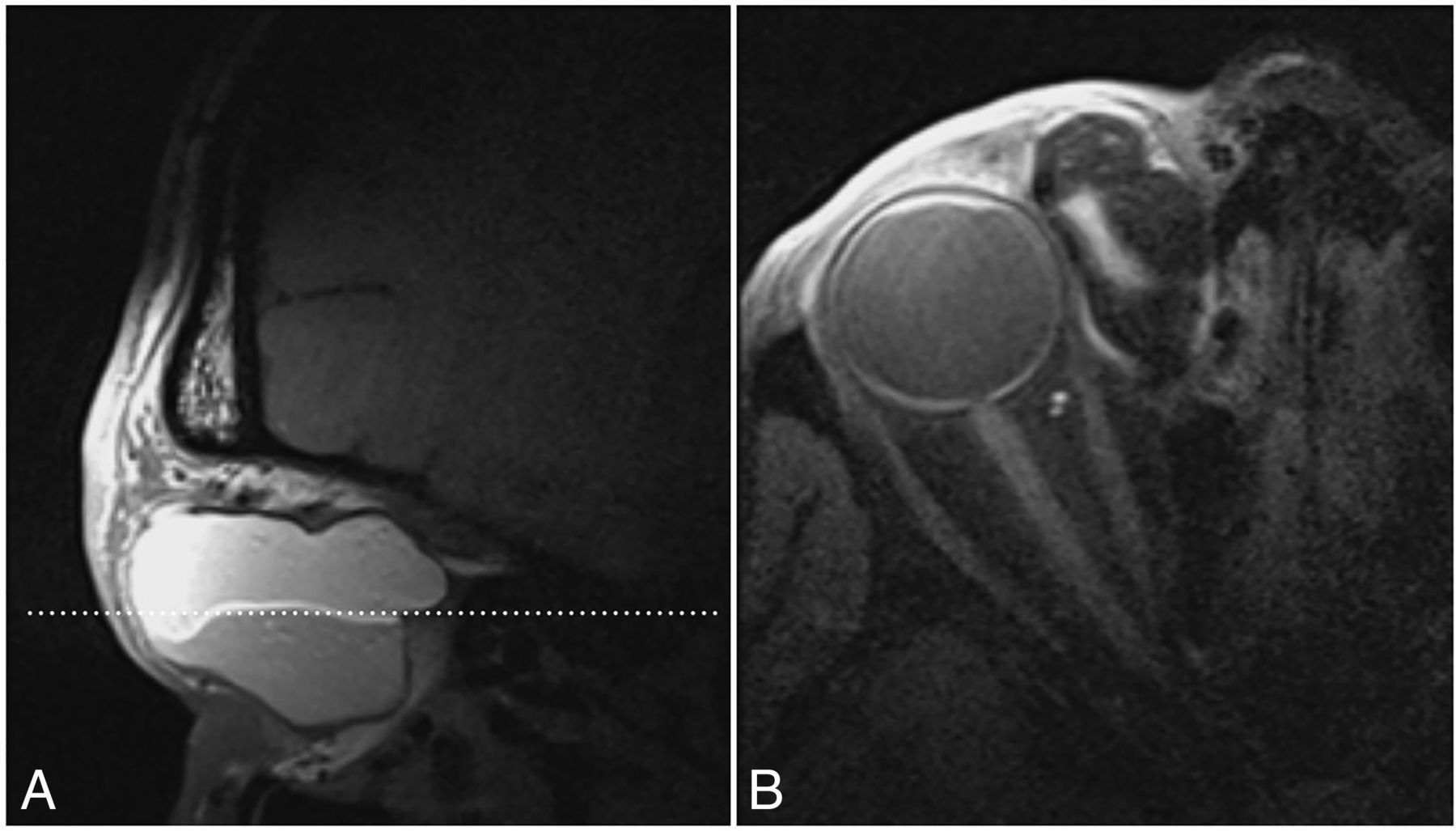
- FIG 2.
Dermoid cyst in the superonasal quadrant of the orbit in a 46-year-old man who had presented with slowly progressive swelling on the medial aspect of the right globe. A, T1-weighted sagittal MC-MR imaging demonstrates a superior layer of high signal intensity and an inferior layer of lower intensity. The acquisition plane of B is denoted by the dotted line. B, Fat-saturated T1-weighted axial MC-MR imaging shows signal drop-out of the superior layer, confirming fat content and reinforcing the preoperative diagnosis of dermoid cyst.
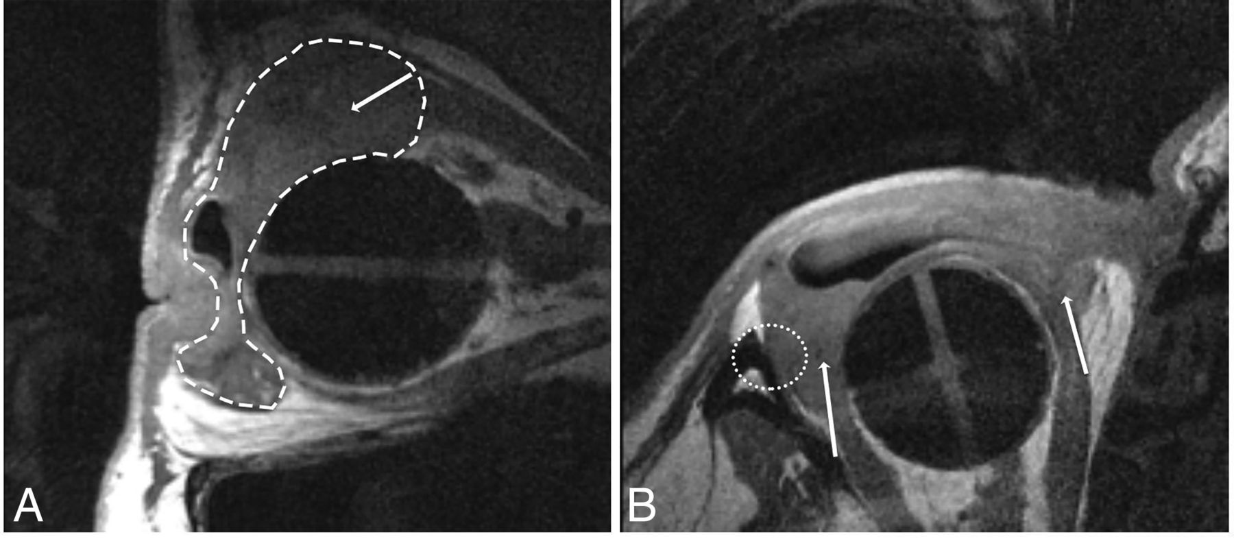
- FIG 3.
Intraorbital melanoma metastasis in an elderly woman with previous enucleation for primary iris melanoma.T1-weighted sagittal MC-MR imaging (A) and T1-weighted axial MC-MR imaging (B) demonstrate a bilobed mass (dashed white line) involving the extraocular muscles (arrows) and abutting the periosteum of the lateral orbital wall (dotted white circle). The tarsal plate was also invaded. Surgical resection margins were planned accordingly, with clear margins confirmed on histologic examination of the resection specimen.
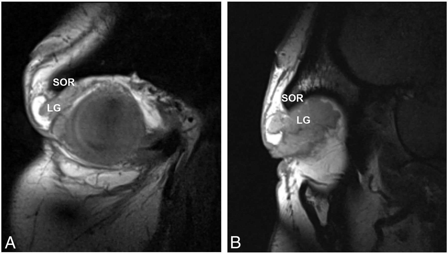
- FIG 4.
Lacrimal gland prolapse in a 27-year-old woman who had presented with palpebral swelling of uncertain origin. T1-weighted coronal MC-MR imaging (A) and T1-weighted sagittal MC-MR imaging (B) demonstrate prolapse of the lacrimal gland (LG) under the superior orbital rim (SOR). The patient was reassured and discharged from the clinic. Should future blepharoplasty be considered, intraoperative repositioning of the gland would be incorporated into the surgical planning.16
- FIG 5.
Venous malformation in a middle-aged woman. T1-weighted sagittal MC-MR imaging T1 (A) and T2-weighted sagittal MC-MR imaging (B) demonstrate a well-defined, lobulated mass (VM) with signal isointense to muscle on T1-weighted acquisitions and signal hyperintense to muscle on T2-weighted acquisitions. A clean fat plane (arrowhead) separates the lesion from the globe (G), in keeping with a fibrous pseudocapsule. Such narrow fat planes would not be resolved on head coil MR imaging. Despite close proximity, neither the rectus muscles nor the tarsal plate was involved, and the lesion was surgically removed intact.
- FIG 6.
A persistent lymphatic malformation in a middle-aged man who had undergone multiple previous operations and steroid injections. MC-MR imaging was performed to guide definitive management. A, Conventional head coil T2-weighted axial MR imaging demonstrates the lymphatic malformation, but the clarity of depiction of the lesion margins and relationships is insufficient to guide treatment. T2-weighted axial MC-MR imaging (B) demonstrates the lesion's characteristic appearance (arrow) and T1-weighted coronal MC-MR imaging (C) demonstrates the lesion (dotted white line) extending from the periorbital soft tissues into the orbit and intraconally, an extent of involvement that precludes safe surgical intervention. Consequently, bleomycin sclerotherapy was performed with pretreatment (D) and posttreatment photography (with the patient’s permission) (E) demonstrating aesthetic improvement.


Jump to section
Related Articles
Cited By...
- No citing articles found.