
Article Figures & Data
Figures
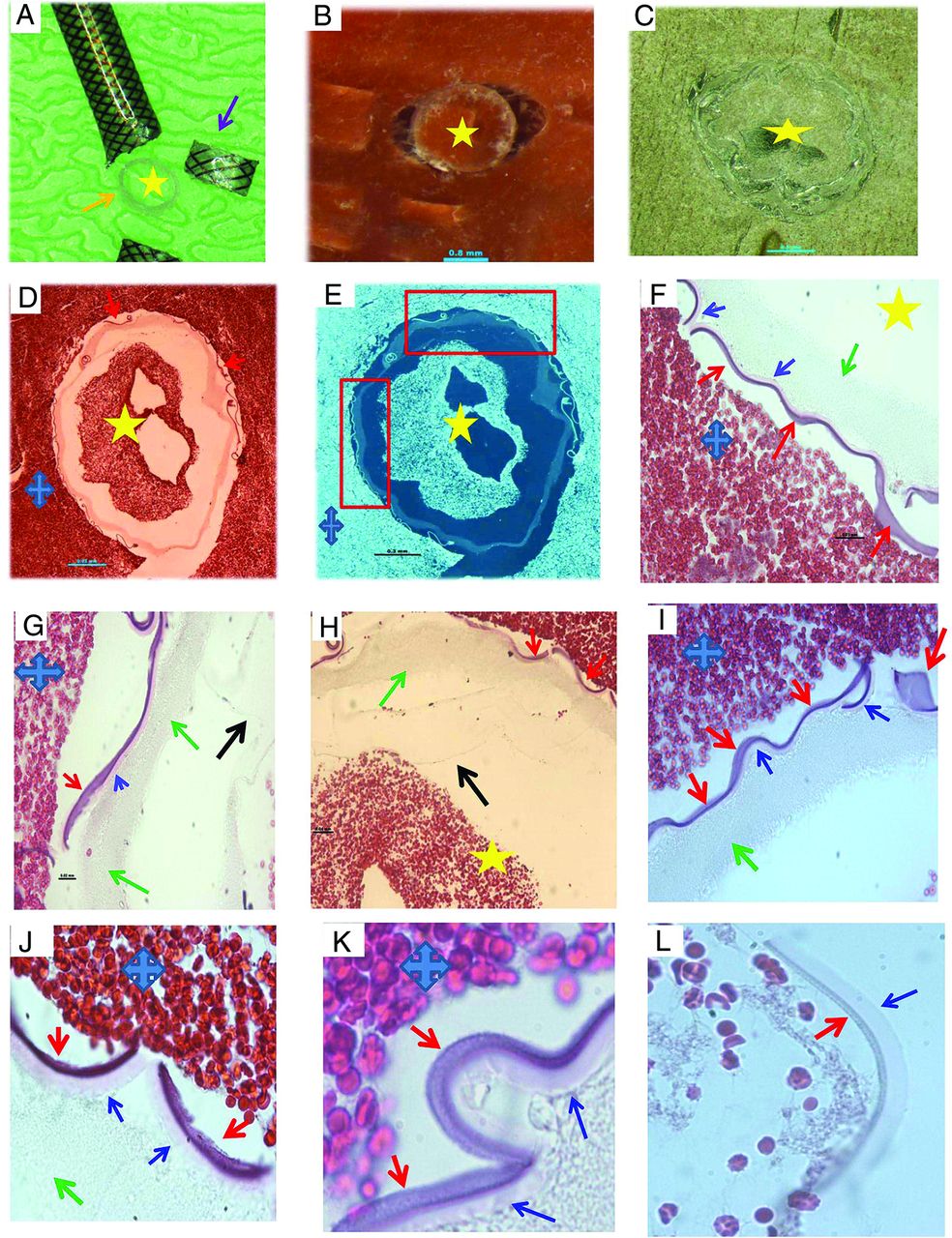
- FIG 1.
Histologic processing of cross-section of MT catheter. A, Macrophotograph of the Sofia catheter, showing the catheter is cut into pieces in coronary plane (arrows), with removal of metal scaffold for further histology processing (orange arrow) (asterisk: lumen side). B, Macrophotograph of catheter piece in image A mixed with clot analog tissue (asterisk: lumen side). C, Macrophotograph showing the catheter piece mixed with clot tissue embedded in paraffin after tissue processing; catheter piece remains in the coronal orientation (asterisk: lumen side). D, Low-magnification microphotograph of H&E-stained catheter piece from image C (blue cross: the surrounding clot; yellow asterisk: the lumen/inner side of catheter; red arrows: outer layer surface-coating material that is next to the surrounding clot tissue). E, Reverse image of image D, showing each layer of the catheter, in relation to the lumen and outside surrounding clot tissue (yellow star: lumen side; blue cross: surrounding clot tissue). F–I, High magnification of microphotograph taken from the red rectangular area in image E, showing each layer of the catheter materials from the outer layer surface (red arrows), middle layer (blue arrows and green arrows) to the inner liner layer (black arrow) (H&E, original magnification ×400; yellow star: the lumen side of catheter; blue cross: the surrounding clot analog tissue). J–L, Microphotographs taken with oil lens, showing the surface-coating material (red arrows), its subsequent layer (blue arrows), and deeper layer (green arrow). The surface-coating material appears to be light gray, gray-pink, or lightly stained; attenuated or loose in texture; and with varied shape, same as the type II foreign material found in the patient clot tissue (Fig 6). The layer that is immediately underneath the coating layer appears to be homogeneous in texture, light pink, or pale in color (blue arrows). It is similar to the type IV foreign material observed in the patient clot tissue (Fig 8) (H&E, oil lens ×100).
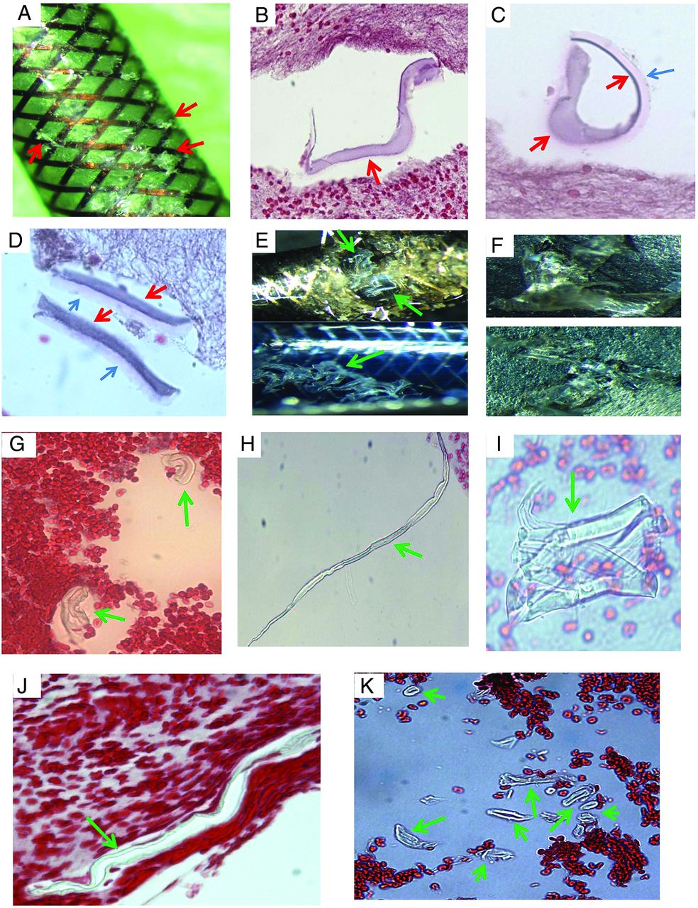
- FIG 2.
Histologic appearance of scratched surface and inner layer of MT catheter. A, Macrophotograph of the Sofia catheter, showing the scratched surface pieces (red arrows). B–D, Microphotograph of scratched pieces from image A associated with clot analog tissue, showing the surface-coating material, which appears to be varied in shape and texture following tissue processing (red arrows), and which is similar to the material seen in the cross-section experiment, Fig 1). The layer immediately underneath the surface-coating material is also present (blue arrow), which appears to be a different shape, light pink/red, and has a homogeneous texture. It is similar to the material seen in the cross-section (Fig 1, blue arrow), and type IV foreign material (Fig 8) found in the patient clot tissue as well. E, Macrophotograph of Sofia and Fubuki catheters, showing the scratched liner layer material in situ (green arrows; upper—Sofia; lower—Fubuki). F, Macrophotograph of the manually torn off liner material pieces from image E, showing the similar gross appearance to that of the PTFE material (Fig 4). These pieces were crushed into small pieces for further routine, histologic processing, similar to the surface scratched material mentioned in Fig 1. G–I, Microphotograph of torn off liner pieces from F (upper), showing the material that appears to be varied in shape, color, and texture because of processing. These could be tubelike, long stripe, or an irregular solid mass, with refraction and light green outline. Those features are the same as what is seen from the PTFE material (Fig 4), and the type I material found in the patient clot (Fig 5). J and K, Representative microphotograph of scratched, torn off liner pieces from F (lower), showing the material that appears to be long, or short, or small tubular in shape, and light green, as shown with the PTFE material (Fig 4) and type I foreign material found in the patient clot tissue (Fig 5).
- FIG 3.
Representative microphotographs of scratched pieces of different layers rather than the surface or inner layer. Microphotographs showing the different materials of MT catheter, rather than the surface-coating material and inner liner materials shown in Figs 1 and 2. The similar foreign material is found in the patients’ retrieved clot tissue (type III, Fig 7) (H&E, original magnification ×400).
- FIG 4.
Histologic features of donated PTFE material. A, Macrophotograph of the PTFE liner material provided by manufacturer sponsor. B–F, Microphotographs of the scratched PTFE pieces in H&E–stained slides. The material appears to have varied shape. It is either a mass with solid, homogeneous texture associated with refraction, and light green edge (B–D), or long strips or tubes with light green outline (E and F). Those features are the same as the scratched inner layer of catheters (Fig 2), and type I material found in the patient clot tissue as well (Fig 5) (B–F, H&E, original magnification ×400).
- FIG 5.
Representative microphotographs of type I foreign material found in patient clot tissue. A, Individual foreign material pieces separately embedded within the clot tissue (arrows). They appear to be small, ringlike, or long tubelike in shape, and light green (H&E, original magnification ×400). B, The cluster of foreign material pieces (arrows). Individual pieces appear to be wormlike with refraction and a light green outline (H&E, original magnification ×400). C, Multiple pieces of material associated with clot tissue. They are long or short stripes or pieces, with refraction, and green outline (arrows) (H&E, original magnification ×200). D, Irregular, solid pieces of material, with refraction, and light green outline (H&E, original magnification ×400). E and F, Large piece of polymer material surrounded with clot tissue (arrows). It appears to be long strips or ringlike circle, and solid homogeneous mass with refraction, with light green outline (H&E, original magnification ×200 [E], ×400 [F]).
- FIG 6.
Representative microphotographs of type II foreign material found in patient clot tissue. A and B, Multiple pieces of foreign material embedded within clot tissue (arrows). They appear to be light gray or dark gray; thin or attenuated in texture (H&E, original magnification ×400). C and D, Foreign material pieces embedded within clot tissue, which appear to be denser and solid in texture (arrows), compared with that in A and B. They are wormlike or irregular in shape, gray or light gray (H&E, original magnification oil lens ×100). E–G, Cluster of the foreign material pieces, as a pool surrounded with clot tissue (H&E, original magnification ×200 [E], ×400 [F and G]).
- FIG 7.
Representative microphotographs of type III foreign material found in patient clot tissue. A, A single, long, oval-like piece of foreign material embedded within clot tissue (arrow). The material is associated with multiple bubblelike particles, which are refracted under light microscopy. B, A single piece of irregular foreign material attaching to the clot tissue (arrow). It appears to be light green, associated dark/black and bubblelike, with refraction particles. C, Irregular piece of foreign material at the gap of clot tissue (arrow). It appears to be solid, homogeneous in texture with refraction, and light green. D, A piece of green, solid foreign material, associated diffused black, dotlike particles (arrow) (A–D, H&E, original magnification ×400). These materials presented in A–D are similar to the materials scratched from the catheters (Fig 3).
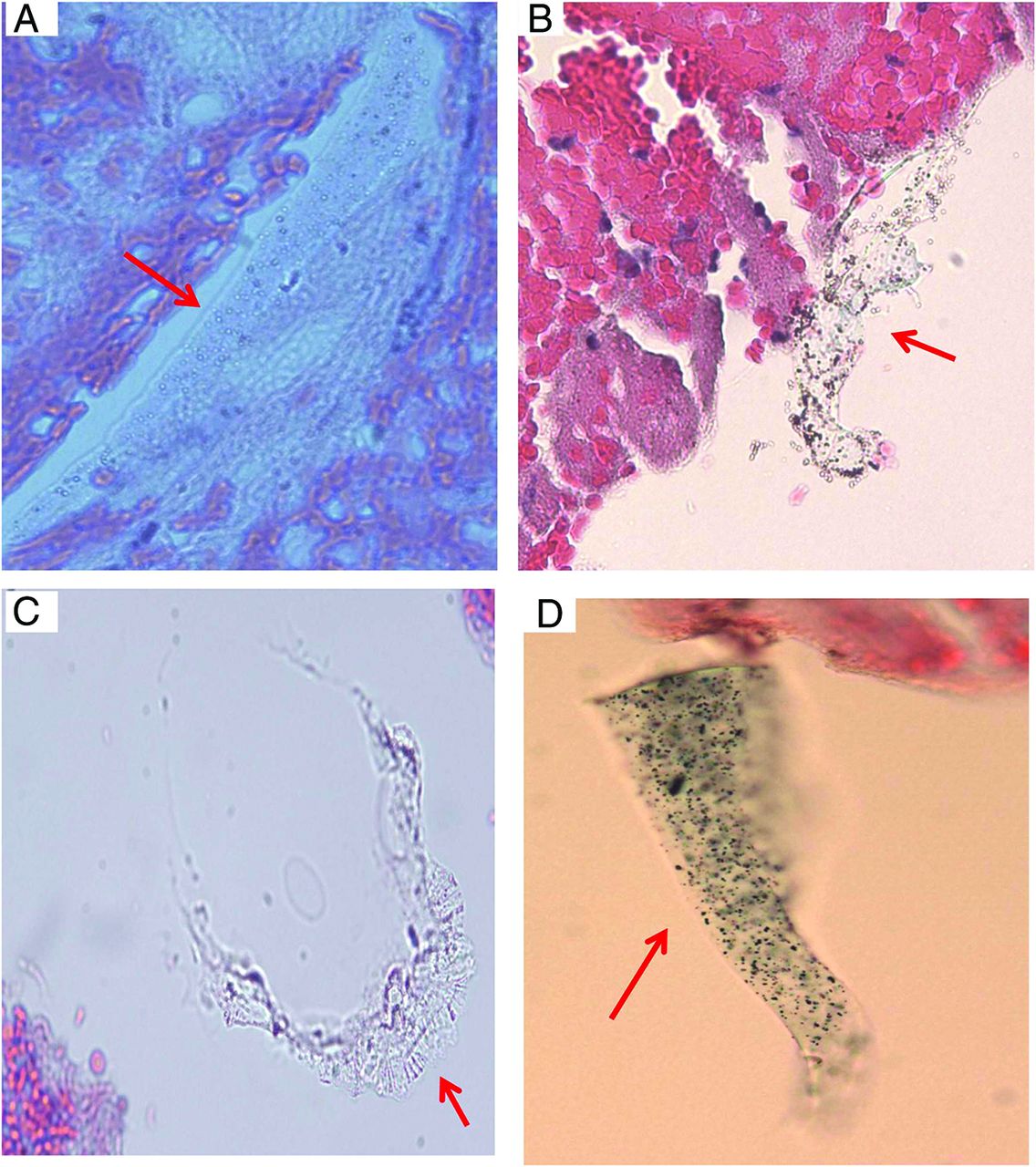
- FIG 8.
Representative microphotographs of type IV foreign material found in patient clot tissue. Different shapes of the type IV foreign material (red arrows) found in the clot tissue retrieved with MT. The foreign material is surrounded with clot tissue, appears to be homogeneous in texture, wormlike or snakelike in shape, and stained lightly. The residual, surface-coating material remains visible (A, black arrows) (A–D, H&E, original magnification ×400).
- FIG 9.
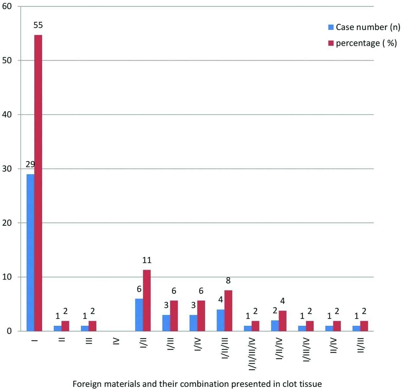
Foreign materials and varied combinations of different types found in patients with stroke treated with MT.
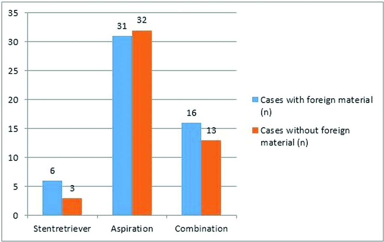
- FIG 10.
The association between foreign materials and MT approach.


Jump to section
Related Articles
Cited By...
- Non-ischemic cerebral enhancing (NICE) lesions after flow diversion for intracranial aneurysms: a multicenter study
- Correspondence on 'Non-ischemic cerebral enhancing (NICE) lesions after flow diversion for intracranial aneurysms: a multicenter study by Richter et al
- Longitudinal radiological follow-up of individual level non-ischemic cerebral enhancing lesions following endovascular aneurysm treatment
- Non-ischemic cerebral enhancing lesions after intracranial aneurysm endovascular repair: a retrospective French national registry