Advertisement

Main menu

  • Home
  • Content
    • Current Issue
    • Accepted Manuscripts
    • Article Preview
    • Past Issue Archive
    • AJNR Case Collection
    • Case of the Week Archive
    • Classic Case Archive
    • Case of the Month Archive
  • Special Collections
    • Spinal CSF Leak Articles (Jan 2020-June 2024)
    • 2024 AJNR Journal Awards
    • Most Impactful AJNR Articles
  • Multimedia
    • AJNR Podcast
    • AJNR Scantastics
    • Video Articles
  • For Authors
    • Submit a Manuscript
    • Author Policies
    • Fast publishing of Accepted Manuscripts
    • Graphical Abstract Preparation
    • Manuscript Submission Guidelines
    • Imaging Protocol Submission
    • Submit a Case for the Case Collection
  • About Us
    • About AJNR
    • Editorial Board
  • More
    • Become a Reviewer/Academy of Reviewers
    • Subscribers
    • Permissions
    • Alerts
    • Feedback
    • Advertisers
    • ASNR Home
  • Other Publications
    • ajnr

User menu

  • Alerts
  • Log in

Search

  • Advanced search
American Journal of Neuroradiology

American Journal of Neuroradiology

ASHNR American Society of Functional Neuroradiology ASHNR American Society of Pediatric Neuroradiology ASSR
  • Alerts
  • Log in

Advanced Search

  • Home
  • Content
    • Current Issue
    • Accepted Manuscripts
    • Article Preview
    • Past Issue Archive
    • AJNR Case Collection
    • Case of the Week Archive
    • Classic Case Archive
    • Case of the Month Archive
  • Special Collections
    • Spinal CSF Leak Articles (Jan 2020-June 2024)
    • 2024 AJNR Journal Awards
    • Most Impactful AJNR Articles
  • Multimedia
    • AJNR Podcast
    • AJNR Scantastics
    • Video Articles
  • For Authors
    • Submit a Manuscript
    • Author Policies
    • Fast publishing of Accepted Manuscripts
    • Graphical Abstract Preparation
    • Manuscript Submission Guidelines
    • Imaging Protocol Submission
    • Submit a Case for the Case Collection
  • About Us
    • About AJNR
    • Editorial Board
  • More
    • Become a Reviewer/Academy of Reviewers
    • Subscribers
    • Permissions
    • Alerts
    • Feedback
    • Advertisers
    • ASNR Home
  • Follow AJNR on Twitter
  • Visit AJNR on Facebook
  • Follow AJNR on Instagram
  • Join AJNR on LinkedIn
  • RSS Feeds

Welcome to the new AJNR, Updated Hall of Fame, and more. Read the full announcements.


AJNR is seeking candidates for the position of Associate Section Editor, AJNR Case Collection. Read the full announcement.


Research ArticleFunctional

Asynchrony in Peritumoral Resting-State Blood Oxygen Level–Dependent fMRI Predicts Meningioma Grade and Invasion

P.B. Wu, D.S. Chow, P.D. Petridis, M.B. Sisti, J.N. Bruce, P.D. Canoll and J. Grinband
American Journal of Neuroradiology July 2021, 42 (7) 1293-1298; DOI: https://doi.org/10.3174/ajnr.A7154
P.B. Wu
aFrom the Vagelos College of Physicians and Surgeons (P.B.W.)
bDepartments of Neurological Surgery (P.B.W., M.B.S., J.N.B.)
  • Find this author on Google Scholar
  • Find this author on PubMed
  • Search for this author on this site
  • ORCID record for P.B. Wu
D.S. Chow
fDepartment of Radiological Sciences (D.S.C.), University of California Irvine, Irvine, California
  • Find this author on Google Scholar
  • Find this author on PubMed
  • Search for this author on this site
  • ORCID record for D.S. Chow
P.D. Petridis
gDepartment of Psychiatry (P.D.P.), New York University, New York, New York.
  • Find this author on Google Scholar
  • Find this author on PubMed
  • Search for this author on this site
  • ORCID record for P.D. Petridis
M.B. Sisti
bDepartments of Neurological Surgery (P.B.W., M.B.S., J.N.B.)
  • Find this author on Google Scholar
  • Find this author on PubMed
  • Search for this author on this site
  • ORCID record for M.B. Sisti
J.N. Bruce
bDepartments of Neurological Surgery (P.B.W., M.B.S., J.N.B.)
  • Find this author on Google Scholar
  • Find this author on PubMed
  • Search for this author on this site
  • ORCID record for J.N. Bruce
P.D. Canoll
cPathology and Cell Biology (P.D.C.)
  • Find this author on Google Scholar
  • Find this author on PubMed
  • Search for this author on this site
  • ORCID record for P.D. Canoll
J. Grinband
dRadiology (J.G.)
ePsychiatry (J.G.), Columbia University, New York, New York
  • Find this author on Google Scholar
  • Find this author on PubMed
  • Search for this author on this site
  • ORCID record for J. Grinband
  • Article
  • Figures & Data
  • Info & Metrics
  • Responses
  • References
  • PDF
Loading

Article Figures & Data

Figures

  • FIG 1.
    • Download figure
    • Open in new tab
    • Download powerpoint
    FIG 1.

    BOLD asynchrony decreases with distance from the tumor. A, Exponential fits were made to BOLD asynchrony values across all voxels within a 3-cm radius as a function of distance from the tumor for each patient. Blue lines represent individual patients with low-grade tumors; red lines represent individual patients with high-grade tumors. B, The mean BOLD asynchrony is greater for high-grade than low-grade tumors at comparable distances from the tumor. Shading represents standard error of the mean. C, Bars represent the median spatial decay for the low- (blue) and high-grade (red) tumors; dots represent decay constants for individual patients. High-grade meningiomas have significantly slower spatial decay rates, indicating that high-grade tumors caused a larger disruption to surrounding vasculature. D–F, FLAIR signal did not differ significantly between groups. Note, the error bars are very large and extend beyond the visible range.
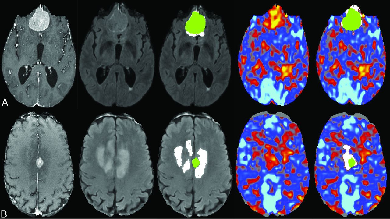
  • FIG 2.
    • Download figure
    • Open in new tab
    • Download powerpoint
    FIG 2.

    Representative axial slices from a 77-year-old male patient (A) with a grade I meningioma and a 75-year-old male patient (B) with a grade II meningioma. The first 3 columns show the T1-weighted postcontrast image, the T2-FLAIR, and the T2-FLAIR with overlaid masks representing the contrast enhancement (green) and the T2-FLAIR hyperintensity outside the tumor (white). The fourth and fifth columns show the BOLD asynchrony map and the BOLD asynchrony map with overlaid masks representing the contrast enhancement (green) and the BOLD asynchrony outside the tumor (white). The high-grade tumor (B) has BOLD asynchrony extending further outside the region of contrast enhancement than the low-grade tumor.

  • FIG 3.
    • Download figure
    • Open in new tab
    • Download powerpoint
    FIG 3.

    Boxplots demonstrate that high-grade meningiomas are characterized by greater BOF values than low-grade meningiomas (A). B, FOF values were higher in high-grade meningiomas, though this did not reach significance. Receiver operating characteristic curves for BOF show high discriminability for grade (C), while FOF demonstrates marginal discriminability (D).

  • FIG 4.
    • Download figure
    • Open in new tab
    • Download powerpoint
    FIG 4.

    A, The weighted mean of BOF and FOF shows a difference comparable with the BOF alone. B, The discriminability of the weighted model is better, though only marginally, than the BOF alone.

PreviousNext
Back to top

In this issue

American Journal of Neuroradiology: 42 (7)
American Journal of Neuroradiology
Vol. 42, Issue 7
1 Jul 2021
  • Table of Contents
  • Index by author
  • Complete Issue (PDF)
Advertisement
Print
Download PDF
Email Article

Thank you for your interest in spreading the word on American Journal of Neuroradiology.

NOTE: We only request your email address so that the person you are recommending the page to knows that you wanted them to see it, and that it is not junk mail. We do not capture any email address.

Enter multiple addresses on separate lines or separate them with commas.
Asynchrony in Peritumoral Resting-State Blood Oxygen Level–Dependent fMRI Predicts Meningioma Grade and Invasion
(Your Name) has sent you a message from American Journal of Neuroradiology
(Your Name) thought you would like to see the American Journal of Neuroradiology web site.
CAPTCHA
This question is for testing whether or not you are a human visitor and to prevent automated spam submissions.
Cite this article
P.B. Wu, D.S. Chow, P.D. Petridis, M.B. Sisti, J.N. Bruce, P.D. Canoll, J. Grinband
Asynchrony in Peritumoral Resting-State Blood Oxygen Level–Dependent fMRI Predicts Meningioma Grade and Invasion
American Journal of Neuroradiology Jul 2021, 42 (7) 1293-1298; DOI: 10.3174/ajnr.A7154

Citation Manager Formats

  • BibTeX
  • Bookends
  • EasyBib
  • EndNote (tagged)
  • EndNote 8 (xml)
  • Medlars
  • Mendeley
  • Papers
  • RefWorks Tagged
  • Ref Manager
  • RIS
  • Zotero
0 Responses
Respond to this article
Share
Bookmark this article
Asynchrony in Peritumoral Resting-State Blood Oxygen Level–Dependent fMRI Predicts Meningioma Grade and Invasion
P.B. Wu, D.S. Chow, P.D. Petridis, M.B. Sisti, J.N. Bruce, P.D. Canoll, J. Grinband
American Journal of Neuroradiology Jul 2021, 42 (7) 1293-1298; DOI: 10.3174/ajnr.A7154
del.icio.us logo Twitter logo Facebook logo Mendeley logo
  • Tweet Widget
  • Facebook Like
  • Google Plus One
Purchase

Jump to section

  • Article
    • Abstract
    • ABBREVIATIONS:
    • MATERIALS AND METHODS
    • RESULTS
    • DISCUSSION
    • CONCLUSIONS
    • Footnotes
    • References
  • Figures & Data
  • Info & Metrics
  • Responses
  • References
  • PDF

Related Articles

  • No related articles found.
  • PubMed
  • Google Scholar

Cited By...

  • No citing articles found.
  • Crossref (2)
  • Google Scholar

This article has been cited by the following articles in journals that are participating in Crossref Cited-by Linking.

  • Asynchronität von BOLD-Signalen bei der fMRT zur Stadieneinteilung bei Meningeomen
    Radiologie up2date 2022 22 04
  • Asynchronität von BOLD-Signalen bei der fMRT zur Stadieneinteilung bei Meningeomen
    Neuroradiologie Scan 2022 12 03

More in this TOC Section

Functional

  • Glutaric Aciduria Type 1: DK vs. Conventional MRI
  • Kurtosis and Epileptogenic Tubers: A Pilot Study
  • WM Neuroaxonal Loss in Type 1 Diabetes
Show more Functional

Adult Brain

  • Diagnostic Neuroradiology of Monoclonal Antibodies
  • ML for Glioma Molecular Subtype Prediction
  • Segmentation of Brain Metastases with BLAST
Show more Adult Brain

Similar Articles

Advertisement

Indexed Content

  • Current Issue
  • Accepted Manuscripts
  • Article Preview
  • Past Issues
  • Editorials
  • Editors Choice
  • Fellow Journal Club
  • Letters to the Editor

Cases

  • Case Collection
  • Archive - Case of the Week
  • Archive - Case of the Month
  • Archive - Classic Case

Special Collections

  • Special Collections

Resources

  • News and Updates
  • Turn around Times
  • Submit a Manuscript
  • Author Policies
  • Manuscript Submission Guidelines
  • Evidence-Based Medicine Level Guide
  • Publishing Checklists
  • Graphical Abstract Preparation
  • Imaging Protocol Submission
  • Submit a Case
  • Become a Reviewer/Academy of Reviewers
  • Get Peer Review Credit from Publons

Multimedia

  • AJNR Podcast
  • AJNR SCANtastic
  • Video Articles

About Us

  • About AJNR
  • Editorial Board
  • Not an AJNR Subscriber? Join Now
  • Alerts
  • Feedback
  • Advertise with us
  • Librarian Resources
  • Permissions
  • Terms and Conditions

American Society of Neuroradiology

  • Not an ASNR Member? Join Now

© 2025 by the American Society of Neuroradiology All rights, including for text and data mining, AI training, and similar technologies, are reserved.
Print ISSN: 0195-6108 Online ISSN: 1936-959X

Powered by HighWire