Advertisement

Main menu

  • Home
  • Content
    • Current Issue
    • Accepted Manuscripts
    • Article Preview
    • Past Issue Archive
    • AJNR Case Collection
    • Case of the Week Archive
    • Classic Case Archive
    • Case of the Month Archive
  • Special Collections
    • Spinal CSF Leak Articles (Jan 2020-June 2024)
    • 2024 AJNR Journal Awards
    • Most Impactful AJNR Articles
  • Multimedia
    • AJNR Podcast
    • AJNR Scantastics
    • Video Articles
  • For Authors
    • Submit a Manuscript
    • Author Policies
    • Fast publishing of Accepted Manuscripts
    • Graphical Abstract Preparation
    • Manuscript Submission Guidelines
    • Imaging Protocol Submission
    • Submit a Case for the Case Collection
  • About Us
    • About AJNR
    • Editorial Board
  • More
    • Become a Reviewer/Academy of Reviewers
    • Subscribers
    • Permissions
    • Alerts
    • Feedback
    • Advertisers
    • ASNR Home
  • Other Publications
    • ajnr

User menu

  • Alerts
  • Log in

Search

  • Advanced search
American Journal of Neuroradiology

American Journal of Neuroradiology

ASHNR American Society of Functional Neuroradiology ASHNR American Society of Pediatric Neuroradiology ASSR
  • Alerts
  • Log in

Advanced Search

  • Home
  • Content
    • Current Issue
    • Accepted Manuscripts
    • Article Preview
    • Past Issue Archive
    • AJNR Case Collection
    • Case of the Week Archive
    • Classic Case Archive
    • Case of the Month Archive
  • Special Collections
    • Spinal CSF Leak Articles (Jan 2020-June 2024)
    • 2024 AJNR Journal Awards
    • Most Impactful AJNR Articles
  • Multimedia
    • AJNR Podcast
    • AJNR Scantastics
    • Video Articles
  • For Authors
    • Submit a Manuscript
    • Author Policies
    • Fast publishing of Accepted Manuscripts
    • Graphical Abstract Preparation
    • Manuscript Submission Guidelines
    • Imaging Protocol Submission
    • Submit a Case for the Case Collection
  • About Us
    • About AJNR
    • Editorial Board
  • More
    • Become a Reviewer/Academy of Reviewers
    • Subscribers
    • Permissions
    • Alerts
    • Feedback
    • Advertisers
    • ASNR Home
  • Follow AJNR on Twitter
  • Visit AJNR on Facebook
  • Follow AJNR on Instagram
  • Join AJNR on LinkedIn
  • RSS Feeds

Welcome to the new AJNR, Updated Hall of Fame, and more. Read the full announcements.


AJNR is seeking candidates for the position of Associate Section Editor, AJNR Case Collection. Read the full announcement.


Research ArticleInterventional

Dural Arteriovenous Fistulas of the Foramen Magnum Region: Clinical Features and Angioarchitectural Phenotypes

M.T. Caton, K.H. Narsinh, A. Baker, C.F. Dowd, R.T. Higashida, D.L. Cooke, S.W. Hetts, V.V. Halbach and M.R. Amans
American Journal of Neuroradiology August 2021, 42 (8) 1486-1491; DOI: https://doi.org/10.3174/ajnr.A7152
M.T. Caton
aFrom the Department of Radiology and Biomedical Imaging, Interventional Neuroradiology Section, University of California, San Francisco, San Francisco, California
  • Find this author on Google Scholar
  • Find this author on PubMed
  • Search for this author on this site
  • ORCID record for M.T. Caton
K.H. Narsinh
aFrom the Department of Radiology and Biomedical Imaging, Interventional Neuroradiology Section, University of California, San Francisco, San Francisco, California
  • Find this author on Google Scholar
  • Find this author on PubMed
  • Search for this author on this site
  • ORCID record for K.H. Narsinh
A. Baker
aFrom the Department of Radiology and Biomedical Imaging, Interventional Neuroradiology Section, University of California, San Francisco, San Francisco, California
  • Find this author on Google Scholar
  • Find this author on PubMed
  • Search for this author on this site
  • ORCID record for A. Baker
C.F. Dowd
aFrom the Department of Radiology and Biomedical Imaging, Interventional Neuroradiology Section, University of California, San Francisco, San Francisco, California
  • Find this author on Google Scholar
  • Find this author on PubMed
  • Search for this author on this site
  • ORCID record for C.F. Dowd
R.T. Higashida
aFrom the Department of Radiology and Biomedical Imaging, Interventional Neuroradiology Section, University of California, San Francisco, San Francisco, California
  • Find this author on Google Scholar
  • Find this author on PubMed
  • Search for this author on this site
  • ORCID record for R.T. Higashida
D.L. Cooke
aFrom the Department of Radiology and Biomedical Imaging, Interventional Neuroradiology Section, University of California, San Francisco, San Francisco, California
  • Find this author on Google Scholar
  • Find this author on PubMed
  • Search for this author on this site
  • ORCID record for D.L. Cooke
S.W. Hetts
aFrom the Department of Radiology and Biomedical Imaging, Interventional Neuroradiology Section, University of California, San Francisco, San Francisco, California
  • Find this author on Google Scholar
  • Find this author on PubMed
  • Search for this author on this site
  • ORCID record for S.W. Hetts
V.V. Halbach
aFrom the Department of Radiology and Biomedical Imaging, Interventional Neuroradiology Section, University of California, San Francisco, San Francisco, California
  • Find this author on Google Scholar
  • Find this author on PubMed
  • Search for this author on this site
  • ORCID record for V.V. Halbach
M.R. Amans
aFrom the Department of Radiology and Biomedical Imaging, Interventional Neuroradiology Section, University of California, San Francisco, San Francisco, California
  • Find this author on Google Scholar
  • Find this author on PubMed
  • Search for this author on this site
  • ORCID record for M.R. Amans
  • Article
  • Figures & Data
  • Info & Metrics
  • Responses
  • References
  • PDF
Loading

Article Figures & Data

Figures

  • Tables
  • FIG 1.
    • Download figure
    • Open in new tab
    • Download powerpoint
    FIG 1.

    Venous anatomy of the FMR from above. The marginal sinus (MS) lines the margin the foramen magnum and connects to the basilar plexus (BP) anteriorly, the anterior condylar vein (ACV) laterally via the hypoglossal canal (HGC), and the suboccipital cavernous sinus inferiorly (not shown). The anterior condylar vein (AVC) connects with the anterior condylar confluence (ACC), which, in turn, communicates with the inferior petrosal sinus (IPS) and inferior petroclival vein (IPCV) and posteriorly with the lateral condylar vein (LCV), and the jugular bulb and internal jugular vein (JB/IJV). The posterior condylar (emissary) vein (PCV) exits via the posterior condylar canal (PCC).
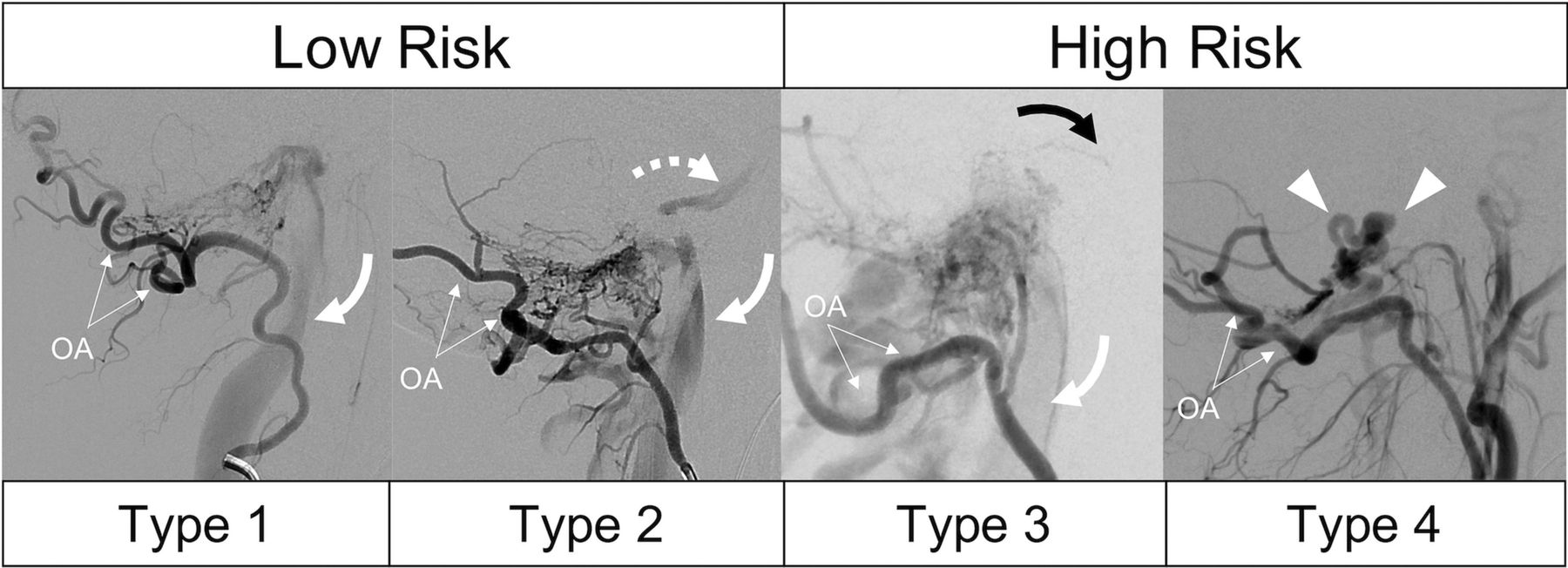
  • FIG 2.
    • Download figure
    • Open in new tab
    • Download powerpoint
    FIG 2.

    Modified grading system for FMR-AVFs shown in 4 patients having undergone lateral occipital artery (OA) injection DSA. Type 1 shows unrestricted drainage via the IJV (curved arrow) without reflux. Type 2 shows both antegrade drainage (curved arrow) and sinus reflux (dashed arrow, inferior petrosal sinus). Type 3 lesions are differentiated by the presence of cortical venous reflux, in this case, pontine perforating veins (black arrow) in addition to sinus drainage (white arrow). Type 4 lesions have restricted, exclusive drainage via cortical veins (arrowheads) without a coexisting sinus drainage pathway.

  • FIG 3.
    • Download figure
    • Open in new tab
    • Download powerpoint
    FIG 3.

    Arterial anatomy of a typical type 1 FMR AVF. TOF-MRA (A) of the skull base shows abnormal flow-related enhancement in the right hypoglossal canal, corresponding to a fistula (asterisk). Arterial supply from the contralateral ascending pharyngeal artery (hypoglossal branch) crosses the foramen magnum and drains via the IJV. Relative anatomy: an anterior-posterior DSA with injection of the left ascending pharyngeal artery (B) shows hypertrophied arterial channels (double-sided arrow). Lateral DSA of the right ICA (C) shows supply to the fistula (asterisk) from the meningohyposphyseal trunk (MHT). D, Lateral DSA of the right vertebral artery (V3) shows direct contribution to fistula (asterisk) and drainage via the IJV. NMT indicates neuromeningeal trunk; MS, marginal sinus.
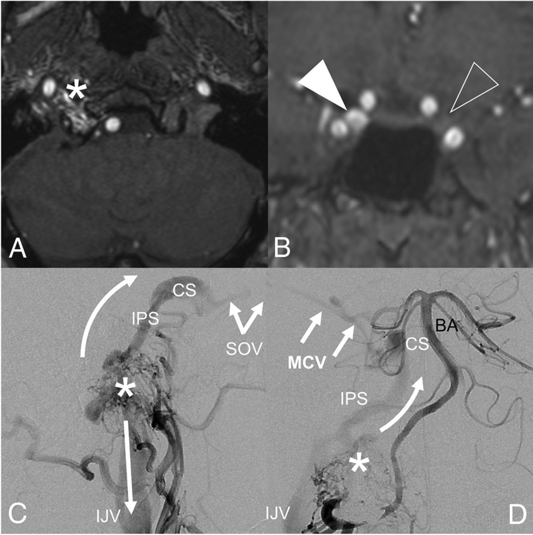
  • FIG 4.
    • Download figure
    • Open in new tab
    • Download powerpoint
    FIG 4.

    A type 3 FMR-AVF with clinical features mimicking a carotid cavernous fistula. MRA of the FMR (A) shows flow-related enhancement in the right hypoglossal canal, corresponding to an anterior condylar vein fistula site. Coronal MRA (B) shows asymmetric flow-related enhancement of the right (white arrowhead) relative to left (clear arrowhead) cavernous sinuses. Lateral-projection DSA of the right ascending pharyngeal artery (C) shows the fistula (asterisk) with antegrade drainage via the IJV (straight arrow) and reflux via the inferior petrosal sinus (IPS, curved arrow) extending to the cavernous sinus (CS) and superior ophthalmic vein (SOV), resulting in proptosis and chemosis. Anterior-posterior DSA of the right vertebral artery (D) shows reflux via the IPS and CS, which continues to the middle cerebral vein (MCV), making this a high-risk type 3 fistula. The asterisk represents the fistulous shunt site. BA indicates the basilar artery.

Tables

  • Figures
  • Venous drainage patterns in FMR AVF

    Venous Angioarchitectural TypeClinical Presentation (% of Cases)
    Pulsatile Tinnitus and/or BruitOrbital SymptomsCN XII PalsyMyelopathyHemorrhageHeadache
    Type 1 (n = 11)90.9%9.1%0.0%0.0%0.0%27.3%
    Type 2 (n = 9)100.0%44.4%22.2%0.0%0.0%44.4%
    Type 3 (n = 6)66.7%33.3%0.0%0.0%16.7%50.0%
    Type 4 (n = 3)33.3%33.3%0.0%33.3%66.7%66.7%
PreviousNext
Back to top

In this issue

American Journal of Neuroradiology: 42 (8)
American Journal of Neuroradiology
Vol. 42, Issue 8
1 Aug 2021
  • Table of Contents
  • Index by author
  • Complete Issue (PDF)
Advertisement
Print
Download PDF
Email Article

Thank you for your interest in spreading the word on American Journal of Neuroradiology.

NOTE: We only request your email address so that the person you are recommending the page to knows that you wanted them to see it, and that it is not junk mail. We do not capture any email address.

Enter multiple addresses on separate lines or separate them with commas.
Dural Arteriovenous Fistulas of the Foramen Magnum Region: Clinical Features and Angioarchitectural Phenotypes
(Your Name) has sent you a message from American Journal of Neuroradiology
(Your Name) thought you would like to see the American Journal of Neuroradiology web site.
CAPTCHA
This question is for testing whether or not you are a human visitor and to prevent automated spam submissions.
Cite this article
M.T. Caton, K.H. Narsinh, A. Baker, C.F. Dowd, R.T. Higashida, D.L. Cooke, S.W. Hetts, V.V. Halbach, M.R. Amans
Dural Arteriovenous Fistulas of the Foramen Magnum Region: Clinical Features and Angioarchitectural Phenotypes
American Journal of Neuroradiology Aug 2021, 42 (8) 1486-1491; DOI: 10.3174/ajnr.A7152

Citation Manager Formats

  • BibTeX
  • Bookends
  • EasyBib
  • EndNote (tagged)
  • EndNote 8 (xml)
  • Medlars
  • Mendeley
  • Papers
  • RefWorks Tagged
  • Ref Manager
  • RIS
  • Zotero
0 Responses
Respond to this article
Share
Bookmark this article
Dural Arteriovenous Fistulas of the Foramen Magnum Region: Clinical Features and Angioarchitectural Phenotypes
M.T. Caton, K.H. Narsinh, A. Baker, C.F. Dowd, R.T. Higashida, D.L. Cooke, S.W. Hetts, V.V. Halbach, M.R. Amans
American Journal of Neuroradiology Aug 2021, 42 (8) 1486-1491; DOI: 10.3174/ajnr.A7152
del.icio.us logo Twitter logo Facebook logo Mendeley logo
  • Tweet Widget
  • Facebook Like
  • Google Plus One
Purchase

Jump to section

  • Article
    • Abstract
    • ABBREVIATIONS:
    • MATERIALS AND METHODS
    • RESULTS
    • DISCUSSION
    • CONCLUSIONS
    • Footnotes
    • References
  • Figures & Data
  • Info & Metrics
  • Responses
  • References
  • PDF

Related Articles

  • No related articles found.
  • PubMed
  • Google Scholar

Cited By...

  • Posterior condylar canal dural arteriovenous fistula: anatomical, symptomatological, and therapeutic considerations in comparison with hypoglossal canal dural arteriovenous fistula
  • Endovascular treatment of dural arteriovenous fistulas involving the vein of Galen: a single-center cohort and meta-analysis
  • Endovascular embolisation of posterior condylar canal dural arteriovenous fistula
  • Angiographically Occult Subarachnoid Hemorrhage: Yield of Repeat Angiography, Influence of Initial CT Bleed Pattern, and Sources of Diagnostic Error in 242 Consecutive Patients
  • Crossref (23)
  • Google Scholar

This article has been cited by the following articles in journals that are participating in Crossref Cited-by Linking.

  • Diagnostic Approach to Pulsatile Tinnitus
    Kazim H. Narsinh, Ferdinand Hui, David Saloner, Adelyn Tu-Chan, Jeffrey Sharon, Andreas M. Rauschecker, Fatima Safoora, Vinil Shah, Karl Meisel, Matthew R. Amans
    JAMA Otolaryngology–Head & Neck Surgery 2022 148 5
  • Endovascular treatment of medullary bridging vein-draining dural arteriovenous fistulas: foramen magnum vs. craniocervical junction lesions
    Dong Hyun Yoo, Young Dae Cho, Tiplada Boonchai, Kang Min Kim, Jeong Eun Kim, Won-Sang Cho, Sung Ho Lee, Chun Kee Chung, Hyun-Seung Kang
    Neuroradiology 2022 64 2
  • Angiographically Occult Subarachnoid Hemorrhage: Yield of Repeat Angiography, Influence of Initial CT Bleed Pattern, and Sources of Diagnostic Error in 242 Consecutive Patients
    I. Nguyen, M.T. Caton, D. Tonetti, A. Abla, A. Kim, W. Smith, S.W. Hetts
    American Journal of Neuroradiology 2022 43 5
  • Treatment of high cervical arteriovenous fistulas in the craniocervical junction region
    Han Su, Jinlu Yu
    Frontiers in Neurology 2023 14
  • Clinical features, angio-architectural phenotypes, and treatment strategy of foramen magnum dural arteriovenous fistulas: a retrospective case series study
    Zhipeng Xiao, Weizhen Gao, Hongyu Zhou, Xiaohua Zhang, Jiong Dai, Jieqing Wan, Liemei Guo
    Frontiers in Neurology 2023 14
  • Enhanced Treatment Options for Dural Arteriovenous Fistulas at the Craniocervical Junction: Endovascular Embolization Versus Microsurgery? A Single-Center 23-Year Experience
    Jiebo Li, Fuxin Lin, Jianyu Zhu, Lingyun Zhuo, Fuxiang Chen, Linsun Dai, Shufa Zheng, Lianghong Yu, Dezhi Kang, Yuanxiang Lin, Dengliang Wang
    World Neurosurgery 2024 182
  • Transvenous Approaches to the Vertebral‐Venous Plexus for Endovascular Treatment of Cervical Epidural Arteriovenous Fistulas: Anatomy and Technique
    Michael T. Caton, Masis Isikbay, Kazim H. Narsinh, Amanda Baker, James Milburn, Daniel L. Cooke, Steven W. Hetts, Christopher F. Dowd, Randall T. Higashida, Matthew R. Amans
    Stroke: Vascular and Interventional Neurology 2022 2 3
  • Endovascular treatment of dural arteriovenous fistulas involving the vein of Galen: a single-center cohort and meta-analysis
    David C Lauzier, Henrik Ullman, Angela Hardi, Colin Derdeyn, Dewitte T Cross, Christopher J Moran
    Journal of NeuroInterventional Surgery 2024 16 12
  • Classification, angioarchitecture and treatment outcomes of medullary bridging vein-draining dural arteriovenous fistulas in the foramen magnum region: a multicenter study
    Tomohiko Ozaki, Masafumi Hiramatsu, Hajime Nakamura, Yasunari Niimi, Shuichi Tanoue, Katsuhiro Mizutani, Ichiro Nakahara, Yuji Matsumaru, Yasushi Matsumoto, Timo Krings, Toshiyuki Fujinaka
    Neuroradiology 2025 67 1
  • Endovascular embolisation of posterior condylar canal dural arteriovenous fistula
    Surya Nandan Prasad, Vivek Singh, Srishti Sharma, Madhuri Kumari
    BMJ Case Reports 2024 17 2

More in this TOC Section

  • SAVE vs. Solumbra Techniques for Thrombectomy
  • CT Perfusion&Reperfusion in Acute Ischemic Stroke
  • Delayed Reperfusion Post-Thrombectomy&Thrombolysis
Show more Interventional

Similar Articles

Advertisement

Indexed Content

  • Current Issue
  • Accepted Manuscripts
  • Article Preview
  • Past Issues
  • Editorials
  • Editors Choice
  • Fellow Journal Club
  • Letters to the Editor

Cases

  • Case Collection
  • Archive - Case of the Week
  • Archive - Case of the Month
  • Archive - Classic Case

Special Collections

  • Special Collections

Resources

  • News and Updates
  • Turn around Times
  • Submit a Manuscript
  • Author Policies
  • Manuscript Submission Guidelines
  • Evidence-Based Medicine Level Guide
  • Publishing Checklists
  • Graphical Abstract Preparation
  • Imaging Protocol Submission
  • Submit a Case
  • Become a Reviewer/Academy of Reviewers
  • Get Peer Review Credit from Publons

Multimedia

  • AJNR Podcast
  • AJNR SCANtastic
  • Video Articles

About Us

  • About AJNR
  • Editorial Board
  • Not an AJNR Subscriber? Join Now
  • Alerts
  • Feedback
  • Advertise with us
  • Librarian Resources
  • Permissions
  • Terms and Conditions

American Society of Neuroradiology

  • Not an ASNR Member? Join Now

© 2025 by the American Society of Neuroradiology All rights, including for text and data mining, AI training, and similar technologies, are reserved.
Print ISSN: 0195-6108 Online ISSN: 1936-959X

Powered by HighWire