
Abstract
BACKGROUND AND PURPOSE: fMRI is a noninvasive tool for predicting postsurgical deficits in candidates with pharmacoresistant temporal lobe epilepsy. We aimed to test an adapted paradigm of the Rey Auditory Verbal Learning Test to evaluate differences in memory laterality indexes between patients and healthy controls and its association with neuropsychological scores.
MATERIALS AND METHODS: We performed a prospective study of 50 patients with temporal lobe epilepsy and 22 healthy controls. Participants underwent a block design language and memory fMRI. Laterality indexes and the hippocampal anterior-posterior index were calculated. Language and memory lateralization was organized into typical and atypical on the basis of laterality indexes. A neuropsychological assessment was performed with a median time from fMRI of 8 months and was compared with fMRI performance.
RESULTS: We studied 40 patients with left temporal lobe epilepsy and 10 with right temporal lobe epilepsy. Typical language occurred in 65.3% of patients and 90.9% of healthy controls (P = .04). The memory fMRI laterality index was obtained in all healthy controls and 92% of patients. The verbal memory laterality index was bilateral (24.3%) more frequently than the language laterality index (7.69%) in patients with left temporal lobe epilepsy. Atypical verbal memory was greater in patients with left temporal lobe epilepsy (56.8%) than in healthy controls (36.4%), and the proportion of bilateral laterality indexes (53.3%) was larger than right laterality indexes (46.7%). Atypical verbal memory might be associated with higher cognitive scores in patients. No relevant differences were seen in the hippocampal anterior-posterior index according to memory impairment.
CONCLUSIONS: The adapted Rey Auditory Verbal Learning Test paradigm fMRI might support verbal memory lateralization. Temporal lobe epilepsy laterality influences hippocampal memory laterality indexes. Left temporal lobe epilepsy has shown a higher proportion of atypical verbal memory compared with language, potentially to memory functional reorganization.
ABBREVIATIONS:
- AVLT
- Rey Auditory Verbal Learning Test
- APi
- hippocampal anterior-posterior index
- ATLR
- anterior temporal lobe resection
- HC
- healthy controls
- IQR
- interquartile range
- LI
- laterality index
- LMI
- immediate Logical Memory
- LMII
- delayed Logical Memory
- SISCOM
- substraction of ictal and interictal SPECT coregistered to MR Imaging
- VMI
- immediate Visual Reproduction
- VMII
- delayed Visual Reproduction
- TLE
- temporal lobe epilepsy
Refractory mesial temporal lobe epilepsy (TLE) usually concurs with progressive cognitive impairment.1-3 Material-specific memory dysfunction has been associated with left and right temporal lobe lesions. Verbal memory processes were hindered by left-sided lesions, whereas visuospatial memory deficits were related to right TLE.4 Focal epilepsies arising in eloquent areas or in the vicinity of the dominant hemisphere have been associated with cerebral plasticity, such as reorganization of adjacent or even distant and contralateral cortical areas to maintain function.5 Factors such as the age of seizure onset, seizure frequency, the presence of hippocampal sclerosis, the duration of the disease, and antiseizure medication may play a relevant role in cognitive function.6,7 Lack of cognitive dysfunction in TLE may also occur, even when patients had similar clinical features to those who do have cognitive deficits.
The left hippocampus has often been associated with verbal memory encoding.8 Its relationship to the seizure-onset zone has been regarded as a determinant factor in changes in verbal memory function.9 From a network perspective, hippocampal sclerosis has been associated with a dysfunction of long-range network connections.10 Neuropsychological testing has been the main diagnostic tool to evaluate memory function before anterior temporal lobe resection (ATLR); however, it is operator-dependent and does not provide information about the underlying functional anatomy.11 fMRI has become a helpful tool to investigate cognitive processes and to predict memory deficits before ATLR.12
To date, memory fMRI studies have methodologic differences, particularly regarding the memory task itself, with complex paradigms difficult to apply on an everyday clinical basis for patients with epilepsy.13 Cognition evaluation in patients with epilepsy is increasingly a multidisciplinary team effort.14 However, neuropsychological tests cannot lateralize memory function before surgical intervention and do not provide information on functional anatomy. fMRI could be a useful noninvasive tool to identify the lateralization and localization of memory functions before surgical intervention in individual patients with TLE.15-19 Prior studies have found that preoperative posterior fMRI activation in the ipsilateral hippocampus during memory encoding was associated with better memory outcome after ATLR.20
In this study, we examined the use of a simple and robust paradigm based on a well-established neuropsychological test on fMRI for verbal memory laterality outcome. We hypothesize that by using this list-learning paradigm we could obtain results in lateralization similar to those in the neuropsychological assessment, incorporating the functional anterior-posterior distribution of the hippocampal formation activity. Our main aim was to test this adapted paradigm for evaluating differences in memory hippocampal anterior-posterior indexes (APis) and laterality indexes (LIs) between healthy controls (HC) and patients with right and left TLE and to search for associations and comparisons in performance with neuropsychological test scores.
MATERIALS AND METHODS
Design and Patients
This was a prospective study of consecutive patients with TLE who were evaluated for presurgical assessment at our multidisciplinary Patient Management Conference. Patients underwent language and memory fMRI based on clinical needs, particularly when there was reasonable doubt about language distribution. Participants were scanned between December 2018 and March 2020.
We included 50 patients with an age range between 18 and 65 years who underwent a presurgical evaluation for potential epilepsy surgery with a neuropsychological assessment. Participants were excluded from the study if diagnosed with neuropsychiatric disorders, were unable to tolerate the fMRI, or when artifacts invalidated the analysis. We also studied 23 HC with no history of neurologic or psychiatric disease using the same paradigm. One subject was excluded because of MR imaging artifacts.
The study was approved by the Ethics Committee of the Hospital Clinic of Barcelona. Informed consent was obtained from all participants in the study. Patients underwent a video-electroencephalography, structural MR imaging, and, additionally in some cases, FDG-PET scan and SISCOM, which established seizure onset location and lateralization. Neuropsychological assessment was obtained for all patients but only for 13 of the 22 HC due to the test being canceled during the pandemic restrictions.
MR Imaging
All scans were performed on a 3T Magnetom Tim Trio (Siemens) scanner or Prisma Fit scanner (Siemens), at the Magnetic Resonance Image Core Facility at August Pi i Sunyer Biomedical Research Institute located in the Diagnostic Imaging Center at Hospital Clinic of Barcelona using a 32- or 64-channel head coil. Each subject underwent a 3D structural high-resolution T1-weighted MPRAGE sequence with a 7.48-minute acquisition, which consisted of a set of 240 adjacent sagittal images with an isometric voxel size of 1 × 1 × 1 mm. A spoiled gradient-echo sequence (TR, 2.30 s; TE, 2.98 s; number of excitations, 1; flip angle, 9°; FOV, 256 × 256) was used to coregister with the fMRI activation maps.
fMRI images were acquired in the axial plane with an echo-planar sequence. For language lateralization, we used 3 different paradigms; and for verbal memory lateralization, a single paradigm: 1) language tasks: TR, 3.00 s; TE, 0.03 s; flip angle, 90°; pixel matrix, 3 × 3 mm; section thickness, 3 mm; 2) memory tasks: TR, 2.00 s; TE, 0.21 s; flip angle, 90°; pixel matrix, 3.8 × 3.8 mm; section thickness, 3 mm.
Paradigms.
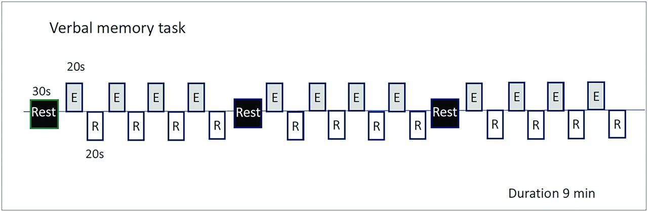
Before a scanning session, an explanation of the memory and language tasks was given to patients. Outside the scanner, for quality assurance, participants were asked about how well they comprehended and performed the commands of the undergone tasks. Paradigms were all block design and are displayed in Fig 1 and the Online Supplemental Data.

Verbal memory paradigm. E indicates encoding; R, retrieval; Min, minutes.
Verbal Memory Paradigm.
We used an adapted version of the Rey Auditory Verbal Learning Test (AVLT)21 that evaluates both encoding and retrieval verbal memory with the aim of achieving a sensitive LI of hippocampal involvement. The control task consisted of the patient hearing a list of incomprehensible words (passive listening), followed by an understandable list of 15 words that the patient must memorize, and silent periods in which the patient must recall as many words from the list as possible. Stimuli were presented in 3 blocks, alternating 30 seconds of the control task (incomprehensible words), followed by 4 cycles consisting of 20 seconds of the encoding task (memorizing the list of words) and 20 seconds of retrieval (recalling the list of words) (Fig 1).
We used 3 different block design paradigms to lateralize language activation: a phonemic verbal fluency task, a semantic verbal fluency task, and an auditory comprehension task (Online Supplemental Data).
All images were processed and analyzed using Statistical Parametric Mapping (SPM12; http://www.fil.ion.ucl.ac.uk/spm/software/spm12) and the MAGIC fMRI toolbox,23 an in-house application developed with MATLAB 2017b (MathWorks) (Online Supplemental Data).
LI for Language and Verbal Memory
Language or verbal memory lateralization was considered typical when the LI was left-lateralized and atypical when it was bilateral or right-lateralized. To determine language lateralization in the case of different results among the 3 language paradigms, we took lateralization from the 2 concordant paradigms.
Activation was classified as “left-sided” if the LI was >0.2, “right-sided” if it was <0.2, and bilateral when the LI ranged between 0.2 and −0.2 (Online Supplemental Data).
Anterior-Posterior Index of the Hippocampus
The APi frames the difference between voxel count in the anterior and posterior parts of the hippocampus. The anterior and posterior segmentation was defined by identifying the section on the Montreal Neurological Institute space where the last part of the uncal apex is appreciated.21 Anterior activation was classified if the APi was >0.2; and posterior activation, if the APi was <0.2. Whole-hippocampal activation was established when the APi fell between 0.2 and −0.2.
Neuropsychological Assessment
Participants underwent a comprehensive neuropsychological assessment with a median time lapse from MR imaging of 8 months. Verbal memory was assessed with the AVLT using total learning and delayed recall to evaluate the patient’s ability to encode, consolidate, and retrieve verbal information.21 T scores for each test and for each subject were calculated on the basis of the normative control group’s means and SDs. Participants with scores of ≤1.5 SD were classified as neurologically impaired.
Wechsler Adult Intelligence Scale-3rd Edition22 and subsets from the Wechsler Memory Scale III such as the immediate Logical Memory (LMI), delayed Logical Memory (LMII), immediate (VMI) and delayed (VMII) Visual Reproduction, and executive functions22 were used for the evaluation (Online Supplemental Data). Naming functions by the Boston Naming Test (BNT)23 were also carried out.
Statistics
The statistical analysis was performed using STATA/IC 14.2 (StataCorp).
A descriptive analysis of the variables collected was performed. Qualitative variables were expressed as a percentage, and the quantitative variables, using standard deviation (SD) or median (interquartile range [IQR]) if they had a normal distribution.
Demographic data and volumetric findings are reported as number (percentage) or median and IQR. To check the impact of the TLE side in the LI, we evaluated it using the Kruskal-Wallis 1-way ANOVA test (χ2 [2]) followed by post hoc analysis with Bonferroni correction (t), and we also used a multivariate ANOVA for influential variables. An analysis of the neuropsychological test scores and the verbal memory LI was performed using the χ2 test or Fisher exact test. Quantitative variables were compared using the Student t test, the Mann Whitney U test, or the Kruskal-Wallis 1-way ANOVA test (χ2 [2]) when appropriate.
A first-level analysis was performed by fitting a general linear model on a voxelwise basis to model the brain activation of each condition. Condition-specific effects were modeled by creating a boxcar function convolved with a canonical hemodynamic response function. Additionally, motion parameters were entered as regressors of no interest. A criterion for statistically significant activation was set at a threshold of P < .001, and a family-wise error cluster threshold of P < .05 was applied to correct for multiple comparisons (Online Supplemental Data).
RESULTS
Clinical and Neuropsychological Profiles
The Table shows clinical variables and the characteristics of the sample. Twenty-three patients had unilateral hippocampal sclerosis, of which 18 (78.3%) were on the left.
Participant characteristics
Intelligence quotients of patients ranged from above average to below average, with no significant differences between groups (P = .92). Of 50 patients, 19 (38%) showed low-average intelligent quotient; 18 (36%), an average intelligence quotient; 12 (24%), above-average intelligence quotient; and 1 (2%), below-average intelligence quotient. HC showed predominantly average scores in 6 (46.1%) participants with 5 (38.5%) having above-average and 2 (15.4%) low-average scores.
Logical memory, visual reproduction, verbal memory learning and recall, attention, and mental flexibility were significantly lower in patients with TLE compared with HC (P < .05) (Online Supplemental Data). Severely impaired verbal learning was more frequent in patients compared with HC (Pearson χ2 [2] = 10.7; Fisher exact test, P = .003). Verbal memory encoding (t = 3.58, P = .002) and retrieval (t = 2.73, P = .025) were significantly worse in left TLE compared with HC but not significantly different from right TLE.
In verbal learning encoding, 29 (58%) patients scored −1 SD or below, and 21 (42%) patients between −1 SD and +1 SD. HC showed significantly higher verbal learning scores (median, 47; IQR 6.1) than patients with left TLE (median, 36.2; IQR, 17.3) (t = 3.58, P = .002), while patients with right TLE (median, 39.9; IQR, 12.0) showed no significant differences compared with HC (t = 1.79; P = .218). Delayed recall was also significantly lower in left TLE (median, 40.47; IQR, 17.32) than in HC (median, 52.86; IQR, 14.64) (t = 2.73, P = .025), while no significant differences were found between HC and patients with right TLE (t = 1.75, P = .235).
fMRI Language Lateralization Profiles
Forty-nine patients showed activation during the language fMRI tasks. One patient did not show activation in any of the 3 paradigms.
The LI was achieved in >94% of patients and HC for the 3 verbal tasks (phonemic verbal fluency, 98% HC/95% patients). Atypical language occurred more frequently in patients (32 of 49 [65.3%]) than in HC (2/22 [9.1%]) (P = .04) (Fig 2A).

A, LI . B, APi. C, Verbal memory LI according to verbal learning impairment of patients and HC.
In both phonemic verbal fluency and semantic verbal fluency tasks, the LI was significantly different depending on handedness only for left TLE. Patients with left TLE who were left-handed had a significantly higher proportion of atypical language (χ2 = 6.62; Fisher exact test, P = .002), but no differences were seen in right TLE or HC regardless of handedness. For the auditory comprehension task this effect was not seen.
Compared with atypical activation, patients with left TLE with typical language showed significantly lower scores in LMI, LMII, VMI, VMII, verbal learning, and Boston Naming Test subtests of the neuropsychological assessment (Online Supplemental Data).
fMRI Verbal Memory Lateralization Profiles
For the verbal memory task, LI was seen in all (100%) HC and 46 of 50 (92.0%) patients. Three patients with left TLE and 1 patient with right TLE did not show hippocampal activation. Of the 4 patients who did not show activation, 3 were left-handed. According to language distribution, we found no hippocampal activation in 2 patients with typical and 2 patients with atypical language. The proportion of patients with each hippocampal activation distribution and handedness is described in the Online Supplemental Data.
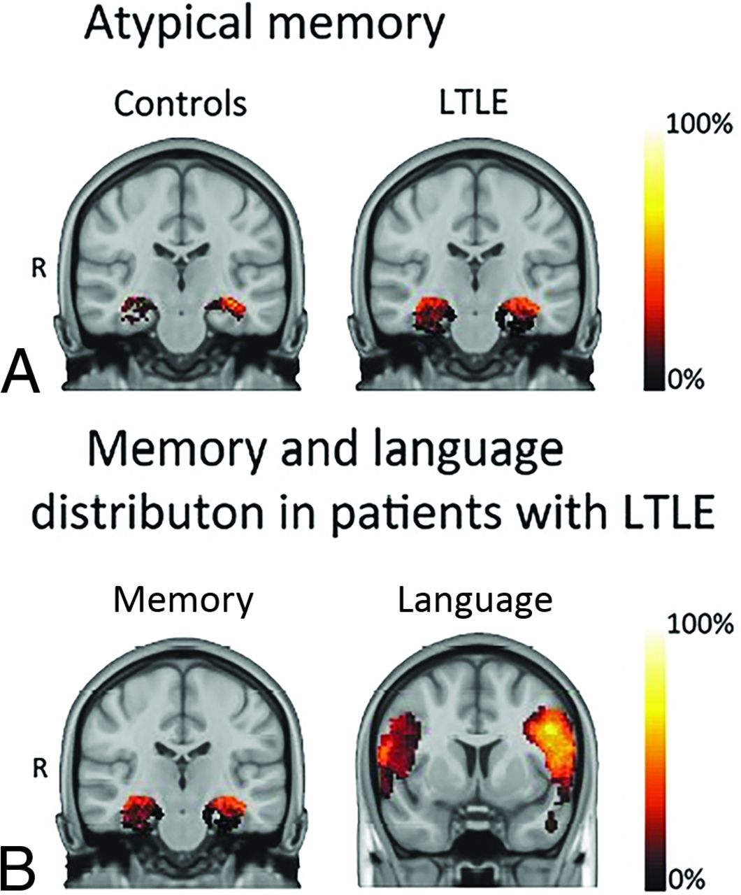
Typical verbal memory was seen in a higher proportion of HC (14/22, 63.6%), compared with patients (21/46, 45.6%). Atypical verbal memory in left TLE (16/37, 56.8%) was greater compared with right TLE (4/9, 44.4%). In patients with atypical verbal memory, the right LI was more frequent than bilateral LI. However, the proportion of bilateral LI was twice as high in patients with left TLE than in those with right TLE (Fig 2B and Fig 3A).

A, Atypical verbal memory was greater in patients with left TLE (56.8%) than in HC (36.4%). B, The verbal memory LI was bilateral (24.3%) more frequently than the language LI (7.7%) in patients with left TLE. The scale on the right indicates the percentage of the overlapping activation of the group (first-level analysis, P = .01), in which 100% represents a zone activated in the whole group. For visual purposes, the zones with activation in <5% of the group were considered noise and excluded. Warmer colors indicate higher overlapping, and darker colors, lesser overlapping. R indicates right.
fMRI Verbal Memory Lateralization Profile in Typical and Atypical Language.
Of 21 HC with typical language, 13/20 (65.0%) demonstrated typical memory lateralization. The verbal memory LI was obtained in 30/32 (93.7%) patients with typical language. Typical verbal memory was seen in 12/30 (40%) of them (Fig 3B). In patients with left TLE, atypical memory was more frequent (15/25; 60.0%). In patients with left TLE with atypical memory, the proportion of bilateral LIs (8/15, 53.3%) was higher than right LIs (7/15, 46.7%).
Only 2 HC showed atypical language. One of them had typical verbal memory, and the other showed atypical verbal memory (right LI). The verbal memory LI was obtained in 15/17 (88.2%) patients with atypical language. Typical verbal memory was seen in 8/15 (53.3%) of them. In patients with left TLE, 11/15 (73.3%), atypical memory was more frequent (6/11, 54.5%). In left TLE with atypical memory, the proportion of right LI (5/11, 45.5%) was higher than bilateral LI (1/11, 9%).
APi Distribution.
The APi was obtained in 19/22 (86.0%) HC and in 46/50 (92.0%) patients. Accounting for both hippocampi, the APi was seen in 20/22 (90.9%) HC and 43/46 (93.5%) patients. Both hippocampi showed activation in 35/40 (87.5%) patients with left TLE and in 8/10 (80%) with right TLE.
The distribution of the APi in both hippocampi according to HC or patients with left TLE and right TLE is shown in Fig 2B.
In the left hippocampus, the posterior APi was the most frequent pattern both in HC and those with left TLE, while posterior and whole activation was most frequently seen in those with right TLE with an equal proportion. In the right hippocampus, anterior activation was most frequently observed in those with right TLE compared with HC and those with left TLE (Fig 2B).
We evaluated potential associations of the anterior-posterior distribution and handedness, age at seizure onset, duration of epilepsy, and learning or delayed verbal recall, but no significant association was found (P > .05). We found a significant association between age and the APi, the older the patient the more anterior activation (β = 0.014, P = .03). The APi was significantly different in the left and right hippocampi of patients with severely impaired verbal learning, but not different in patients with moderate and slight impairment.
Association of Language and Verbal Memory fMRI with Neuropsychological Performance
Patients with atypical language scored −0.5 SD or below their age-matched peers in 12 (70.6%) cases, between −0.5 and +0.5 SDs in 4 (23.5%) cases, and +0.5 SD or above in 1 (5.9%) case.
The Online Supplemental Data show neuropsychological tests scores related to typical and atypical verbal memory according to HC and patients with left and right TLE. Immediate and delayed logical memory, visual reproduction, and naming scores (BNT) were significantly lower in patients with left TLE with typical verbal memory compared with HC. LMII and VMI were also significantly lower in patients with right TLE with typical verbal memory compared with HC. Patients with left TLE with atypical verbal memory showed a tendency toward higher scores in every subtest of the Wechsler Memory Scale, but significance was not achieved.
HC and patients with left TLE with a bilateral verbal memory LI showed a nonsignificant tendency toward higher scores in verbal memory encoding and retrieval compared with those participants who had more unilateral lateralization.
Patients with left TLE showed a nonsignificant tendency toward bilateral memory independent of verbal memory impairment (Fig 2C). No significant differences were seen in APis in memory impairment (Online Supplemental Data).
The proportion of patients with right-handedness was significantly lower in those with atypical language (χ = 11.40; Fisher exact test, P = .001), but it was not significantly different in patients with atypical memory (χ = 0.61; Fisher exact test, P = .568).
DISCUSSION
ATLR is the most established treatment for drug-refractory TLE.24 A surgical approach in refractory TLE has been associated with a 5-year increase in life expectancy.25 Prediction of cognitive decline after ATLR is essential to counsel patients regarding potential memory loss. To our knowledge, there is not a highly sensitive verbal learning paradigm standardized to use during memory fMRI. This prospective study focused on testing a verbal memory paradigm based on an established memory test to evaluate differences in memory LI and its association with neuropsychological impairment and language. The AVLT-adapted paradigm was able to elicit hippocampal activation in 100% of HC and 92% of patients. Our results suggest a distinctive redistribution of verbal memory and language systems in left TLE.
Verbal Memory Paradigm in Epilepsy
American and European task forces from the American College of Radiology and European Society of Neuroradiology have worked diligently to standardize the use of language fMRI in clinical practice.26 However, the nature of memory is complex, and activation depends on the task, paradigm design, data acquisition, and analysis. Tasks have varied across memory fMRI studies, leading to unsuccessful result replication. Thus, a prominent center such as UCL-Queen Square has used a task that involves making a judgment on whether each presented stimuli is pleasant, with an event-related analysis. We found that an AVLT-adapted paradigm based on word encoding or a recall task and analyzed with a blocked analysis elicited hippocampal activation. Event-related analysis was associated with a more reliable activation of anterior hippocampal activity.27 However, this approach is less powerful at detecting activation, more vulnerable to hemodynamic response function, time-consuming, and more demanding for patients and staff in a clinical setting.
Language and Verbal Memory Lateralization and Clinical Repercussions
Dominant TLE has been associated with extensive effects on the language systems. Patients have been associated with higher atypical language representation.28,29 In our study, atypical language occurred significantly more often in patients than in HC, similar to results in previous work (34.7% versus 9%).5 In left TLE, language was more lateralized than verbal memory, which was more bilateral. Patients with atypical language and slight verbal memory impairment showed a more frequent right verbal memory LI compared with a more bilateral LI in moderately and severely impaired patients. Memory and language systems seem to differ in their functional redistribution when affected by the epileptogenic network in TLE.30 Our findings agreed with the hypothesis that temporal pathology might first affect close areas, leading to more dysfunction during tasks specifically relying on them.31 Also, patients with TLE were associated with reduced flexibility and increased intra- and interregional communication involved in the cognitive task.32 These findings might suggest functional regulation to other regions capable of better meeting the current cognitive goal. Prior reports described a tendency of a left-to-right shift of language activation in left-hemisphere pathology.33,34 Atypical dominance in adults with left-hemispheric lesions was associated with both poorer35 and better cognitive abilities.29,36 We found that patients with left TLE and better memory scores showed a more atypical activation in comparison with those with right TLE and HC. Also, verbal learning and delayed recall were significantly lower in left TLE compared with right TLE and HC. Hand dominance did not significantly influence the memory LI compared to the language LI with the semantic and phonemic fluency tasks.
APi
A posterior hippocampal remnant might support postoperative memory in patients with TLE37 We found that half of the patients with left TLE showed predominant posterior hippocampal activation in both the left and right hippocampi. Prior work described distinct connections of hippocampal functionally. The anterior hippocampus is connected to the entorhinal cortex, temporal pole, and orbitofrontal cortex and is associated with verbal memory. The posterior hippocampus is linked to memory retrieval and polysynaptic-pathway propagation.38-41 The posterior remnant of the ipsilateral hippocampus, rather than the functional reserve of the contralateral hippocampus, was designated as the relevant structure for maintaining verbal memory function after ATLR.23,35,41 In addition, it was postulated that patients with left TLE with retained verbal memory function had an adaptive functional reorganization mediated by the right temporal lobe to keep higher memory scores.6,27,37,41 In our sample, patients with left TLE showed a higher proportion of whole activation in the right hippocampus compared with HC, which could be related to complex functional network plasticity with increased intrahemispheric connectivity that extends to the contralateral hippocampus. Additionally, patients with right TLE also showed a high proportion of whole activation and a higher proportion of posterior activation in the left compared with the right hippocampus. We found a tendency toward higher verbal learning scores in patients with a more posterior activation, which favors this hypothesis.
Neurophysiologic connectivity studies during a cognitive task found significant reductions of high-frequency oscillations rates in epileptic hippocampi.42 We hypothesize that a complete verbal memory transfer to the contralateral hippocampus might be associated with less severe memory impairment. This hypothesis could explain why some patients with TLE have memory impairment, while others do not. Complete transfer might increase the chance of preserving memory function after epilepsy surgery.
Limitations
Memory fMRI in the temporal lobe is prone to geometric distortions and blood oxygen level–dependent signal drop-out related to susceptibility of the echo-planar imaging sequence and to the field strength of the magnet, fMRI pulse sequence, and the level of cooperation and education of the participants.43 We used a block design approach to maximize signal changes. One limitation is the small number of patients with right TLE and that half of them were left-handed because scanning was based on clinical uncertainty of language distribution, which might bias results. Also, the acquisition time is relatively long for patients with cognitive decline and might cause fatigue that could influence performance. Additionally, all neuropsychological test scores were collected for patients but were available for a proportion of healthy controls. Other factors such as the amount of interictal activity on the day of testing or antiseizure medication might also have had an impact. This is a single-center study, moderate in size, and it only addresses patients with TLE. Larger databases of patients with epilepsy with temporal and extratemporal epilepsy with various pathologies would provide with a deeper knowledge.
CONCLUSIONS
The AVLT-adapted paradigm was able to elicit hippocampal activation. The laterality of a dysfunctional hippocampus in TLE induces specific processes of functional reorganization that affect verbal memory and language systems differently. Left TLE shows a higher proportion of bilateral LI in verbal memory compared with language, potentially secondary to reorganization in memory function, which happens more frequently than language function. That may lead to a discrepancy between language and memory laterality, which could potentially be relevant for predicting memory function after surgery. Right and especially bilateral memory activation patterns are much more frequent in patients with left TLE compared with HC and are generally associated with better memory function. Future studies will clarify whether this paradigm could be used to predict memory function after surgery.
Acknowledgments
The authors would also like to thank the patients and their caregivers for their contribution.
Footnotes
This study was supported by project PI19/00348, financed by the Instituto de Salud Carlos III and the European Regional Development Fund. This research did not receive any specific grant from commercial or not-for-profit sectors. E.C.-B. was supported by the fellowship grant RH041910.
Disclosure forms provided by the authors are available with the full text and PDF of this article at www.ajnr.org.
Indicates open access to non-subscribers at www.ajnr.org
References
- Received March 6, 2022.
- Accepted after revision July 1, 2022.
- © 2022 by American Journal of Neuroradiology


Jump to section
Related Articles
Cited By...
- No citing articles found.