Advertisement

Main menu

  • Home
  • Content
    • Current Issue
    • Accepted Manuscripts
    • Article Preview
    • Past Issue Archive
    • AJNR Case Collection
    • Case of the Week Archive
    • Classic Case Archive
    • Case of the Month Archive
  • Special Collections
    • Spinal CSF Leak Articles (Jan 2020-June 2024)
    • 2024 AJNR Journal Awards
    • Most Impactful AJNR Articles
  • Multimedia
    • AJNR Podcast
    • AJNR Scantastics
    • Video Articles
  • For Authors
    • Submit a Manuscript
    • Author Policies
    • Fast publishing of Accepted Manuscripts
    • Graphical Abstract Preparation
    • Manuscript Submission Guidelines
    • Imaging Protocol Submission
    • Submit a Case for the Case Collection
  • About Us
    • About AJNR
    • Editorial Board
  • More
    • Become a Reviewer/Academy of Reviewers
    • Subscribers
    • Permissions
    • Alerts
    • Feedback
    • Advertisers
    • ASNR Home
  • Other Publications
    • ajnr

User menu

  • Alerts
  • Log in

Search

  • Advanced search
American Journal of Neuroradiology

American Journal of Neuroradiology

ASHNR American Society of Functional Neuroradiology ASHNR American Society of Pediatric Neuroradiology ASSR
  • Alerts
  • Log in

Advanced Search

  • Home
  • Content
    • Current Issue
    • Accepted Manuscripts
    • Article Preview
    • Past Issue Archive
    • AJNR Case Collection
    • Case of the Week Archive
    • Classic Case Archive
    • Case of the Month Archive
  • Special Collections
    • Spinal CSF Leak Articles (Jan 2020-June 2024)
    • 2024 AJNR Journal Awards
    • Most Impactful AJNR Articles
  • Multimedia
    • AJNR Podcast
    • AJNR Scantastics
    • Video Articles
  • For Authors
    • Submit a Manuscript
    • Author Policies
    • Fast publishing of Accepted Manuscripts
    • Graphical Abstract Preparation
    • Manuscript Submission Guidelines
    • Imaging Protocol Submission
    • Submit a Case for the Case Collection
  • About Us
    • About AJNR
    • Editorial Board
  • More
    • Become a Reviewer/Academy of Reviewers
    • Subscribers
    • Permissions
    • Alerts
    • Feedback
    • Advertisers
    • ASNR Home
  • Follow AJNR on Twitter
  • Visit AJNR on Facebook
  • Follow AJNR on Instagram
  • Join AJNR on LinkedIn
  • RSS Feeds

Welcome to the new AJNR, Updated Hall of Fame, and more. Read the full announcements.


AJNR is seeking candidates for the position of Associate Section Editor, AJNR Case Collection. Read the full announcement.


Research ArticleInterventional

Successful Reperfusion is Associated with Favorable Functional Outcome despite Vessel Perforation during Thrombectomy: A Case Series and Systematic Review

C. Ducroux, W. Boisseau, A.Y. Poppe, N. Daneault, Y. Deschaintre, J.D.B. Diestro, J. Eneling, L.C. Gioia, D. Iancu, B. Maier, B. Nauche, L. Nico, C. Odier, J. Raymond, D. Roy, C. Stapf, A. Weill and G. Jacquin
American Journal of Neuroradiology November 2022, 43 (11) 1633-1638; DOI: https://doi.org/10.3174/ajnr.A7650
C. Ducroux
aFrom the Department of Neurosciences (C.D., A.Y.P., N.D., Y.D. L.C.G., C.O., C.S., G.J.), Faculté de médecine, Université de Montréal, Montréal, Quebec. Canada
bDepartments of Medicine (Neurology) (C.D., A.Y.P., N.D., Y.D., L.C.G., C.O., C.S., G.J.)
eDivision of Neurology (C.D.), Department of Medicine, The Ottawa Hospital, Ottawa, Ontario, Canada
  • Find this author on Google Scholar
  • Find this author on PubMed
  • Search for this author on this site
  • ORCID record for C. Ducroux
W. Boisseau
cRadiology (W.B., J.D.B.D., J.E., D.I., B.M., L.N., J.R., D.R., A.W.)
gDepartment of Interventional Neuroradiology (W.B., B.M.), Hopital Fondation A. De Rothschild, Paris, France
  • Find this author on Google Scholar
  • Find this author on PubMed
  • Search for this author on this site
  • ORCID record for W. Boisseau
A.Y. Poppe
aFrom the Department of Neurosciences (C.D., A.Y.P., N.D., Y.D. L.C.G., C.O., C.S., G.J.), Faculté de médecine, Université de Montréal, Montréal, Quebec. Canada
bDepartments of Medicine (Neurology) (C.D., A.Y.P., N.D., Y.D., L.C.G., C.O., C.S., G.J.)
dNeurovascular Group (A.Y.P., N.D., Y.D., L.C.G., C.O., C.S., G.J.), Neurosciences Axis, Centre de Recherche du Centre Hospitalier de l’Université de Montréal, Montréal, Quebec, Canada
  • Find this author on Google Scholar
  • Find this author on PubMed
  • Search for this author on this site
  • ORCID record for A.Y. Poppe
N. Daneault
aFrom the Department of Neurosciences (C.D., A.Y.P., N.D., Y.D. L.C.G., C.O., C.S., G.J.), Faculté de médecine, Université de Montréal, Montréal, Quebec. Canada
bDepartments of Medicine (Neurology) (C.D., A.Y.P., N.D., Y.D., L.C.G., C.O., C.S., G.J.)
dNeurovascular Group (A.Y.P., N.D., Y.D., L.C.G., C.O., C.S., G.J.), Neurosciences Axis, Centre de Recherche du Centre Hospitalier de l’Université de Montréal, Montréal, Quebec, Canada
  • Find this author on Google Scholar
  • Find this author on PubMed
  • Search for this author on this site
Y. Deschaintre
aFrom the Department of Neurosciences (C.D., A.Y.P., N.D., Y.D. L.C.G., C.O., C.S., G.J.), Faculté de médecine, Université de Montréal, Montréal, Quebec. Canada
bDepartments of Medicine (Neurology) (C.D., A.Y.P., N.D., Y.D., L.C.G., C.O., C.S., G.J.)
dNeurovascular Group (A.Y.P., N.D., Y.D., L.C.G., C.O., C.S., G.J.), Neurosciences Axis, Centre de Recherche du Centre Hospitalier de l’Université de Montréal, Montréal, Quebec, Canada
  • Find this author on Google Scholar
  • Find this author on PubMed
  • Search for this author on this site
  • ORCID record for Y. Deschaintre
J.D.B. Diestro
cRadiology (W.B., J.D.B.D., J.E., D.I., B.M., L.N., J.R., D.R., A.W.)
  • Find this author on Google Scholar
  • Find this author on PubMed
  • Search for this author on this site
  • ORCID record for J.D.B. Diestro
J. Eneling
cRadiology (W.B., J.D.B.D., J.E., D.I., B.M., L.N., J.R., D.R., A.W.)
  • Find this author on Google Scholar
  • Find this author on PubMed
  • Search for this author on this site
L.C. Gioia
aFrom the Department of Neurosciences (C.D., A.Y.P., N.D., Y.D. L.C.G., C.O., C.S., G.J.), Faculté de médecine, Université de Montréal, Montréal, Quebec. Canada
bDepartments of Medicine (Neurology) (C.D., A.Y.P., N.D., Y.D., L.C.G., C.O., C.S., G.J.)
dNeurovascular Group (A.Y.P., N.D., Y.D., L.C.G., C.O., C.S., G.J.), Neurosciences Axis, Centre de Recherche du Centre Hospitalier de l’Université de Montréal, Montréal, Quebec, Canada
  • Find this author on Google Scholar
  • Find this author on PubMed
  • Search for this author on this site
  • ORCID record for L.C. Gioia
D. Iancu
cRadiology (W.B., J.D.B.D., J.E., D.I., B.M., L.N., J.R., D.R., A.W.)
  • Find this author on Google Scholar
  • Find this author on PubMed
  • Search for this author on this site
  • ORCID record for D. Iancu
B. Maier
cRadiology (W.B., J.D.B.D., J.E., D.I., B.M., L.N., J.R., D.R., A.W.)
gDepartment of Interventional Neuroradiology (W.B., B.M.), Hopital Fondation A. De Rothschild, Paris, France
hUniversité Paris-Cité (B.M.), Paris, France
  • Find this author on Google Scholar
  • Find this author on PubMed
  • Search for this author on this site
  • ORCID record for B. Maier
B. Nauche
fBibliothèque du Centre Hospitalier de l’Université de Montréal (B.N.), Montreal, Quebec, Canada
  • Find this author on Google Scholar
  • Find this author on PubMed
  • Search for this author on this site
  • ORCID record for B. Nauche
L. Nico
cRadiology (W.B., J.D.B.D., J.E., D.I., B.M., L.N., J.R., D.R., A.W.)
  • Find this author on Google Scholar
  • Find this author on PubMed
  • Search for this author on this site
  • ORCID record for L. Nico
C. Odier
aFrom the Department of Neurosciences (C.D., A.Y.P., N.D., Y.D. L.C.G., C.O., C.S., G.J.), Faculté de médecine, Université de Montréal, Montréal, Quebec. Canada
bDepartments of Medicine (Neurology) (C.D., A.Y.P., N.D., Y.D., L.C.G., C.O., C.S., G.J.)
dNeurovascular Group (A.Y.P., N.D., Y.D., L.C.G., C.O., C.S., G.J.), Neurosciences Axis, Centre de Recherche du Centre Hospitalier de l’Université de Montréal, Montréal, Quebec, Canada
  • Find this author on Google Scholar
  • Find this author on PubMed
  • Search for this author on this site
J. Raymond
cRadiology (W.B., J.D.B.D., J.E., D.I., B.M., L.N., J.R., D.R., A.W.)
  • Find this author on Google Scholar
  • Find this author on PubMed
  • Search for this author on this site
  • ORCID record for J. Raymond
D. Roy
cRadiology (W.B., J.D.B.D., J.E., D.I., B.M., L.N., J.R., D.R., A.W.)
  • Find this author on Google Scholar
  • Find this author on PubMed
  • Search for this author on this site
  • ORCID record for D. Roy
C. Stapf
aFrom the Department of Neurosciences (C.D., A.Y.P., N.D., Y.D. L.C.G., C.O., C.S., G.J.), Faculté de médecine, Université de Montréal, Montréal, Quebec. Canada
bDepartments of Medicine (Neurology) (C.D., A.Y.P., N.D., Y.D., L.C.G., C.O., C.S., G.J.)
dNeurovascular Group (A.Y.P., N.D., Y.D., L.C.G., C.O., C.S., G.J.), Neurosciences Axis, Centre de Recherche du Centre Hospitalier de l’Université de Montréal, Montréal, Quebec, Canada
  • Find this author on Google Scholar
  • Find this author on PubMed
  • Search for this author on this site
  • ORCID record for C. Stapf
A. Weill
cRadiology (W.B., J.D.B.D., J.E., D.I., B.M., L.N., J.R., D.R., A.W.)
  • Find this author on Google Scholar
  • Find this author on PubMed
  • Search for this author on this site
  • ORCID record for A. Weill
G. Jacquin
aFrom the Department of Neurosciences (C.D., A.Y.P., N.D., Y.D. L.C.G., C.O., C.S., G.J.), Faculté de médecine, Université de Montréal, Montréal, Quebec. Canada
bDepartments of Medicine (Neurology) (C.D., A.Y.P., N.D., Y.D., L.C.G., C.O., C.S., G.J.)
dNeurovascular Group (A.Y.P., N.D., Y.D., L.C.G., C.O., C.S., G.J.), Neurosciences Axis, Centre de Recherche du Centre Hospitalier de l’Université de Montréal, Montréal, Quebec, Canada
  • Find this author on Google Scholar
  • Find this author on PubMed
  • Search for this author on this site
  • ORCID record for G. Jacquin
  • Article
  • Figures & Data
  • Supplemental
  • Info & Metrics
  • Responses
  • References
  • PDF
Loading

Abstract

BACKGROUND: Arterial perforation is a potentially serious complication during endovascular thrombectomy.

PURPOSE: Our aim was to describe interventional approaches after arterial perforation during endovascular thrombectomy and to determine whether reperfusion remains associated with favorable outcome despite this complication.

DATA SOURCES: Data from consecutive patients with acute stroke undergoing endovascular thrombectomy were retrospectively collected between 2015 to 2020 from a single-center cohort, and a systematic review was performed using PubMed, EMBASE, and Ovid MEDLINE up to June 2020.

STUDY SELECTION: Articles reporting functional outcome after arterial perforation during endovascular thrombectomy were selected.

DATA ANALYSIS: Functional outcomes of patients achieving successful reperfusion (TICI 2b/3) were compared with outcomes of those with unsuccessful reperfusion in our single-center cohort. We then summarized the literature review to describe interventional approaches and outcomes after arterial perforation during endovascular thrombectomy.

DATA SYNTHESIS: In our single-center cohort, 1419 patients underwent endovascular thrombectomy, among whom 32 (2.3%) had vessel perforation and were included in the analysis. The most common hemostatic strategy was watchful waiting (71% of cases). Patients with successful reperfusion had a higher proportion of favorable 90-day mRS scores (60% versus 12.5%; P = .006) and a lower mortality rate (13.3% versus 56.3%, P = .01) than patients without successful reperfusion. Thirteen articles were included in the systematic review. Successful reperfusion also appeared to be associated with better outcomes.

LIMITATIONS: Given the low number of published reports, we performed only a descriptive analysis.

CONCLUSIONS: Arterial perforation during endovascular thrombectomy is rare but is associated with high mortality rates and poor outcome. However, successful reperfusion remains correlated with favorable outcome in these patients.

ABBREVIATIONS:

EVT
endovascular thrombectomy
IQR
interquartile range
mTICI
modified TICI
sICH
symptomatic intracerebral hemorrhage

Endovascular therapy (EVT) is the standard of care for the treatment of patients with acute ischemic stroke due to large-vessel occlusion.1 Technologic advances have allowed development of new devices that have greatly improved the efficacy and safety of EVT compared with older-generation tools.2 Despite these improvements, as well as greater comfort and expertise among interventionalists across time, the procedure is still not exempt from severe complications.3,4 Arterial perforation, defined as contrast extravasation noticed during a procedural angiographic run, is one of the most serious and feared complications during EVT because it has been associated with poor functional outcomes and death.5 In large EVT trials, the rate of procedural arterial perforations varied between 0.6% and 4.9%.1

When a vessel perforation occurs, some operators might be tempted to abort the procedure, with the risk of leaving the culprit occlusion untreated. Others may decide to pursue EVT once hemostasis is achieved, risking further arterial wall damage. However, little is known regarding the best approach when facing an arterial perforation during EVT and whether reperfusion still represents a reasonable goal in the context of such a complication.5

Therefore, we aimed to describe rescue therapies, management, and clinical outcomes after intraprocedural arterial perforation during EVT, and we sought to determine whether reperfusion is still associated with favorable outcome in this setting.

MATERIALS AND METHODS

This single-center observational study retrospectively analyzed clinical and imaging data prospectively gathered from a quality-improvement database: the Montreal Neurovascular and STrokE Repository (MONSTER), maintained by a high-volume comprehensive stroke center in Montreal, Canada (Center Hospitalier de l’Université de Montréal).

Patient Population

All consecutive adult patients (18 years of age or older) treated with EVT for a large-vessel occlusion between March 1, 2015, and December 31, 2020, in whom arterial perforation occurred during EVT were included. Patients were included when an arterial perforation, defined as contrast extravasation noticed during a procedural angiographic run, was reported in radiology reports or clinical notes. Baseline demographics, stroke characteristics, and procedural details were collected, including information regarding the procedural technique, location of arterial occlusion, type of devices used (stent retriever, contact aspiration, and combined technique), number of passes, procedural duration, and the adopted rescue therapy after vessel perforation. All angiograms were reviewed by 2 interventional neuroradiologists (D.R. and B.M.). We then performed a descriptive analysis of the main steps undertaken after arterial perforation was observed.

Outcomes

Patients were divided in 2 groups with either successful reperfusion or unsuccessful reperfusion to correlate the angiographic results with functional outcomes. Successful reperfusion was defined as a modified TICI (mTICI) ≥ 2b on the final intracranial run. Favorable functional outcome was defined as an mRS score of 0–2, as determined by a certified assessor at 90-day telephone or in-person follow-up. Proportions of hemorrhagic transformation, symptomatic intracerebral hemorrhage (sICH, defined as any hemorrhagic transformation associated with a 4-point increase in the baseline NIHSS score6), and death at 90 days were also collected. All patients had immediate brain imaging following the end of their procedure (plain CT or DynaCT [Siemens] in the angio suite) and a 24-hour follow-up CT. The 24-hour CT was used to identify hemorrhagic transformation to better distinguish true hemorrhagic transformation from contrast staining.

Systematic Review of the Literature

We also performed a systematic review to identify articles reporting on outcomes of patients with acute ischemic stroke with arterial perforation during EVT. With the help of a medical librarian having expertise in systematic reviews (B.N.), we searched the PubMed database, EMBASE database, and Ovid MEDLINE with search terms including “perforation,” “complication,” “stent retriever,” “contact aspiration,” “thrombectomy,” and “stroke” (see the Online Supplemental Data for the full search strategy). We included any study published between January 1, 2015, and June 30, 2020, specifically describing functional outcomes of patients who had arterial perforation observed during EVT. This date range was used to capture studies primarily completed in the modern EVT era. We selected only the references that met all of the following criteria: full-text articles in French or English, studies reporting 3-month clinical outcomes using the mRS score, and studies of patients with stroke undergoing EVT with a procedural arterial perforation. Using a free Web-based application,7 two reviewers (C.D., G.J.) independently screened all titles and abstracts. Full texts were obtained for any article considered relevant for the research question. Disagreements about eligibility were resolved by consensus. Data collection was completed by one author (C.D.) and reviewed by a second (G.J.). Collected data for each study included, when available, the following: baseline NIHSS score, thrombus location, ASPECTS, intravenous thrombolysis, type of anesthesia, EVT technique (stent retriever, contact aspiration, and a combined technique), mTICI score, causes of perforation, sICH, mRS score, and death at 90-day follow-up. The review was performed according to the Preferred Reporting Items for Systematic Reviews and Meta-Analysis (PRISMA) guidelines.8

Statistics

Differences between groups were tested using the χ2 test of independence or the Fisher exact test for categoric variables and the Mann-Whitney U test for continuous variables. We described the population according to reperfusion status. Reperfusion status was dichotomized as either successful reperfusion (mTICI ≥ 2b) or unsuccessful reperfusion (mTICI < 2b). The statistical level of significance was set at P < .05. Data were analyzed using SPSS (Version 26.0.0.1; IBM). Given the expected low number of previously published reports, we did not plan to perform a statistical meta-analysis of the systematic review results and only performed a descriptive analysis.

The study was approved by our local institutional ethics board for retrospective data collection and review (project No. 20.028). The data supporting the findings of this study are available on reasonable request by a qualified investigator to the corresponding author.

RESULTS

From March 1, 2015, to December 31, 2020, a total of 1419 patients underwent EVT for acute ischemic stroke in our comprehensive stroke center, among whom 32 (2.3%) experienced a vessel perforation during EVT and were included in the study. The annual proportion of EVT procedures complicated by a perforation appeared to decrease during the study period (Online Supplemental Data). Follow-up at 90 days was unavailable for 1 (3%) patient. Baseline demographics and stroke characteristics are shown in the Online Supplemental Data.

Procedural Details

We extracted data from radiology reports and review of source angiographic images, though only 29 complete angiographic studies were available for analysis.

Endovascular thrombectomy was performed with the patient under general anesthesia in 5 patients (15.6%) (including 2 cases with conversion to general anesthesia after arterial perforation), under conscious sedation in 12 patients (37.5%), and under local anesthesia only in the remaining 15 patients (46.9%). The median number of device passes was 1 (interquartile range [IQR], 0–1.75). Vessel perforation occurred more often with the use of a microwire (n = 14, 43.8%) than with a microcatheter (n = 9, 28.1%). The cause of vessel perforation was unknown in the remaining 9 patients (28.1%) (Online Supplemental Data.). Patients with perforation due to a microwire had numerically better 3-month outcomes than patients with microcatheter perforation (6/14 patients, 46.2%, versus 3/9 patients, 33.3%, respectively).

After review of available angiograms, we found that 16 patients had a perforation identified only on microcatheter injection (16/29; 55.2%), 13 patients (44.8%) had a perforation noticed after ICA injection, and 13 patients (44.8%) had a perforation viewed on multiple runs. Outcomes of these patients were similar regardless of how perforation was identified or the number of runs on which the perforation was viewed (Online Supplemental Data).

Most intraprocedural vessel perforations occurred at a distal location: the MCA M2 in 18 patients (56.3%) and the anterior cerebral artery in 3 patients (9.4%). The remainder occurred proximally: the MCA M1 in 7 patients (21.9%) and the ICA in 2 patients (6.3%). Among patients with proximal perforation (ICA and M1), rescue therapy was performed in 4/9 patients (intermittent balloon inflation in 2 patients and parent vessel occlusion in 1 patient). In the last case, the microcatheter was left in place and cut and sewn at the groin in an attempt to seal the intracranial arterial perforation point. Contrast extravasation stopped spontaneously in the 5 remaining cases (Fig 2). Among patients with distal perforation, contrast extravasation was self-resolving in most (17/22 patients), parent vessel occlusion was performed in 4 cases, and intermittent balloon inflation was performed in the last case. “Self-resolving” is defined as contrast extravasation stopping without a specific intervention. The median number of runs before concluding that a bleed was self-resolving was 1 (IQR, 1–2).

Outcomes and Association with Reperfusion

Of 31 patients with an available 90-day mRS, 11 (34.4%) patients had favorable outcomes and 11 (34.4%) patients died (5 sICH, 3 cases of malignant edema, 2 cases of aspiration pneumonia, and 1 from multiple causes).

Reperfusion (mTICI 2b/3) was successful in 16 (50%) patients and unsuccessful in 16 (50%) others, with baseline characteristics being similar in these 2 groups (Online Supplemental Data).

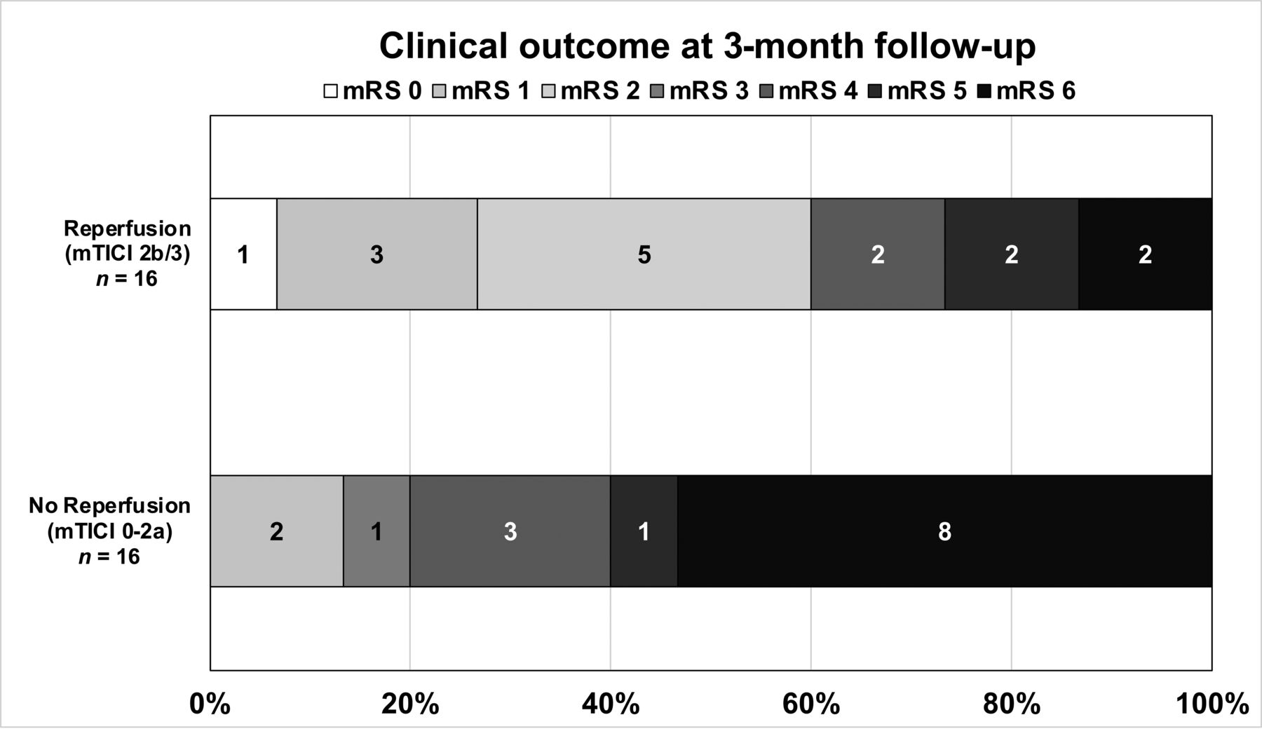
As shown in Fig 1, patients with successful reperfusion had a higher proportion of favorable functional outcomes at 90 days than patients without successful reperfusion (60% versus 12.5%, P = .006) as well significantly lower mortality rates (13.3% versus 56.3%, P = .01) (Table).

FIG 1.
  • Download figure
  • Open in new tab
  • Download powerpoint
FIG 1.

The mRS score distribution at 90 days for patients with successful reperfusion (TICI 2b/3) and no reperfusion (TICI 0-2a), despite vessel perforation during endovascular thrombectomy.

FIG 2.
  • Download figure
  • Open in new tab
  • Download powerpoint
FIG 2.

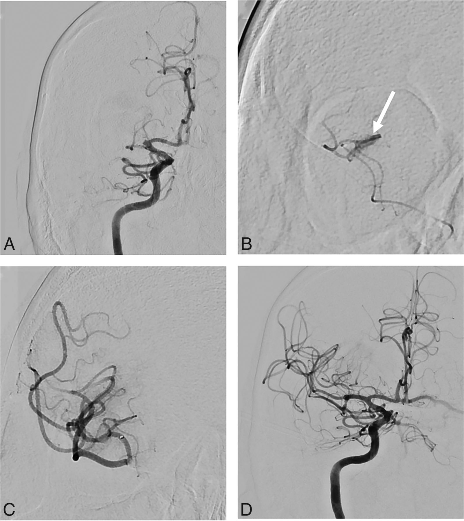
Illustrative case. A 69-year-old man with a proximal right MCA occlusion (A). B, After catheterization of M1, contrast extravasation (white arrow) was identified on microcatheter injection. C, After 10 minutes, contrast extravasation stopped spontaneously. D, Thrombectomy was pursued without further complications, and successful reperfusion was achieved. The patient had a favorable outcome at 3 months (mRS 1).

View this table:
  • View inline
  • View popup

Outcomes of patients with procedural perforation with successful reperfusion (mTICI ≥ 2b/3) and with unsuccessful reperfusion (mTICI < 2b/3)a

Among patients with successful reperfusion, vessel perforation occurred before achieving reperfusion in 13 cases (13/16, 81.3%) and after achieving reperfusion in 3 cases (18.7%). In these last 3 cases, a secondary medium vessel occlusion (A2 and M3) was targeted and recanalized, despite a reperfusion status of ≥TICI 2b.

Among patients with unsuccessful reperfusion, the procedure was aborted after vessel perforation in 5 patients, while in the remaining 11 cases, the procedure was stopped because of failure to achieve successful recanalization. Among these patients, 9 had unsuccessful thrombectomy despite several passes and 2 patients had persistent distal emboli.

Patients with self-resolving bleeding did not have a greater proportion of favorable outcomes than patients needing hemostatic intervention (14/23 patients [60.9%] versus 6/9 patients [66.7%]).

Intracerebral Hemorrhage

The median delay between the groin puncture and control imaging was 18 hours (IQR, 13–24 hours). One patient died within 24 hours before any follow-up imaging was performed. All patients had an SAH on their 24-hour CT. Intraparenchymal hemorrhage of any type occurred in 14 patients (37.5%), of whom 6 (18.8%) had sICH (Table). Among patients with sICH, 4 patients had a distal perforation (distal M2, M3, or A2) and 2 patients had a proximal perforation (M1 segment).

Systematic Review

The initial literature search yielded 6052 articles. After all titles and abstracts were screened by C.D. and G.J., 5969 articles were considered irrelevant. Of the remaining 83 articles, 70 were excluded for incorrect outcome assessments, wrong population, wrong study design, and wrong publication type (abstract only, language other than French or English), leaving a total of 13 studies eligible for analysis (n = 37 patients; Online Supplemental Data: PRISMA diagram). Eight studies were cohort studies, and 5 were case reports (Online Supplemental Data). The median baseline NIHSS score was 14.5 (IQR, 11.75–19). A stent retriever was used in most cases (28/37 cases); contact aspiration, in 13/37 cases; and combined therapy, in 1 case. Nine patients had multiples passes using different techniques. Vessel perforation occurred more often in distal branches (16/37 cases; 43%), while 14 patients (38%) had proximal vessel perforation, and the exact location was not detailed in the remainder.

In most cases (23/37, 62%), the cause of vessel perforation was not described. For those in whom the cause was mentioned, 9 patients (24%) had vessel perforation due to a microwire; 4 patients (11%), due to a microcatheter; and 1 patient (0.03%), due to a strong contrast injection. Different hemostatic strategies were described for 26 patients: 1) procedural abandonment without any additional maneuver despite a persistent intracranial occlusion (7 cases); 2) inflation of an intracranial balloon (8 cases); 3) parent vessel occlusion using coils or glue (6 cases); or 4) watchful waiting (no intervention performed because of spontaneous regression of bleeding) in 2 cases. In 1 case, despite inflation of an intracranial balloon catheter, bleeding persisted, and therapeutic occlusion using coils was performed.

In all studies included in this literature review, the functional outcome at 3 months was available for 35 of 37 patients. The outcomes were poor (median mRS, 6 [IQR, 2.5–6]): Only 9 patients (9/35, 26%) had a favorable outcome (mRS, 0–2) at 3 months, and mortality was high (20/35 cases, 57%). Both reperfusion status and outcome at 3 months were available for 34 patients. Although formal statistical analysis was not possible due to an overall paucity of data in the published reports, the median mRS appeared slightly better in patients with successful reperfusion (median mRS, 5; IQR, 2–6, versus 6; IQR, 4–6) as did mortality, which was numerically lower in these patients (7/14, 50%, versus 13/20, 65%).

DISCUSSION

Our findings demonstrate that vessel perforation in the context of EVT for acute stroke is infrequent but may result in a high rate of mortality and poor functional outcome. In our series, the proportion of patients having this complication was similar to that reported in recent EVT randomized trials (0.6%–4%), registries,1,4 and a previous cohort study by Mokin et al5 (1%). However, our results suggest that patients might, nevertheless, do better despite perforation if successful reperfusion is achieved.

Vessel perforation is arguably one of the most serious and feared complications during EVT and is associated with high rates of mortality.3⇓-5,9,10 The risk of vessel perforation is increased during ‘‘blind maneuvering’’ while trying to gain access to occluded intracranial vessels with a microwire or microcatheter11⇓⇓⇓⇓⇓-17 and while withdrawing a stent retriever.5,18,19

Vessel perforation tends to involve more distal vessel segments: 65% of cases in our study and 61% in our literature review. This finding is particularly relevant because the field is moving toward more aggressive recanalization of distal vessel occlusions,20 with upcoming trials evaluating the role of EVT in such patients (NCT05029414). However, the clinical impact of distal perforation might be less severe than that of proximal perforation (ICA or M1). In our cohort, rescue therapy was performed more often in patients with proximal perforation, but outcomes were similar regardless of perforation site.

It is usually thought that intraprocedural vessel perforation requires immediate action to achieve hemostatic control, such as blood pressure reduction and/or the interruption or reversal of any ongoing thrombolytic agent or blood thinners,10 temporary intracranial balloon occlusion,5,16 or parent vessel occlusion by injection of either liquid embolic agents16,21 or detachable coils.10,22 These maneuvers were used in most patients found in our systematic review, other than cases in which the procedure was simply aborted prematurely. However, in our cohort, most cases of contrast extravasation resolved spontaneously (71.9%) after the device or microcatheter was withdrawn. In some cases, the offending thrombus itself may provide effective hemostasis when a perforation occurs in the vasculature distal to the occlusion. On the other hand, parent vessel occlusion may potentially result in major stroke with severe disability and could increase the risk of poor clinical outcome and mortality.3,22,23

In our series, as well as in previously published reports, procedural perforations during EVT were associated with overall high mortality rates and low rates of good clinical outcome.5,24 Compared with outcomes found in the major EVT trials1 and in most large EVT registries,25 functional outcomes of patients with vessel perforation remain relatively poor. However, such poor clinical outcomes might be due to the absence of reperfusion26 rather than cerebral hemorrhage. The decision to resume or abort EVT should be based on stroke severity, hemostatic control, technical difficulty, and the safety of pursuing thrombectomy, while always being aware that successful reperfusion appears to be associated with better outcomes and lower mortality in this population.5,27

There are several suspected risk factors for vessel perforation, notably a distal occlusion, the presence of arterial tortuosity, and intracranial atherosclerosis.18 Moreover, some clot types can be more difficult to cross with a microcatheter and microwire, thus increasing the risk of traumatic vessel injury and perforation.5,23 Contact aspiration does not always require crossing the clot with a microcatheter and might intuitively seem safer than stent retrievers in this regard. However, a randomized trial comparing both techniques did not show a significant difference in vessel perforation.28 Some authors have suggested that crossing the clot with a wireless microcatheter may reduce the risk of vessel perforation.29

In our cohort, functional outcome and mortality at 3 months were better than in the literature review. Most studies reporting procedural complications, including vessel perforation, described only severe cases with poor outcomes. Conversely, in our study, we reported all cases of perforation, which could be more representative of real life than smaller series or case reports. Since the widespread adoption of EVT following the major randomized trials,1 the tools and techniques available have continuously improved,2 and in our center, the rate of vessel perforation decreased from 3% in 2015 to 1.6% in 2020.

Despite being a larger cohort than those in previously published reports, our study has several limitations, including its single-center, retrospective design, a relatively small sample size, and the heterogeneity in the management of complications. This last limitation reflects the known variety of practices among neurointerventionalists and stroke physicians regarding the management of vessel perforation during EVT.3,5 Because this complication is, fortunately, rare, the limited number of cases precluded the use of statistical analysis to reliably determine which clinical or technical variables were associated with favorable outcome. We did not assign degrees of severity to the perforations, given the lack of a standardized angiographic definition and because procedural imaging of the perforation varied widely among cases. Finally, the 90-day mRS was assessed by raters who were not necessarily blinded to perforation status, and although performed by certified assessors, the mRS is known to have only fair-to-moderate interobserver reliability.30

CONCLUSIONS

Procedural vessel perforation during EVT is a rare-but-serious complication, associated with high mortality and poor clinical outcome. However, successful reperfusion remains correlated with favorable outcome in these patients and can be pursued when technically safe and feasible.

Footnotes

  • Disclosure forms provided by the authors are available with the full text and PDF of this article at www.ajnr.org.

References

  1. 1.↵
    1. Goyal M,
    2. Menon BK,
    3. van Zwam WH, et al
    . Endovascular thrombectomy after large-vessel ischaemic stroke: a meta-analysis of individual patient data from five randomised trials. Lancet 2016;387:1723–31 doi:10.1016/S0140-6736(16)00163-X pmid:26898852
    CrossRefPubMed
  2. 2.↵
    1. Yoo AJ,
    2. Andersson T
    . Thrombectomy in acute ischemic stroke: challenges to procedural success. J Stroke 2017;19:121–30 doi:10.5853/jos.2017.00752 pmid:28592779
    CrossRefPubMed
  3. 3.↵
    1. Balami JS,
    2. White PM,
    3. McMeekin PJ, et al
    . Complications of endovascular treatment for acute ischemic stroke: prevention and management. Int J Stroke 2018;13:348–61 doi:10.1177/1747493017743051 pmid:29171362
    CrossRefPubMed
  4. 4.↵
    1. Happi Ngankou E,
    2. Gory B,
    3. Marnat G, et al
    . Thrombectomy complications in large vessel occlusions: incidence, predictors, and clinical impact in the ETIS Registry. Stroke 2021;52:e764–68 doi:10.1161/STROKEAHA.121.034865 pmid:34706564
    CrossRefPubMed
  5. 5.↵
    1. Mokin M,
    2. Fargen KM,
    3. Primiani CT, et al
    . Vessel perforation during stent retriever thrombectomy for acute ischemic stroke: technical details and clinical outcomes. J Neurointerv Surg 2017;9:922–28 doi:10.1136/neurintsurg-2016-012707 pmid:27688267
    Abstract/FREE Full Text
  6. 6.↵
    1. von Kummer R,
    2. Broderick JP,
    3. Campbell BC, et al
    . The Heidelberg Bleeding Classification: classification of bleeding events after ischemic stroke and reperfusion therapy. Stroke 2015;46:2981–86 doi:10.1161/STROKEAHA.115.010049 pmid:26330447
    FREE Full Text
  7. 7.↵
    1. Ouzzani M,
    2. Hammady H,
    3. Fedorowicz Z, et al
    . Rayyan: a web and mobile app for systematic reviews. Syst Rev 2016;5:210 doi:10.1186/s13643-016-0384-4 pmid:27919275
    CrossRefPubMed
  8. 8.↵
    1. Moher D,
    2. Liberati A,
    3. Tetzlaff J, et al
    ; PRISMA Group. Preferred reporting items for systematic reviews and meta-analyses: the PRISMA statement. Int J Surg 2010;8:336–41 doi:10.1016/j.ijsu.2010.02.007 pmid:20171303
    CrossRefPubMed
  9. 9.↵
    1. Nguyen TN,
    2. Lanthier S,
    3. Roy D
    . Iatrogenic arterial perforation during acute stroke interventions. AJNR Am J Neuroradiol 2008;29:974–75 doi:10.3174/ajnr.A0958 pmid:18258704
    CrossRefPubMed
  10. 10.↵
    1. Akpinar SH,
    2. Yilmaz G
    . Periprocedural complications in endovascular stroke treatment. Br J Radiol 2016;89:20150267 doi:10.1259/bjr.20150267 pmid:26529228
    CrossRefPubMed
  11. 11.↵
    1. Frahm D,
    2. Wunderlich S,
    3. Schubert MI, et al
    . Mechanical thrombectomy in acute occlusion of the carotid-T: a retrospective single centre study in 51 patients. Clin Neuroradiol 2016;26:23–99 doi:10.1007/s00062-014-0322-6 pmid:25060064
    CrossRefPubMed
  12. 12.↵
    1. Matsumoto H,
    2. Nishiyama H,
    3. Tetsuo Y, et al
    . Initial clinical experience using the two-stage aspiration technique (TSAT) with proximal flow arrest by a balloon guiding catheter for acute ischemic stroke of the anterior circulation. J Neurointerv Surg 2017;9:1160–65 doi:10.1136/neurintsurg-2016-012787 pmid:27899519
    Abstract/FREE Full Text
  13. 13.↵
    1. Raz E,
    2. Dehkharghani S,
    3. Shapiro M, et al
    . Possible empirical evidence of glymphatic system on computed tomography after endovascular perforations. World Neurosurg 2020;134:e400–04 doi:10.1016/j.wneu.2019.10.089 pmid:31655242
    CrossRefPubMed
  14. 14.↵
    1. Seo JH,
    2. Jeong HW,
    3. Kim ST, et al
    . Adjuvant tirofiban injection through deployed Solitaire stent as a rescue technique after failed mechanical thrombectomy in acute stroke. Neurointervention 2015;10:22–27 doi:10.5469/neuroint.2015.10.1.22 pmid:25763294
    CrossRefPubMed
  15. 15.↵
    1. Wong JH,
    2. Do HM,
    3. Telischak NA, et al
    . Initial experience with SOFIA as an intermediate catheter in mechanical thrombectomy for acute ischemic stroke. J Neurointerv Surg 2017;9:1103–06 doi:10.1136/neurintsurg-2016-012750 pmid:27789787
    Abstract/FREE Full Text
  16. 16.↵
    1. Xu H,
    2. Guan S,
    3. Liu C, et al
    . Rescue glue embolization of vessel perforation during mechanical thrombectomy for acute ischemic stroke: technical note. World Neurosurg 2019;121:19–23 doi:10.1016/j.wneu.2018.09.131 pmid:30266702
    CrossRefPubMed
  17. 17.↵
    1. Balkan B,
    2. Gezmiş A,
    3. Erol AT, et al
    . Cushing reflex: a sign of vessel perforation during mechanical thrombectomy performed under general anesthesia. Turkish Journal of Cerebrovascular Diseases 2018;24:82–86 doi:10.5505/tbdhd.2018.50251
    CrossRef
  18. 18.↵
    1. Leishangthem L,
    2. Satti SR
    . Vessel perforation during withdrawal of Trevo ProVue stent retriever during mechanical thrombectomy for acute ischemic stroke. J Neurosurg 2014;121:995–98 doi:10.3171/2014.4.JNS132187 pmid:24926655
    CrossRefPubMed
  19. 19.↵
    1. Kallenberg K,
    2. Solymosi L,
    3. Taschner CA, et al
    . Endovascular stroke therapy with the Aperio thrombectomy device. J Neurointerv Surg 2016;8:834–89 doi:10.1136/neurintsurg-2015-011678 pmid:26220408
    Abstract/FREE Full Text
  20. 20.↵
    1. Ospel JM,
    2. Goyal M
    . A review of endovascular treatment for medium vessel occlusion stroke. J Neurointerv Surg 2021;13:623–30 doi:10.1136/neurintsurg-2021-017321 pmid:33637570
    Abstract/FREE Full Text
  21. 21.↵
    1. Davis MC,
    2. Deveikis JP,
    3. Harrigan MR
    . Clinical presentation, imaging, and management of complications due to neurointerventional procedures. Semin Intervent Radiol 2015;32:98–107 doi:10.1055/s-0035-1549374 pmid:26038618
    CrossRefPubMed
  22. 22.↵
    1. Watanabe S,
    2. Oda J,
    3. Nakahara I, et al
    . Experimental analysis of intra-luminal pressure by contrast injection during mechanical thrombectomy: simulation of rupture risk of hidden cerebral aneurysm in tandem occlusion with blind alley. Neurol Med Chir (Tokyo) 2020;60:286–92 doi:10.2176/nmc.oa.2019-0265 pmid:32448828
    CrossRefPubMed
  23. 23.↵
    1. Dobrocky T,
    2. Piechowiak E,
    3. Cianfoni A, et al
    . Thrombectomy of calcified emboli in stroke. Does histology of thrombi influence the effectiveness of thrombectomy? J Neurointerv Surg 2018;10:345–50 doi:10.1136/neurintsurg-2017-013226 pmid:28798266
    Abstract/FREE Full Text
  24. 24.↵
    1. Haussen DC,
    2. Ferreira IM,
    3. Barreira C, et al
    . Active reperfusion hemorrhage during thrombectomy: angiographic findings and real-time correlation with the CT “spot sign.” Interv Neurol 2018;7:370–77 doi:10.1159/000488084 pmid:30410514
    CrossRefPubMed
  25. 25.↵
    1. Jansen IG,
    2. Mulder M,
    3. Goldhoorn RB
    ; MR CLEAN Registry Investigators. Endovascular treatment for acute ischaemic stroke in routine clinical practice: prospective, observational cohort study (MR CLEAN Registry). BMJ 2018;360:k949 doi:10.1136/bmj.k949 pmid:29523557
    Abstract/FREE Full Text
  26. 26.↵
    1. Dargazanli C,
    2. Fahed R,
    3. Blanc R, et al
    ; ASTER Trial Investigators. Modified thrombolysis in cerebral infarction 2c/thrombolysis in cerebral infarction 3 reperfusion should be the aim of mechanical thrombectomy: insights from the ASTER trial (Contact Aspiration Versus Stent Retriever for Successful Revascularization). Stroke 2018;49:1189–96 doi:10.1161/STROKEAHA.118.020700 pmid:29626134
    Abstract/FREE Full Text
  27. 27.↵
    1. Massari F,
    2. Henninger N,
    3. Lozano JD, et al
    . ARTS (Aspiration-Retriever Technique for Stroke): Initial clinical experience. Interv Neuroradiol 2016;22:325–32 doi:10.1177/1591019916632369 pmid:26908591
    CrossRefPubMed
  28. 28.↵
    1. Lapergue B,
    2. Blanc R,
    3. Gory B, et al
    ; ASTER Trial Investigators. Effect of endovascular contact aspiration vs stent retriever on revascularization in patients with acute ischemic stroke and large vessel occlusion: the ASTER randomized clinical trial. JAMA 2017;318:443–52 doi:10.1001/jama.2017.9644 pmid:28763550
    CrossRefPubMed
  29. 29.↵
    1. Keulers A,
    2. Nikoubashman O,
    3. Mpotsaris A, et al
    . Preventing vessel perforations in endovascular thrombectomy: feasibility and safety of passing the clot with a microcatheter without microwire: the wireless microcatheter technique. J Neurointerv Surg 2019;11:653–58 doi:10.1136/neurintsurg-2018-014267 pmid:30530771
    Abstract/FREE Full Text
  30. 30.↵
    1. Quinn TJ,
    2. Dawson J,
    3. Walters MR, et al
    . Exploring the reliability of the modified Rankin scale. Stroke 2009;40:762–66 doi:10.1161/STROKEAHA.108.522516 pmid:19131664
    Abstract/FREE Full Text
  • Received February 17, 2022.
  • Accepted after revision July 17, 2022.
  • © 2022 by American Journal of Neuroradiology
PreviousNext
Back to top

In this issue

American Journal of Neuroradiology: 43 (11)
American Journal of Neuroradiology
Vol. 43, Issue 11
1 Nov 2022
  • Table of Contents
  • Index by author
  • Complete Issue (PDF)
Advertisement
Print
Download PDF
Email Article

Thank you for your interest in spreading the word on American Journal of Neuroradiology.

NOTE: We only request your email address so that the person you are recommending the page to knows that you wanted them to see it, and that it is not junk mail. We do not capture any email address.

Enter multiple addresses on separate lines or separate them with commas.
Successful Reperfusion is Associated with Favorable Functional Outcome despite Vessel Perforation during Thrombectomy: A Case Series and Systematic Review
(Your Name) has sent you a message from American Journal of Neuroradiology
(Your Name) thought you would like to see the American Journal of Neuroradiology web site.
CAPTCHA
This question is for testing whether or not you are a human visitor and to prevent automated spam submissions.
Cite this article
C. Ducroux, W. Boisseau, A.Y. Poppe, N. Daneault, Y. Deschaintre, J.D.B. Diestro, J. Eneling, L.C. Gioia, D. Iancu, B. Maier, B. Nauche, L. Nico, C. Odier, J. Raymond, D. Roy, C. Stapf, A. Weill, G. Jacquin
Successful Reperfusion is Associated with Favorable Functional Outcome despite Vessel Perforation during Thrombectomy: A Case Series and Systematic Review
American Journal of Neuroradiology Nov 2022, 43 (11) 1633-1638; DOI: 10.3174/ajnr.A7650

Citation Manager Formats

  • BibTeX
  • Bookends
  • EasyBib
  • EndNote (tagged)
  • EndNote 8 (xml)
  • Medlars
  • Mendeley
  • Papers
  • RefWorks Tagged
  • Ref Manager
  • RIS
  • Zotero
0 Responses
Respond to this article
Share
Bookmark this article
Reperfusion and Outcome After Thrombectomy
C. Ducroux, W. Boisseau, A.Y. Poppe, N. Daneault, Y. Deschaintre, J.D.B. Diestro, J. Eneling, L.C. Gioia, D. Iancu, B. Maier, B. Nauche, L. Nico, C. Odier, J. Raymond, D. Roy, C. Stapf, A. Weill, G. Jacquin
American Journal of Neuroradiology Nov 2022, 43 (11) 1633-1638; DOI: 10.3174/ajnr.A7650
del.icio.us logo Twitter logo Facebook logo Mendeley logo
  • Tweet Widget
  • Facebook Like
  • Google Plus One
Purchase

Jump to section

  • Article
    • Abstract
    • ABBREVIATIONS:
    • MATERIALS AND METHODS
    • RESULTS
    • DISCUSSION
    • CONCLUSIONS
    • Footnotes
    • References
  • Figures & Data
  • Supplemental
  • Info & Metrics
  • Responses
  • References
  • PDF

Related Articles

  • PubMed
  • Google Scholar

Cited By...

  • Incidence and outcome of perforations during medium vessel occlusion compared with large vessel occlusion thrombectomy
  • Clinical consequence of vessel perforations during endovascular treatment of acute ischemic stroke
  • Crossref (6)
  • Google Scholar

This article has been cited by the following articles in journals that are participating in Crossref Cited-by Linking.

  • Incidence and clinical outcomes of perforations during mechanical thrombectomy for medium vessel occlusion in acute ischemic stroke: A retrospective, multicenter, and multinational study
    Adam A Dmytriw, Basel Musmar, Hamza Salim, Sherief Ghozy, James E Siegler, Hassan Kobeissi, Hamza Shaikh, Jane Khalife, Mohamad Abdalkader, Piers Klein, Thanh N Nguyen, Jeremy J Heit, Robert W Regenhardt, Nicole M Cancelliere, Joshua D Bernstock, Kareem El Naamani, Abdelaziz Amllay, Lukas Meyer, Anne Dusart, Flavio Bellante, Géraud Forestier, Aymeric Rouchaud, Suzana Saleme, Charbel Mounayer, Jens Fiehler, Anna Luisa Kühn, Ajit S Puri, Christian Dyzmann, Peter T Kan, Marco Colasurdo, Gaultier Marnat, Jérôme Berge, Xavier Barreau, Igor Sibon, Simona Nedelcu, Nils Henninger, Thomas R Marotta, Christopher J Stapleton, James D Rabinov, Takahiro Ota, Shogo Dofuku, Leonard LL Yeo, Benjamin YQ Tan, Anil Gopinathan, Juan Carlos Martinez-Gutierrez, Sergio Salazar-Marioni, Sunil Sheth, Leonardo Renieri, Carolina Capirossi, Ashkan Mowla, Lina Chervak, Achala Vagal, Nimer Adeeb, Hugo H Cuellar-Saenz, Stavropoula I Tjoumakaris, Pascal Jabbour, Priyank Khandelwal, Arundhati Biswas, Frédéric Clarençon, Mahmoud Elhorany, Kevin Premat, Iacopo Valente, Alessandro Pedicelli, João Pedro Filipe, Ricardo Varela, Miguel Quintero-Consuegra, Nestor R Gonzalez, Markus A Möhlenbruch, Jessica Jesser, Vincent Costalat, Adrien ter Schiphorst, Vivek Yedavalli, Pablo Harker, Yasmin Aziz, Benjamin Gory, Christian Paul Stracke, Constantin Hecker, Ramanathan Kadirvel, Monika Killer-Oberpfalzer, Christoph J Griessenauer, Ajith J Thomas, Cheng-Yang Hsieh, David S Liebeskind, Răzvan Alexandru Radu, Andrea M Alexandre, Illario Tancredi, Tobias D Faizy, Robert Fahed, Charlotte Weyland, Boris Lubicz, Aman B Patel, Vitor Mendes Pereira, Adrien Guenego
    European Stroke Journal 2024 9 2
  • Incidence and outcome of perforations during medium vessel occlusion compared with large vessel occlusion thrombectomy
    Victor Schulze-Zachau, Alex Brehm, Nikolaos Ntoulias, Nadja Krug, Ioannis Tsogkas, Kristine Ann Blackham, Markus A Möhlenbruch, Jessica Jesser, Amedeo Cervo, Kornelia Kreiser, Katharina Althaus, Errikos Maslias, Patrik Michel, Guillaume Saliou, Christoph Riegler, Christian H Nolte, Ilko Maier, Ala Jamous, Riitta Rautio, Pauli Ylikotila, Kyle M Fargen, Stacey Q Wolfe, Davide Castellano, Andrea Boghi, Daniel P O Kaiser, Ani Cuberi, Jan S Kirschke, Julian Schwarting, Nicola Limbucci, Leonardo Renieri, Sami Al Kasab, Alejandro M Spiotta, Isabel Fragata, Tania Rodriquez-Ares, Christoph Johannes Maurer, Ansgar Berlis, Manuel Moreu, Alfonso López-Frías, Carlos Pérez-García, Christian Commodaro, Marco Pileggi, Justin Mascitelli, Flavio Giordano, Walter Casagrande, Cynthia P Purves, Maxim Bester, Fabian Flottmann, Peter T Kan, Gautam Edhayan, Jeremy Hofmeister, Paolo Machi, Marius Kaschner, Daniel Weiss, Mira Katan, Urs Fischer, Marios-Nikos Psychogios
    Journal of NeuroInterventional Surgery 2024 16 8
  • Prediction of delayed reperfusion in patients with incomplete reperfusion following thrombectomy
    Adnan Mujanovic, Robin Brigger, Christoph C Kurmann, Felix Ng, Mattia Branca, Tomas Dobrocky, Thomas R Meinel, Daniel Windecker, William Almiri, Lorenz Grunder, Morin Beyeler, David J Seiffge, Sara Pilgram-Pastor, Marcel Arnold, Eike I Piechowiak, Bruce Campbell, Jan Gralla, Urs Fischer, Johannes Kaesmacher
    European Stroke Journal 2023 8 2
  • Clinical consequence of vessel perforations during endovascular treatment of acute ischemic stroke
    P. Matthijs van der Sluijs, R. Su, S. A. P. Cornelissen, A. C. G. M. van Es, G. Lycklama a Nijeholt, B. Roozenbeek, P. J. van Doormaal, J. Hofmeijer, A. van der Lugt, T. van Walsum, Diederik W. J. Dippel, Charles B. L. M. Majoie, Yvo B. W. E. M. Roos, Robert J. van Oostenbrugge, Wim H. van Zwam, Jelis Boiten, Jan Albert Vos, Ivo G. H. Jansen, Maxim J. H. L. Mulder, Robert-Jan B. Goldhoorn, Kars C. J. Compagne, Manon Kappelhof, Josje Brouwer, Sanne J. den Hartog, Wouter H. Hinsenveld, Bart J. Emmer, Jonathan M. Coutinho, Wouter J. Schonewille, Marieke J. H. Wermer, Marianne A. A. van Walderveen, Julie Staals, Jasper M. Martens, Sebastiaan F. de Bruijn, Lukas C. van Dijk, H. Bart van der Worp, Rob H. Lo, Ewoud J. van Dijk, Hieronymus D. Boogaarts, J. de Vries, Paul L. M. de Kort, Julia van Tuijl, Jo P. Peluso, Puck Fransen, Jan S. P. van den Berg, Boudewijn A. A. M. van Hasselt, Leo A. M. Aerden, René J. Dallinga, Maarten Uyttenboogaart, Omid Eschgi, Reinoud P. H. Bokkers, Tobien H. C. M. L. Schreuder, Roel J. J. Heijboer, Koos Keizer, Lonneke S. F. Yo, Heleen M. den Hertog, Tomas Bulut, Paul J. A. M. Brouwers, Marieke E. S. Sprengers, Sjoerd F. M. Jenniskens, René van den Berg, Albert J. Yoo, Ludo F. M. Beenen, Alida A. Postma, Stefan D. Roosendaal, Bas F. W. van der Kallen, Ido R. van den Wijngaard, Joost Bot, Anton Meijer, Elyas Ghariq, Marc P. van Proosdij, G. Menno Krietemeijer, Wouter Dinkelaar, Auke P. A. Appelman, Bas Hammer, Sjoert Pegge, Anouk van der Hoorn, Saman Vinke, H. Zwenneke Flach, Hester F. Lingsma, Naziha el Ghannouti, Martin Sterrenberg, Wilma Pellikaan, Rita Sprengers, Marjan Elfrink, Michelle Simons, Marjolein Vossers, Joke de Meris, Tamara Vermeulen, Annet Geerlings, Gina van Vemde, Tiny Simons, Gert Messchendorp, Nynke Nicolaij, Hester Bongenaar, Karin Bodde, Sandra Kleijn, Jasmijn Lodico, Hanneke Droste, Maureen Wollaert, Sabrina Verheesen, D. Jeurrissen, Erna Bos, Yvonne Drabbe, Michelle Sandiman, Nicoline Aaldering, Berber Zweedijk, Jocova Vervoort, Eva Ponjee, Sharon Romviel, Karin Kanselaar, Denn Barning, Esmee Venema, Vicky Ralph R. ChalosGeuskens, Tim van Straaten, Saliha Ergezen, Roger R. M. Harmsma, Daan Anouk Muijresde Jong, Olvert A. Berkhemer, Anna M. M. J. BoersHuguet, P. F. C. Groot, Marieke A. Mens, Katinka R. van Kranendonk, Kilian M. Treurniet, Manon L. Tolhuisen, Heitor Alves, Annick J. Eleonora L. F. WeteringsKirkels, Eva J. H. F. Voogd, Lieve M. Schupp, Sabine L. Collette, Adrien E. D. Groot, Natalie E. LeCouffe, Praneeta R. Konduri, Haryadi Prasetya, Nerea Arrarte-Terreros, Lucas A. Ramos
    Neuroradiology 2024 66 2
  • Insights into vessel perforations during thrombectomy: Characteristics of a severe complication and the effect of thrombolysis
    Victor Schulze-Zachau, Nikki Rommers, Nikolaos Ntoulias, Alex Brehm, Nadja Krug, Ioannis Tsogkas, Matthias Mutke, Thilo Rusche, Amedeo Cervo, Claudia Rollo, Markus Möhlenbruch, Jessica Jesser, Kornelia Kreiser, Katharina Althaus, Manuel Requena, Marc Rodrigo-Gisbert, Tomas Dobrocky, Bettina L Serrallach, Christian H Nolte, Christoph Riegler, Jawed Nawabi, Errikos Maslias, Patrik Michel, Guillaume Saliou, Nathan Manning, Alexander McQuinn, Alon Taylor, Christoph J Maurer, Ansgar Berlis, Daniel PO Kaiser, Ani Cuberi, Manuel Moreu, Alfonso López-Frías, Carlos Pérez-García, Riitta Rautio, Ylikotila Pauli, Nicola Limbucci, Leonardo Renieri, Isabel Fragata, Tania Rodriguez-Ares, Jan S Kirschke, Julian Schwarting, Sami Al Kasab, Alejandro M Spiotta, Ahmad Abu Qdais, Adam A Dmytriw, Robert W Regenhardt, Aman B Patel, Vitor Mendes Pereira, Nicole M Cancelliere, Carsten Schmeel, Franziska Dorn, Malte Sauer, Grzegorz M Karwacki, Jane Khalife, Ajith J Thomas, Hamza A Shaikh, Christian Commodaro, Marco Pileggi, Roland Schwab, Flavio Bellante, Anne Dusart, Jeremy Hofmeister, Paolo Machi, Edgar A Samaniego, Diego J Ojeda, Robert M Starke, Ahmed Abdelsalam, Frans van den Bergh, Sylvie De Raedt, Maxim Bester, Fabian Flottmann, Daniel Weiss, Marius Kaschner, Peter T Kan, Gautam Edhayan, Michael R Levitt, Spencer L Raub, Mira Katan, Urs Fischer, Marios-Nikos Psychogios
    European Stroke Journal 2025 10 1
  • Cerebrolysin as an adjuvant therapy after mechanical thrombectomy in large vessel occlusion cardioembolic stroke: a propensity score matching analysis
    Ahmed ElBassiouny, Mohamed S. A. Shehata, Amr S. Zaki, Rady Y. Bedros, Ayman Hassan El-Sudany, Azza Abdel Nasser
    Frontiers in Neurology 2025 16

More in this TOC Section

  • SAVE vs. Solumbra Techniques for Thrombectomy
  • CT Perfusion&Reperfusion in Acute Ischemic Stroke
  • Delayed Reperfusion Post-Thrombectomy&Thrombolysis
Show more Interventional

Similar Articles

Advertisement

Indexed Content

  • Current Issue
  • Accepted Manuscripts
  • Article Preview
  • Past Issues
  • Editorials
  • Editors Choice
  • Fellow Journal Club
  • Letters to the Editor

Cases

  • Case Collection
  • Archive - Case of the Week
  • Archive - Case of the Month
  • Archive - Classic Case

Special Collections

  • Special Collections

Resources

  • News and Updates
  • Turn around Times
  • Submit a Manuscript
  • Author Policies
  • Manuscript Submission Guidelines
  • Evidence-Based Medicine Level Guide
  • Publishing Checklists
  • Graphical Abstract Preparation
  • Imaging Protocol Submission
  • Submit a Case
  • Become a Reviewer/Academy of Reviewers
  • Get Peer Review Credit from Publons

Multimedia

  • AJNR Podcast
  • AJNR SCANtastic
  • Video Articles

About Us

  • About AJNR
  • Editorial Board
  • Not an AJNR Subscriber? Join Now
  • Alerts
  • Feedback
  • Advertise with us
  • Librarian Resources
  • Permissions
  • Terms and Conditions

American Society of Neuroradiology

  • Not an ASNR Member? Join Now

© 2025 by the American Society of Neuroradiology All rights, including for text and data mining, AI training, and similar technologies, are reserved.
Print ISSN: 0195-6108 Online ISSN: 1936-959X

Powered by HighWire