
On Friday November 8, 1895, Wilhelm Conrad Roentgen revolutionized the field of medicine with the discovery of x-rays, by creating a photographic image of the bones of his wife Bertha’s hand wearing her wedding ring. Within weeks, Roentgen reported his preliminary findings, “On a New Kind of Rays,” in the Proceedings of the Würzburg Physico-Medical Society, which was subsequently published in the journal Science.1 Just a few months later in February 1896, the first clinical x-ray was performed at Dartmouth College in Hanover, New Hampshire, which diagnosed a Colles fracture; early efforts at angiography soon followed. For his pioneering work, Roentgen was awarded the first Nobel Prize in Physics in 1901.
Applications of Roentgen’s discovery to imaging of the skull and nervous system quickly followed, and by the early 1900s, the term “Neuro-Röntgenologie” was coined by an Austrian doctor at Vienna University, Arthur Schüller (December 28, 1874, to October 13, 1957, of Hand-Schüller-Christian disease fame).2,3 Schüller published Die Schädelbasis im Röntgenbilde (The Skull Base on the Radiogram), the first systematic survey of head x-rays, in 1905. Although his 1912 book Röntgendiagnostik der Erkrankungen des Kopfes (X-ray Diagnosis of Diseases of the Head) became the then standard text on neuroimaging, Schüller, owing to his Jewish origins, was expelled from Austria in 1938 after it was annexed by the Nazis, ultimately relocating to Melbourne, Australia (his sons Franz and Hans were killed in concentration camps in 1943 and 1944). In 1949, Schüller was elected as honorary president (in absentia) of the second Symposium Neuroradiologicum.
As early as 1902, Antoine Béclère studied acromegaly on skull x-rays and was an early leader in the development of radiation therapy.4 In 1908, G.E. Pfahler, who studied the use of x-rays in oral surgery, made several technical advances in x-ray tube technology, such as focal spot optimization.5 Head and neck radiology, as an early subspecialty within neuroradiology, further expanded with the description of the Waters view of the paranasal sinuses in 1914 (Fig 1).

Waters projection of the paranasal sinuses. Image created by Saurabh Patil, 2012 and reproduced under Creative Commons Attribution-SA license.
The next set of innovations that moved the field of neuroimaging forward related to the use of contrast media. Before the advent of cross-sectional imaging, x-ray projections could only reliably distinguish the density differences among bone, air, fat, and soft-tissue water, markedly limiting assessment of gray matter, white matter, and supporting vascular and neural parenchymal tissues. The first big advance in this regard was the seminal development of air contrast ventriculography and pneumoencephalography in 1918 by Dr Walter Edward Dandy (April 6, 1886, to April 19, 1946, of Dandy-Walker syndrome fame), an American neurosurgeon and scientist, considered one of the founding fathers of neurosurgery.6,7 In 1921, Julius Sicard published the first use of iodized oil as an intrathecal myelography contrast material.8 Further refinement of the myelographic technique continued during the next several decades, including with the development of safer, more effective water-soluble nonionic contrast agents. Soon after the widespread adoption of CT scanning in the mid-1970s, this technique evolved into CT myelography, first published in 1976 by Di Chiro and Schellinger.9 Indeed, today myelography remains an essential tool in our diagnostic armamentarium for selected patients, even in this current age of MR imaging as a cornerstone of neurodiagnosis.10
The adoption of cerebral angiography in advancing both neurodiagnosis and neurotherapeutics cannot be overstated. In 1927, António Caetano de Abreu Freire Egas Moniz (November 29, 1874, to December 13, 1955), a Portuguese neurologist and creator of cerebral angiography, presented his findings at the Neurological Society in Paris and the French Academy of Medicine.11 He was the first person to successfully visualize the brain vasculature by using radiopaque substances, which, together with contrast-enhanced myelography and pneumoencephalography, formed the basis of all advanced neurodiagnosis until the advent of CT (and later MR) cross-sectional imaging. Additional refinements included those by Dr Bernard George Ziedses des Plantes, who was first to publish the methodology for both plain film tomography (1931) and DSA (1934).12
Indeed, the origin of the North American subspecialization in radiology in general and of neuroradiology in particular can largely be traced to the efforts of Dr Juan Taveras and Dr Ernest Wood at the Neurologic Institute of New York City’s Columbia Presbyterian Medical Center, beginning in the early 1950s. Taveras was successful in creating the first National Institutes of Health–funded neuroradiology fellowship in North America (Fig 2),13 as well as establishing radiologist expertise for both performance and interpretation of arteriograms, pneumoencephalograms, and myelograms, a practice already well-established in Europe.

Advertisement for first neuroradiology fellowship program in North America, funded by the National Institute of Neurological Diseases and Blindness around March, 1960. The “stipend” was negotiable on the basis of the “individual needs of the applicant.” Reproduced with permission of the Radiological Society of North America from: Fellowship in Neuroradiology Columbia University, New York. Radiology 1960 74:3, 491.
Moreover, and perhaps more important, Taveras and Wood collated, codified, and disseminated their decades of collective experience with neuroangiography, pneumoencephalography, and myelography diagnostic techniques to write their magnum opus, state-of-the-art, 2-volume, almost 2000-page textbook, titled Diagnostic Neuroradiology (Fig 3). This work was recognized as “monumental” in a 1964 Archives of Neurology book review, which also acknowledged “the heritage of Dr. Cornelius Dyke” (for whom a major American Society of Neuroradiology [ASNR] research award is named) regarding the depth and breadth of material covered:14

Cover of volume 1, Diagnostic Neuroradiology, by Taveras and Wood (Courtesy of Eric Russell, MD). Reproduced with permission of Wolters Kluwer (Williams and Wilkins) from JM Taveras and EH Wood: Diagnostic Radiology 2 Volume Set, 1976.
“It is, indeed, a rare privilege to review the volume on ‘Diagnostic Neuroradiology’ by Drs. Taveras and Wood, as one at once recognizes the monumental character of this work. With great humility the authors, both leaders in this relatively new field of radiology, present this book as a text for students, a handbook for graduates in the field, and a record of working principles found useful in their cumulative experience. The book, not only achieves these objectives, but, based on almost 20 years of consecutive service, first by Dr. Wood and then by Dr. Taveras as Directors of Radiology at the Neurological Institute of New York, it reflects the heritage of Dr. Cornelius Dyke and the author's wealth of clinical exposure in the depth and richness of the material presented.
“The work is divided into 4 major areas: (1) The Skull, (2) Intracranial Pneumography, (3) Angiography, and (4) Diseases of the Spinal Cord ….”
Dr. Taveras established one of the first annual postgraduate courses in neuroradiology in 1956, and in 1961, New York City was selected to host the VII Symposium Neuroradiologicum, to be held in 1964 (Fig 4). History was made, however, on April 19, 1962, at a dinner organized by Dr Taveras at Keen’s English Chop House in New York City, during which the ASNR was founded.15 Per Dr Gordon Potts, Dr Taveras’ motivation for forming a society was 2-fold:

Photo of a medallion from the VIII Symposium Neuroradiologicum, which took place in Paris in 1967. This front side shows individuals considered to be pivotal in the development of neuroradiology; the opposite side lists the locations and dates of previous meetings, including the VII Symposium in New York City (courtesy of Jonathan D. Clemente, MD).
“First, he [Juan] felt that there should be a society that would bring together all neuroradiologists in North America to exchange ideas and to have some unity of purpose as we planned the future of the specialty. Second, he wanted those neuroradiologists to participate in the planning of the symposium, which he felt had the potential to enhance the international recognition of neuroradiology in North America.”
Invitations were extended to radiologists from strong programs, with broad geographic diversity, who were full-time dedicated neuroimagers; participants included Ernest Wood (Dr. Taveras’ predecessor and successor at the Neurologic Institute), Norman Chase and Gordon Potts (also Neurologic Institute staff), Norman Leeds (first National Institutes of Health–funded neuroradiology fellow in North America), Mannie Schecter (St. Vincent’s Hospital, trained at Queen Square London), Giovanni di Chiro (Director of National Institutes of Health Neuroradiology), Eugene Leslie (University of Buffalo and first National Institutes of Health–funded neuroradiology fellowship for training outside the US), Donald McRae (from the Montreal Neurological Institute, the only Canadian, and considered the “father of Canadian neuroradiology”), Fred Hodges III (Mallinckrodt Institute of Radiology in St. Louis), Harold O. Peterson and Colin B. Holman (University of Minnesota and organizer of the first neuroradiology postgraduate course in 1939, and Mayo Clinic Minnesota, respectively),16⇓-18 and finally, Hans Newton and William Hanafee (both west coast angiographers at University of California, San Francisco, and University of California, Los Angeles, respectively, the latter of Bentson-Hanafee catheter fame). Most interesting, Keen’s English Chop House, founded in 1885 as a “gentlemen’s only” restaurant until it was “invaded” in 1901 by “infamous” British actress Lilly Langtry, is still in operation, just northeast of Macy’s Herald Square on 36th Street in New York City; its Web presence continues to highlight “enormous steaks and signature mutton chops served in a maze of clubby, wood-paneled rooms.”
From this small group of 14 all-male neuroradiologists, sitting at a single long table eating a meal of beef in a wood-paneled room, the ASNR grew rapidly, maturing to its current diverse and inclusive membership of more than 6000 neuroimagers and neurointerventionalists in 2022. Its first annual meeting, less than 6 months later, on October 7, 1963, was held in Montreal, Canada at the Queen Elizabeth Hotel. A little more than 1 year later in February 1, 1965, the ASNR was incorporated in the state of New York, and during the next decade, it continued to grow at an accelerating pace (Fig 5). Moreover, although its name states “American,” the ASNR has had an increasingly international membership and presence, with its 10th annual meeting held in Mexico City at the Maria-Isabel Sheraton Hotel. Indeed, 54 years after the “dinner at Keen’s,” the ASNR International Imaging Series was founded, providing speakers for global programs, with the first meeting held in Bangkok in May 2016. The next year, 2017, saw the launch of the Anne G. Osborn ASNR International Outreach Professor Program.

Growth of ASNR membership and scientific presentations during its first 25 years.
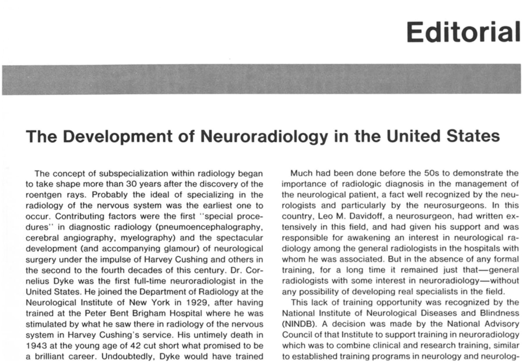
Through the American Journal of Neuroradiology (AJNR), the ASNR has maintained a leadership role in the advancement and dissemination of research, reviews, guidelines, and expert opinion related to neuroimaging and neurointervention. On July 27, 1978, Norman Leeds formed a committee to consider publication of an ASNR journal, culminating in the first issue of the AJNR appearing on January 1, 1980; an editorial by Dr Taveras appeared on page 1, volume 1 of that inaugural issue (Fig 6). Although initially the AJNR was published as a bimonthly joint venture between the American Roentgen Ray Society and the ASNR, with Juan Taveras as the Editor-in-Chief,16,19 in 1986, ownership of the AJNR officially passed solely to the ASNR; the only changes at that time were to the front cover, including both a minor change in color shading and insertion of the words, “Official Journal, American Society of Neuroradiology.”20 Periodic changes in cover design and shading have continued during the next several decades (Fig 7).

AJNR, page 1/volume 1, January/February 1980; editorial by Juan Taveras, Editor-in-Chief.

Cover design and shading changes have continued periodically for the AJNR (Courtesy of Eric Russell, MD).
The 20th, 25th, and 30th annual meetings were held at the Washington, DC Hilton (1982), New York City Hilton (1987), and St. Louis Adam’s Mark Hotel (1992), respectively.21⇓-23 Another major milestone in 1992 was the adoption of a neuroradiology “Certificate of Added Qualification” (CAQ) examination by the American Board of Radiology; Dr Taveras participated in that first neuro-CAQ oral board examination, held in Louisville, Kentucky. The Foundation of the ASNR, the philanthropic arm of the American Society of Neuroradiology, a 501 (c) (6) professional society for neuroradiologists, was founded in 1995. The first ASNR website went live on December 20, 1996; it contained only two pages of information. ASNR joined Twitter in 2010. The 40th annual meeting was held in 2002 at the Vancouver, Canada Convention & Exhibition Center, and the 50th annual meeting again was held at the New York City Hilton on 6th Avenue (Fig 8). Golden anniversary celebration events included a dinner at Keen’s for the ASNR leadership.

Part of the brochure for the ASNR 50th “Golden Anniversary” Annual Meeting and Foundation of the ASNR Symposium in New York, the city in which the ASNR was founded.
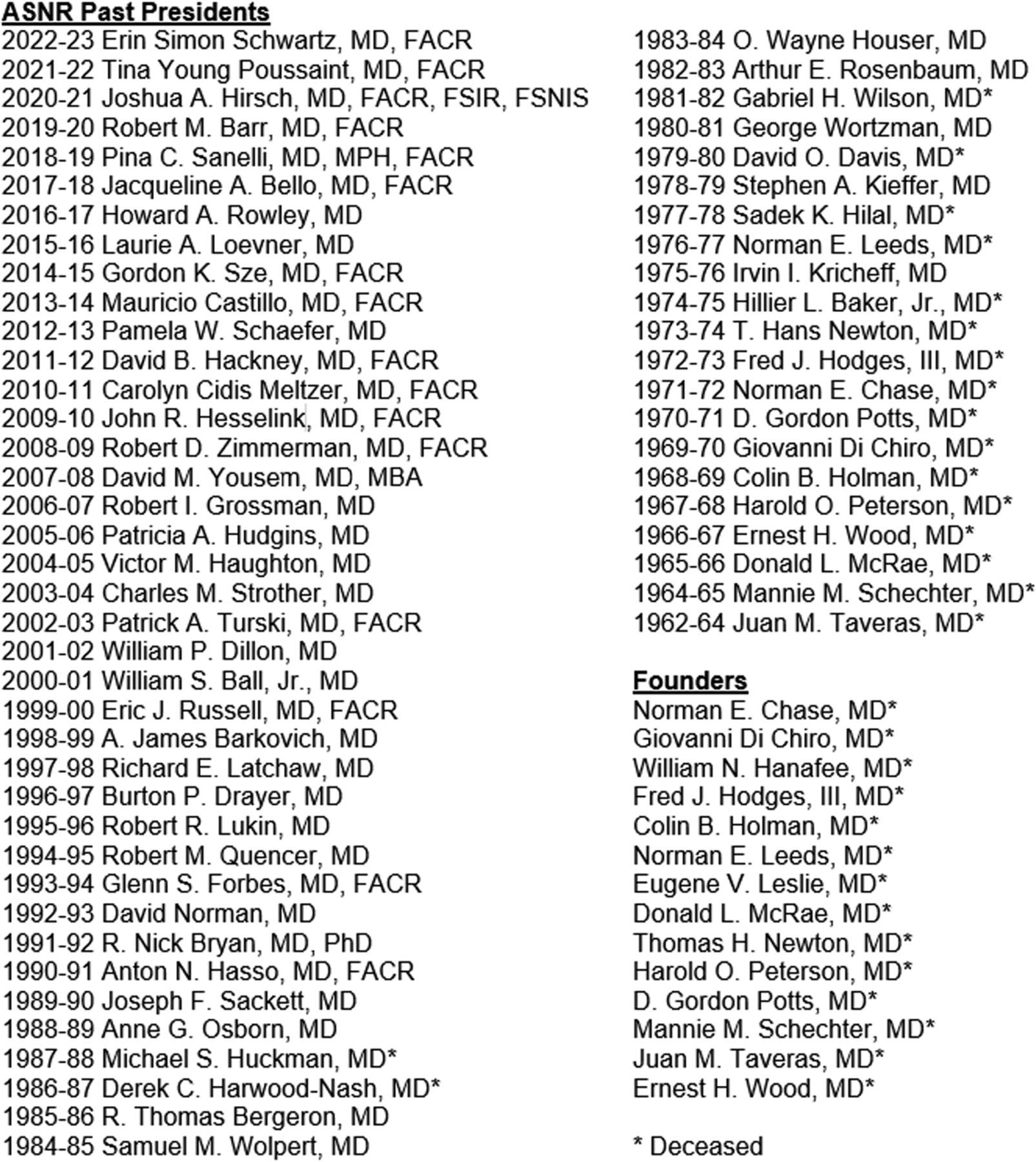
The ASNR Past Presidents Association was established in 2019; a roster of all ASNR presidents since the society’s founding 60 years ago (Fig 9) as well as a listing of the officers during the initial, early years of the society (Fig 10) are presented in Figures 9 and 10. Across the years, several of the pioneering founding and early leaders of the society have been memorialized in the pages of the AJNR.24⇓⇓⇓-28 An oral history of the ASNR is currently in preparation, which will include numerous personal recollections, along with amusing anecdotes. One example, recounted by Ralph Heinz, one of Dr Taveras’ first trainees, to his friend Dr Jim Provenzale concerns Mannie Schechter, first Vice President and second President of the ASNR (Fig 10). Mannie, who in original photos is often seen sporting an impish grin, was asked by Dr Taveras to describe a skull radiograph during teaching rounds. Schechter replied, “I see abnormal calcification near the vertex; the patient has a vertex meningioma.” Being his playful self, Juan replied, “Well, what if I told you that this patient had just been walking under a construction site and a brick fell on the top of his head? What would you say then?” Without missing a beat, Schechter replied, “I’d say ‘God damn’, it fell right on that meningioma!”

Sixtieth anniversary roster of ASNR presidents, 1962–2022.

Slate of officers from the early, founding years of the ASNR, 1962–1970.
The ASNR’s first fully virtual meeting was held remotely from May 30 to June 4, 2020, during the first year of coronavirus disease (COVID) social distancing. A virtual meeting was again held in 2021, with “social events” that ranged from online wine-tasting (led by ASNR senior member and Master Sommelier Alisa Gean) to “wellness” yoga and “relaxation response” sessions. Our 2022, Sixtieth Anniversary ASNR meeting was again held at the New York City Hilton as our first hybrid live/remote annual meeting, 3 years into COVID; this was a joint meeting with the Symposium Neuroradiologicum XXII.
CONCLUSIONS
An update of the 20-year-old numbers cited in the final paragraph of Michael Huckman’s 2001 AJNR historical retrospective, Dinner at Keen’s: The Founding of the American Society of Neuroradioloy15 is the following:
“From an intimate meeting of 14 radiologists seated at a single table (in 1962), the ASNR (in 2022) has grown to a membership of nearly 6000, publishes a journal with worldwide circulation of 7102, holds an annual meeting with attendance of approximately 1800, oversees a well-funded foundation to support the research efforts of young investigators, and has among its membership numerous department chairs and major officeholders in prestigious national and international medical organizations. It was the first organized subspecialty society in radiology and, during its 60-year existence, has set an example of establishing training standards and educational programs that have been emulated many times by younger radiologic subspecialty societies.”
In 2022, the year of our 60th “Diamond” Anniversary, the ASNR has matured into a global society that manages 6 regional and specialty societies, including the American Society of Functional Neuroradiology, American Society of Head and Neck Radiology, American Society of Spine Radiology, Eastern Neuroradiological Society, and Western Neuroradiological Society, plus the World Federation of Neuroradiological Societies, and is run by 18 full-time staff members. The society remains true to its founding mission statement of April 19, 1962:
To develop and support standards for the training in the practice of neuroradiology
To foster independent research in neuroradiology
To promote a closer fellowship and exchange of ideas among neuroradiologists.
The ASNR has been leading the cutting edge in the development, clinical translation, and dissemination of novel, game-changing imaging technologies since its earliest days, including-but-not-limited-to CT scanning in the early/mid-1970s and MR imaging in the late 1970s/early 1980s, both Nobel Prize–winning, and details of which are well-described throughout the literature.29⇓⇓⇓-33 We look forward to continued growth, as well as to the exciting new innovations certain to occur in the next 60 years, confident that the future holds promise.34,35
References
- © 2022 by American Journal of Neuroradiology


Related Articles
Cited By...
- No citing articles found.