
Article Figures & Data
Figures
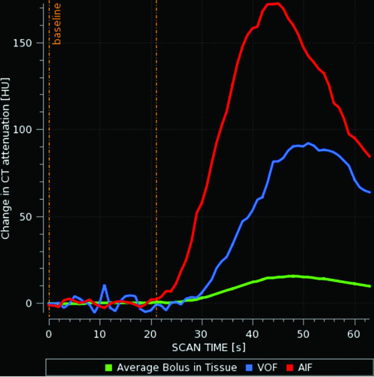
- FIG 1.
An example of early cutoff (truncation) of the arterial and venous time-density curves. VOF indicates venous output function; AIF, arterial input function.
- FIG 2.
This case serves as an example of the potential pitfalls of truncation. A 78-year-old woman presented as a code stroke due to altered mental status. Contrast-enhancement curves demonstrate early termination of the venous output measurements. Perfusion maps erroneously suggest that nearly the entire brain is penumbra. Subsequent MR imaging demonstrated only a few scattered punctate infarcts.
- FIG 3.
An example of the longer LCO protocol, triggered after the patient’s time-to-enhancement upswing rise was >15 seconds. VOF indicates venous output function; AIF, arterial input function.
- FIG 4.
Example of the CTA test-dose curve; in this case the time-to-enhancement upswing rise was <15 seconds; therefore, the standard protocol was used. Enh indicates enhancement, measured in Hounsfield Units.
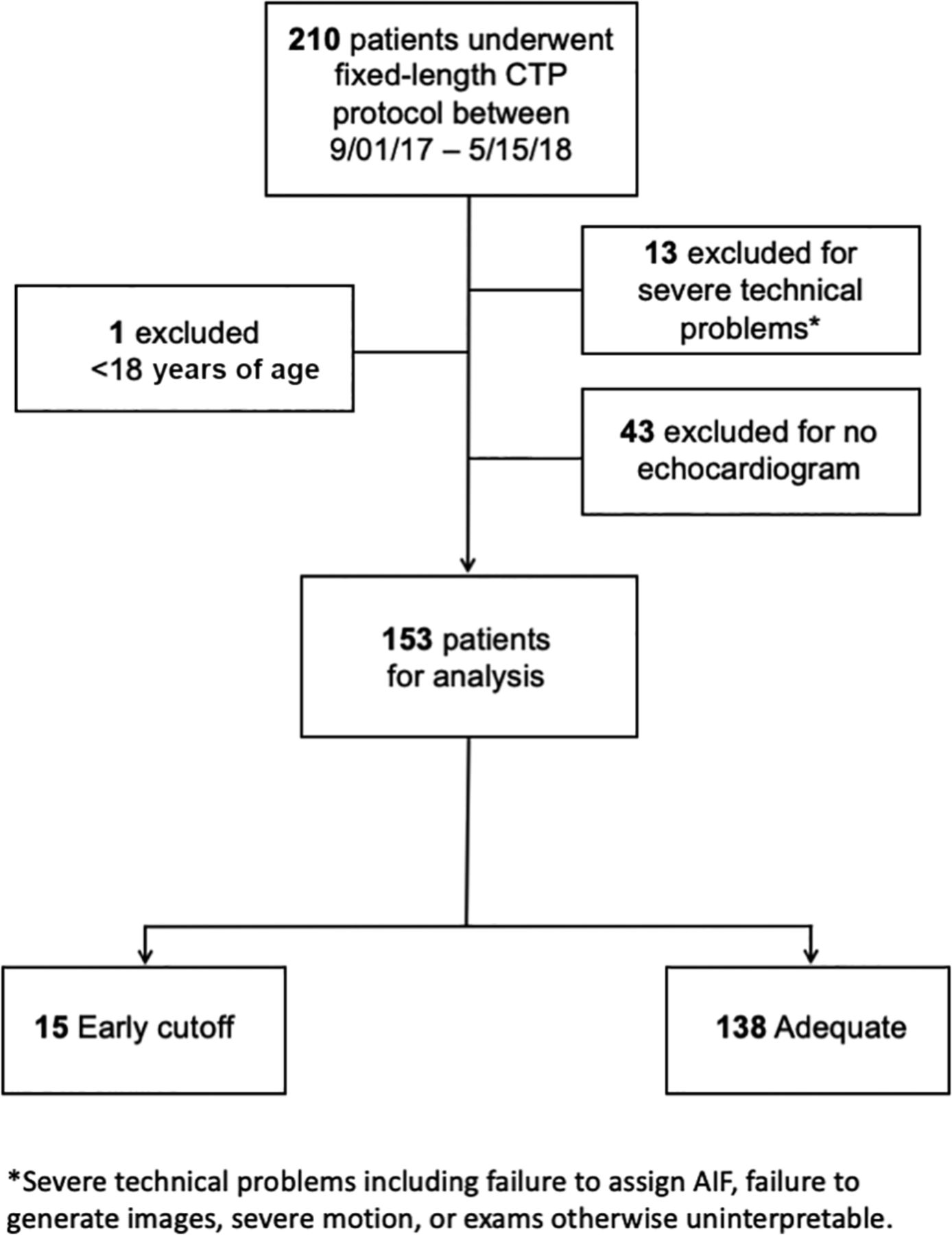
- FIG 5.
Flow chart for the fixed-timing cohort. AIF indicates arterial input function.
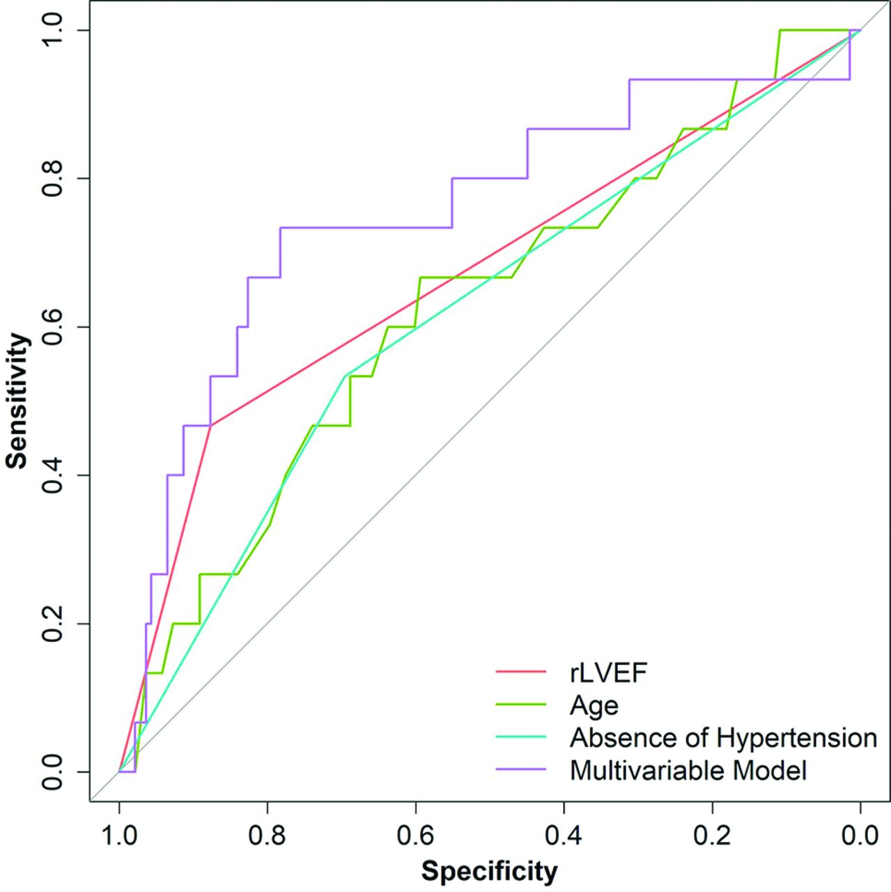
- FIG 6.
Receiver operating characteristic curves for the factors associated with truncation in the early fixed-timing cohort (n = 153). The AUC value for age is 0.63, with optimized cutoff value of 69 (sensitivity of 67% and specificity of 59%). The AUC value for rLVEF is 0.67 (sensitivity of 47% and specificity of 88%). The AUC value for the absence of hypertension is 0.61 (sensitivity of 53% and specificity of 70%). When the 3 factors are combined, the AUC value increases to 0.75 (sensitivity of 73% and specificity of 78%). AIF indicates arterial input function.
- FIG 7.
Flow chart for the case-specific cohort. AIF indicates arterial input function.


Tables
Variable Truncation Univariable Models Multivariable Model Yes (n = 15) No (n = 138) ORa 95% CI P Value ORa 95% CI P Value Age (yr) Mean, 72 (SD, 13) Mean, 66 (SD, 15) 1.42 0.95–2.14 .09 1.82 1.10–3.01 .02 rLVEF 7 (47%) 17 (12%) 6.23 2.00–19.36 .002 9.23 2.53–33.69 .001 Atrial fibrillation 10 (67%) 55 (40%) 3.02 0.98–9.31 .05 Hypertension 7 (47%) 96 (70%) 0.38 0.13–1.12 .08 0.32 0.10–1.05 .06 Diabetes mellitus 5 (33%) 45 (33%) 1.03 0.33–3.20 .95 Hyperlipidemia 6 (40%) 65 (47%) 0.75 0.25–2.22 .60 ↵a Odds ratio for age is presented per 10-year increase; the intercept term in the multivariable model was −6.33 on the log-odds scale.
Variable Sensitivity Specificity AUC 95% CI No. % 95% CI No. % 95% CI Agea 10/15 67% 38%–88% 82/138 59% 51%–68% 0.63 0.47–0.78 rLVEF 7/15 47% 21%–73% 121/138 88% 81%– 93% 0.67 0.54– 0.81 Atrial fibrillation 10/15 67% 38%–88% 83/138 60% 51%–68% 0.63 0.50– 0.76 Absence of hypertension 8/15 53% 27%–79% 96/138 70% 61%–77% 0.61 0.48– 0.75 Diabetes mellitus 5/15 33% 12%–62% 93/138 67% 59%–75% 0.50 0.37– 0.63 Absence of hyperlipidemia 9/15 60% 32%–84% 65/138 47% 39%–56% 0.54 0.40– 0.67 Multivariable model (age, rLVEF, hypertension)b 11/15 73% 45%–92% 108/138 78% 70%–85% 0.75 0.60– 0.90


