
Article Figures & Data
Figures
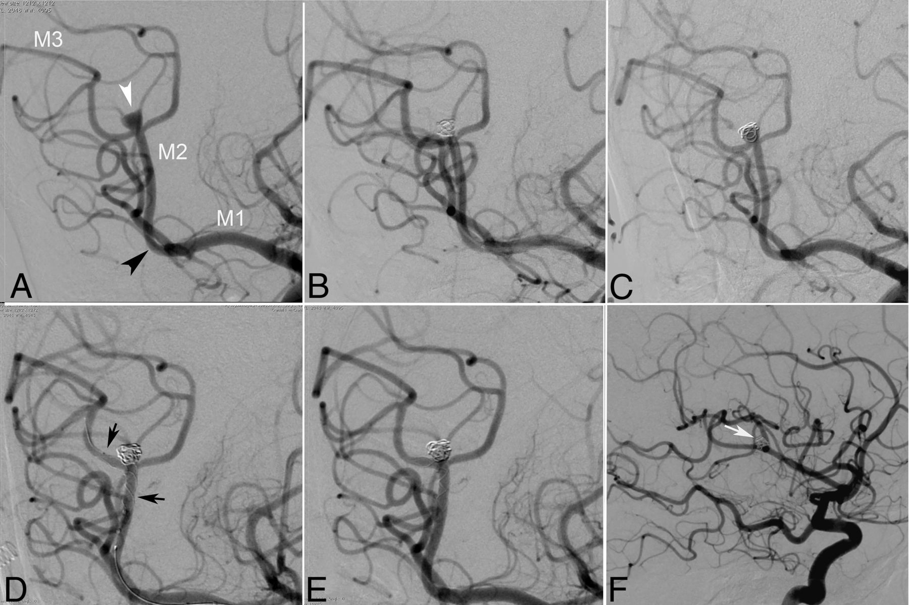
- FIG 1.
An adult patient with a recurrent right insular segment MCA aneurysm. A, DSA image shows a saccular aneurysm located distal to the MCA genu (arrowhead) in the insular segment of the right MCA. Two cortical branches arise from its neck. B, Postprocedural DSA image after the primary coiling procedure reveals complete occlusion of the aneurysm. C, Follow-up DSA after the primary coiling shows coil compaction and recanalization of the sac. D, The immediate postprocedural DSA image that was obtained following the stent-assisted coiling shows complete occlusion of the aneurysm. A self-expandable stent is deployed into one of the cortical branches (arrows). E and F, 36-month follow-up DSA images demonstrate the stable occlusion of the aneurysm (white arrow) and the patency of the cortical branches arising from its neck.
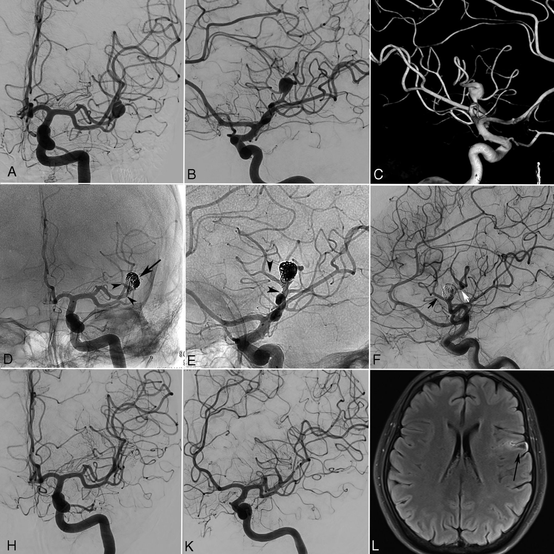
- FIG 2.
An adult patient with a left insular segment MCA aneurysm. A–C, Preprocedural DSA and 3D reconstructed angiography images show an insular segment MCA aneurysm with a very complex morphology. Two cortical stem arteries of the superior trunk originate from the neck and the superior-medial wall of the aneurysm sac. D and E, Immediate postprocedural nonsubstracted angiography images show the deployment of a self-expandable braided stent (arrowheads) into the stem artery that arises from the neck, extending proximally to the superior trunk. The sac of the aneurysm is partially coiled. Only the lateral compartment of the aneurysm sac is coiled to sustain the patency of the stem artery arising from the superior-medial wall of the sac. The arrow shows the coil mesh inside the aneurysm sac. F, Immediate postprocedural DSA image shows the patency of 2 stem arteries that arise from the neck (black arrow) and sac of the aneurysm (white arrow). H and K, Thirty-six-month follow-up DSA images demonstrate complete occlusion of the aneurysm. L, Follow-up MR image (FLAIR) shows a small cortical infarction (arrow) in the left frontal operculum. The mRS score of the patient was 1 during the clinical follow-up.
- FIG 3.
A 35-year-old male patient with a right insular segment MCA aneurysm. A and B, Preprocedural 3D-reconstructed angiography and DSA images show a wide-neck saccular aneurysm (arrow) located at the superior trunk of the right MCA. Three stem arteries arise from the aneurysm neck. C, Intraprocedural DSA shows that one of the branches arising from the aneurysm neck is catheterized for stent placement (black arrowhead) and another microcatheter is jailed inside the aneurysm for coiling (white arrowhead). D and E, Immediate postprocedural DSA and nonsubstracted angiography images show that the aneurysm is coiled (white arrow) after the deployment of an open-cell self-expandable Neuroform Atlas stent into one of the stem arteries arising from the neck, which extends proximally to the superior trunk (arrowheads). F, A 24-month follow-up DSA image demonstrates the complete occlusion of the aneurysm and patency of all the MCA branches.


Tables
Demographics Mean age (yr) 53.3 (SD, 11.3) Sex Female 19 (70.4%) Male 8 (29.6%) Aneurysm location Right superior MCA trunk 6 (22.2%) Right inferior MCA trunk 8 (29.6%) Left superior MCA trunk 9 (33.3%) Left inferior MCA trunk 4 (14.8%) Aneurysm size (maximal diameter) 2–4 mm 7 (25.9%) 5–7 mm 14 (51.9%) 8–12 mm 5 (18.5%) 13–15 mm 1 (3.7%) Aneurysm neck diameter (mm)a 3.8 (SD, 1.1) (2.1–6.7) Diameter of the artery at the proximal end of stent (mm)a 2.1 (SD, 0.22) (1.7–2.8) Diameter of the artery at the distal end of stent (mm)a 1.6 (SD, 0.17) (1.3–2.0) ↵a Data are given as the mean value (SD) (minimum-maximum values).
Deployed Stent No. of Patients LEO Baby (Balt) 20 (74.1%) Neuroform Atlas (Stryker) 5 (18.5%) LVIS EVO (MicroVention) 1 (3.7%) Accero (Accandis) 1 (3.7%)


Jump to section
Related Articles
Cited By...
- No citing articles found.