Advertisement

Main menu

  • Home
  • Content
    • Current Issue
    • Accepted Manuscripts
    • Article Preview
    • Past Issue Archive
    • Video Articles
    • AJNR Case Collection
    • Case of the Week Archive
    • Case of the Month Archive
    • Classic Case Archive
  • Special Collections
    • AJNR Awards
    • Low-Field MRI
    • Alzheimer Disease
    • ASNR Foundation Special Collection
    • Photon-Counting CT
    • View All
  • Multimedia
    • AJNR Podcasts
    • AJNR SCANtastic
    • Trainee Corner
    • MRI Safety Corner
    • Imaging Protocols
  • For Authors
    • Submit a Manuscript
    • Submit a Video Article
    • Submit an eLetter to the Editor/Response
    • Manuscript Submission Guidelines
    • Statistical Tips
    • Fast Publishing of Accepted Manuscripts
    • Graphical Abstract Preparation
    • Imaging Protocol Submission
    • Author Policies
  • About Us
    • About AJNR
    • Editorial Board
    • Editorial Board Alumni
  • More
    • Become a Reviewer/Academy of Reviewers
    • Subscribers
    • Permissions
    • Alerts
    • Feedback
    • Advertisers
    • ASNR Home

User menu

  • Alerts
  • Log in

Search

  • Advanced search
American Journal of Neuroradiology

American Journal of Neuroradiology

ASHNR American Society of Functional Neuroradiology ASHNR American Society of Pediatric Neuroradiology ASSR
  • Alerts
  • Log in

Advanced Search

  • Home
  • Content
    • Current Issue
    • Accepted Manuscripts
    • Article Preview
    • Past Issue Archive
    • Video Articles
    • AJNR Case Collection
    • Case of the Week Archive
    • Case of the Month Archive
    • Classic Case Archive
  • Special Collections
    • AJNR Awards
    • Low-Field MRI
    • Alzheimer Disease
    • ASNR Foundation Special Collection
    • Photon-Counting CT
    • View All
  • Multimedia
    • AJNR Podcasts
    • AJNR SCANtastic
    • Trainee Corner
    • MRI Safety Corner
    • Imaging Protocols
  • For Authors
    • Submit a Manuscript
    • Submit a Video Article
    • Submit an eLetter to the Editor/Response
    • Manuscript Submission Guidelines
    • Statistical Tips
    • Fast Publishing of Accepted Manuscripts
    • Graphical Abstract Preparation
    • Imaging Protocol Submission
    • Author Policies
  • About Us
    • About AJNR
    • Editorial Board
    • Editorial Board Alumni
  • More
    • Become a Reviewer/Academy of Reviewers
    • Subscribers
    • Permissions
    • Alerts
    • Feedback
    • Advertisers
    • ASNR Home
  • Follow AJNR on Twitter
  • Visit AJNR on Facebook
  • Follow AJNR on Instagram
  • Join AJNR on LinkedIn
  • RSS Feeds

AJNR Awards, New Junior Editors, and more. Read the latest AJNR updates

Research ArticleAdult Brain

Nonlesional Sources of Contrast Enhancement on Postgadolinium “Black-Blood” 3D T1-SPACE Images in Patients with Multiple Sclerosis

L. Danieli, L. Roccatagliata, D. Distefano, E. Prodi, G.C. Riccitelli, A. Diociasi, L. Carmisciano, A. Cianfoni, T. Bartalena, A. Kaelin-Lang, C. Gobbi, C. Zecca and E. Pravatà
American Journal of Neuroradiology June 2022, 43 (6) 872-880; DOI: https://doi.org/10.3174/ajnr.A7529
L. Danieli
aForm the Department of Neuroradiology (L.D., E. Prodi, A.C., E. Pravatà), Neurocenter of Southern Switzerland, Ente Ospedaliero Cantonale, Lugano, Switzerland
  • Find this author on Google Scholar
  • Find this author on PubMed
  • Search for this author on this site
  • ORCID record for L. Danieli
L. Roccatagliata
bDipartimento di Scienze della Salute (L.R., A.D.), Università degli Studi di Genova, Genoa, Italy
  • Find this author on Google Scholar
  • Find this author on PubMed
  • Search for this author on this site
  • ORCID record for L. Roccatagliata
D. Distefano
  • Find this author on Google Scholar
  • Find this author on PubMed
  • Search for this author on this site
  • ORCID record for D. Distefano
E. Prodi
aForm the Department of Neuroradiology (L.D., E. Prodi, A.C., E. Pravatà), Neurocenter of Southern Switzerland, Ente Ospedaliero Cantonale, Lugano, Switzerland
  • Find this author on Google Scholar
  • Find this author on PubMed
  • Search for this author on this site
  • ORCID record for E. Prodi
G.C. Riccitelli
cDepartment of Neurology (G.C.R., A.K.-L., C.G., C.Z., E. Pravatà), Neurocenter of Southern Switzerland, Ente Ospedaliero Cantonale, Lugano, Switzerland
dFaculty of Biomedical Sciences (G.C.R., A.C., A.K.-L., C.G., C,Z., E. Pravatà), Università della Svizzera Italiana, Lugano, Switzerland
  • Find this author on Google Scholar
  • Find this author on PubMed
  • Search for this author on this site
  • ORCID record for G.C. Riccitelli
A. Diociasi
bDipartimento di Scienze della Salute (L.R., A.D.), Università degli Studi di Genova, Genoa, Italy
  • Find this author on Google Scholar
  • Find this author on PubMed
  • Search for this author on this site
  • ORCID record for A. Diociasi
L. Carmisciano
eDepartment of Health Sciences, Section of Biostatistics (L.C.), Università degli Studi di Genova, Genoa, Italy
  • Find this author on Google Scholar
  • Find this author on PubMed
  • Search for this author on this site
  • ORCID record for L. Carmisciano
A. Cianfoni
aForm the Department of Neuroradiology (L.D., E. Prodi, A.C., E. Pravatà), Neurocenter of Southern Switzerland, Ente Ospedaliero Cantonale, Lugano, Switzerland
dFaculty of Biomedical Sciences (G.C.R., A.C., A.K.-L., C.G., C,Z., E. Pravatà), Università della Svizzera Italiana, Lugano, Switzerland
  • Find this author on Google Scholar
  • Find this author on PubMed
  • Search for this author on this site
  • ORCID record for A. Cianfoni
T. Bartalena
gDepartment of Radiology (T.B.), Pol. Zappi Bartalena, Imola, Italy.
  • Find this author on Google Scholar
  • Find this author on PubMed
  • Search for this author on this site
  • ORCID record for T. Bartalena
A. Kaelin-Lang
cDepartment of Neurology (G.C.R., A.K.-L., C.G., C.Z., E. Pravatà), Neurocenter of Southern Switzerland, Ente Ospedaliero Cantonale, Lugano, Switzerland
dFaculty of Biomedical Sciences (G.C.R., A.C., A.K.-L., C.G., C,Z., E. Pravatà), Università della Svizzera Italiana, Lugano, Switzerland
  • Find this author on Google Scholar
  • Find this author on PubMed
  • Search for this author on this site
  • ORCID record for A. Kaelin-Lang
C. Gobbi
cDepartment of Neurology (G.C.R., A.K.-L., C.G., C.Z., E. Pravatà), Neurocenter of Southern Switzerland, Ente Ospedaliero Cantonale, Lugano, Switzerland
dFaculty of Biomedical Sciences (G.C.R., A.C., A.K.-L., C.G., C,Z., E. Pravatà), Università della Svizzera Italiana, Lugano, Switzerland
  • Find this author on Google Scholar
  • Find this author on PubMed
  • Search for this author on this site
  • ORCID record for C. Gobbi
C. Zecca
cDepartment of Neurology (G.C.R., A.K.-L., C.G., C.Z., E. Pravatà), Neurocenter of Southern Switzerland, Ente Ospedaliero Cantonale, Lugano, Switzerland
dFaculty of Biomedical Sciences (G.C.R., A.C., A.K.-L., C.G., C,Z., E. Pravatà), Università della Svizzera Italiana, Lugano, Switzerland
  • Find this author on Google Scholar
  • Find this author on PubMed
  • Search for this author on this site
  • ORCID record for C. Zecca
E. Pravatà
aForm the Department of Neuroradiology (L.D., E. Prodi, A.C., E. Pravatà), Neurocenter of Southern Switzerland, Ente Ospedaliero Cantonale, Lugano, Switzerland
dFaculty of Biomedical Sciences (G.C.R., A.C., A.K.-L., C.G., C,Z., E. Pravatà), Università della Svizzera Italiana, Lugano, Switzerland
  • Find this author on Google Scholar
  • Find this author on PubMed
  • Search for this author on this site
  • ORCID record for E. Pravatà
  • Article
  • Figures & Data
  • Supplemental
  • Info & Metrics
  • Responses
  • References
  • PDF
Loading

Article Figures & Data

Figures

  • Tables
  • FIG 1.
    • Download figure
    • Open in new tab
    • Download powerpoint
    FIG 1.

    Flow chart shows the included and excluded participants and the TP and FP determination procedure including blinded readers' and senior readers' image review. CIS indicates clinically isolated syndrome; RIS, radiologically isolated syndrome.

  • FIG 2.
    • Download figure
    • Open in new tab
    • Download powerpoint
    FIG 2.

    Illustrative case of nodular vFP enhancement. A tiny juxtacortical enhancement source on SPACE (circle in A), corresponding to a triangle-shaped hypointensity on the high-resolution susceptibility-weighted image (A1), is not detected on MPRAGE and VIBE images (circles in B and C, respectively). The corresponding FLAIR T2-weighed image (D) shows no evidence of demyelinating lesions at this level. This vFP was reported by ER 1 and the BR.

  • FIG 3.
    • Download figure
    • Open in new tab
    • Download powerpoint
    FIG 3.

    The upper panel shows an open-ring vFP enhancement consistent with a developmental venous anomaly and mimicking a TP enhancing juxtacortical CEL on SPACE images. Characteristic arcuate enhancement on SPACE image with subtle margins (circles) in the juxtacortical white matter (A), associated with faint hyperintensity on FLAIR T2 image (B), is barely visible on MPRAGE (C) and VIBE (D) images. This vFP was reported by the BR only. TP CELs with morphology similar to that of A on SPACE image (E) exhibits characteristic T2-FLAIR hyperintensity (F). In this case, CEL enhancement is clearly detected on MPRAGE (G) and VIBE (H) images as well.
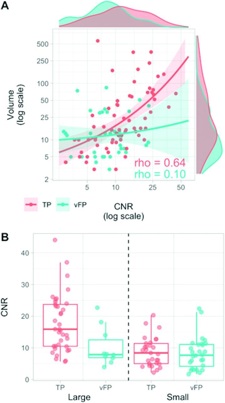
  • FIG 4
    • Download figure
    • Open in new tab
    • Download powerpoint
    FIG 4

    A, Relationships between CNR and volume variables in TP (red) and vFP (cyan) findings. Higher CNR and larger volume values are more frequently represented in TP than in vFP (marginal density plots). The variables are significantly correlated in TP (ρ = 0.644, P < .001), but not in vFP (ρ = 0.096, P = .568). B, Boxplots represent CNR value-distribution differences between TP and vFP after median split categorization into small (≤14 mm3) and large (>14 mm3). CNR values are lower in vFP compared with TP in the large category only, but similar in the small category.

Tables

  • Figures
    • View popup
    Table 1:

    Included patients' main demographics, clinical characteristics, and DMT

    Demographics
    No. of evaluable patients232
    Relapsing-emitting MS177 (76.3%)
    Secondary-progressive MS20 (8.6%)
    Primary-progressive MS10 (4.3%)
    CIS (%)21 (9.1%)
    RIS (%)4 (1.7%)
    Age (mean) (yr)46 (SD, 12.65)
    Sex (men/women)81:151
    EDSSa (median) (interquartile range)2.5/2
    Disease durationa (median) (interquartile range)11/11
    DMT efficacyb
        Moderatec126 (60.9%)
        Highd9 (4.3%)
        Very highe36 (17.4%)
        No DMT36 (17.4%)
    • Note:—CIS indicates clinically isolated syndrome; RIS, radiologically isolated syndrome; EDSS, Expanded Disability Status Scale; DMT, disease-modifying treatment.

    • ↵a Patients with relapsing-remitting, secondary-progressive, or primary-progressive MS only.

    • ↵b According to Dobson et al.28

    • ↵c Includes dimethyl fumarate, glatiramer acetate, interferon-β 1-α, interferon-β-1b, and teriflunomide.

    • ↵d Includes fingolimod, siponimod, and ozanimod.

    • ↵e Includes alemtuzumab, cladribine, mitoxantrone, natalizumab, and ocrelizumab.

    • View popup
    Table 2:

    Frequency of TP and FP sources of enhancement for each SPACE, MPRAGE, VIBE sequence, and blind readers

    SPACEMPRAGEVIBEPa (SPACE vs MPRAGE)Pa (SPACE vs VIBE)
    TP enhancement sources (No. of patients)
        ER 150 (19)44 (16)42 (15).070.042b
        ER 253 (15)42 (15)41 (15).026b.008b
        BR42 (14)34 (13)36 (12).006b.016b
    FP enhancement sources (No. of patients)
        ER 122 (20)8 (6)8 (7).004b.008b
        ER 224 (23)7 (7)9 (8).004b.006b
        BR31 (28)6 (6)11 (11)<.001b.006b
    • ↵a McNemar test.

    • ↵b Statistically significant results.

    • View popup
    Table 3:

    Patient-wise diagnostic accuracy results by OR analysisa

    SPACE vs MPRAGESPACE vs VIBEP (SPACE vs MPRAGE)P (SPACE vs VIBE)
    Chance of detecting more patients with TP
        ER 11.20 (0.6–2.4)1.29 (0.6–2.6).598.478
        ER 21.00 (0.48–2.09)1.00 (0.48–2.09)1.0001.000
        BR1.08 (0.5–2.4)1.18 (0.53–2.6).686.687
    Chance of detecting more patients with FP
        ER 13.55 (1.4–9.1)3.03 (1.25–7.3).015b.027b
        ER 23.53 (1.5–8.4)3.08 (1.3–7.1).008b.015b
        BR5.17 (2.1–12.7)2.76 (1.3–5.7)<.001b.012b
    • ↵a OR and 95% CI are provided for SPACE versus MPRAGE and SPACE versus VIBE comparisons and for each reader.

    • ↵b Statistically significant results.

    • View popup
    Table 4:

    Quantitative characteristics of vFP and TP contrast-enhancement sources on SPACE imagesa

    vFPTPPvFPsmallTPsmallPvFPlargeTPlargeP
    No.386628291037
    Volume (mm3)
        Median11.516.027b,d7.59.981b3449.730b
        Range3–843–5653–143–1430–8415–565
    CNR
        Median7.7311.18.003b,d7.668.38.544b7.8515.87.015b,d
        Range1.67–22.672.18–44.061.67–22.372.18–20.283.92–22.675.67–44.06
    Morphology (No.) (%)
        Nodular26 (68.4%)52 (78.8%).078c22 (78.6%)26 (89.7%).292c4 (40%)26 (70.3%).036c,d
        Tubular4 (10.5%)0 (0%)2 (7.1%)0 (0%)2 (20%)0 (0%)
        Ring/open-ring8 (28.1%)14 (21.2%)4 (14.3%)3 (10.3%)4 (40%)11 (29.7%)
    • ↵a Large and small TPs and vFPs are categorized according to the median of their volume-distribution frequency (14 mm3).

    • ↵b Mann-Whitney U test.

    • ↵c Pearson χ2.

    • ↵d Statistically significant results.

PreviousNext
Back to top

In this issue

American Journal of Neuroradiology: 43 (6)
American Journal of Neuroradiology
Vol. 43, Issue 6
1 Jun 2022
  • Table of Contents
  • Index by author
  • Complete Issue (PDF)
Advertisement
Print
Download PDF
Email Article

Thank you for your interest in spreading the word on American Journal of Neuroradiology.

NOTE: We only request your email address so that the person you are recommending the page to knows that you wanted them to see it, and that it is not junk mail. We do not capture any email address.

Enter multiple addresses on separate lines or separate them with commas.
Nonlesional Sources of Contrast Enhancement on Postgadolinium “Black-Blood” 3D T1-SPACE Images in Patients with Multiple Sclerosis
(Your Name) has sent you a message from American Journal of Neuroradiology
(Your Name) thought you would like to see the American Journal of Neuroradiology web site.
CAPTCHA
This question is for testing whether or not you are a human visitor and to prevent automated spam submissions.
Cite this article
L. Danieli, L. Roccatagliata, D. Distefano, E. Prodi, G.C. Riccitelli, A. Diociasi, L. Carmisciano, A. Cianfoni, T. Bartalena, A. Kaelin-Lang, C. Gobbi, C. Zecca, E. Pravatà
Nonlesional Sources of Contrast Enhancement on Postgadolinium “Black-Blood” 3D T1-SPACE Images in Patients with Multiple Sclerosis
American Journal of Neuroradiology Jun 2022, 43 (6) 872-880; DOI: 10.3174/ajnr.A7529

Citation Manager Formats

  • BibTeX
  • Bookends
  • EasyBib
  • EndNote (tagged)
  • EndNote 8 (xml)
  • Medlars
  • Mendeley
  • Papers
  • RefWorks Tagged
  • Ref Manager
  • RIS
  • Zotero
0 Responses
Respond to this article
Share
Bookmark this article
Nonlesional Contrast Enhancement in MS
L. Danieli, L. Roccatagliata, D. Distefano, E. Prodi, G.C. Riccitelli, A. Diociasi, L. Carmisciano, A. Cianfoni, T. Bartalena, A. Kaelin-Lang, C. Gobbi, C. Zecca, E. Pravatà
American Journal of Neuroradiology Jun 2022, 43 (6) 872-880; DOI: 10.3174/ajnr.A7529
del.icio.us logo Twitter logo Facebook logo Mendeley logo
  • Tweet Widget
  • Facebook Like
  • Google Plus One
Purchase

Jump to section

  • Article
    • Abstract
    • ABBREVIATIONS:
    • MATERIALS AND METHODS
    • RESULTS
    • DISCUSSION
    • CONCLUSIONS
    • ACKNOWLEDGMENTS
    • Footnotes
    • References
  • Figures & Data
  • Supplemental
  • Info & Metrics
  • Responses
  • References
  • PDF

Related Articles

  • PubMed
  • Google Scholar

Cited By...

  • Improved detection of multiple sclerosis gadolinium-enhancing lesions: 3D T1 Turbo-Spin-Echo Outperforms 3D T1 Turbo-Field Echo MRI
  • Crossref
  • Google Scholar

This article has not yet been cited by articles in journals that are participating in Crossref Cited-by Linking.

More in this TOC Section

  • Diagnostic Neuroradiology of Monoclonal Antibodies
  • Clinical Outcomes After Chiari I Decompression
  • Segmentation of Brain Metastases with BLAST
Show more Adult Brain

Similar Articles

Advertisement

Indexed Content

  • Current Issue
  • Accepted Manuscripts
  • Article Preview
  • Past Issues
  • Editorials
  • Editor's Choice
  • Fellows' Journal Club
  • Letters to the Editor
  • Video Articles

Cases

  • Case Collection
  • Archive - Case of the Week
  • Archive - Case of the Month
  • Archive - Classic Case

More from AJNR

  • Trainee Corner
  • Imaging Protocols
  • MRI Safety Corner
  • Book Reviews

Multimedia

  • AJNR Podcasts
  • AJNR Scantastics

Resources

  • Turnaround Time
  • Submit a Manuscript
  • Submit a Video Article
  • Submit an eLetter to the Editor/Response
  • Manuscript Submission Guidelines
  • Statistical Tips
  • Fast Publishing of Accepted Manuscripts
  • Graphical Abstract Preparation
  • Imaging Protocol Submission
  • Evidence-Based Medicine Level Guide
  • Publishing Checklists
  • Author Policies
  • Become a Reviewer/Academy of Reviewers
  • News and Updates

About Us

  • About AJNR
  • Editorial Board
  • Editorial Board Alumni
  • Alerts
  • Permissions
  • Not an AJNR Subscriber? Join Now
  • Advertise with Us
  • Librarian Resources
  • Feedback
  • Terms and Conditions
  • AJNR Editorial Board Alumni

American Society of Neuroradiology

  • Not an ASNR Member? Join Now

© 2025 by the American Society of Neuroradiology All rights, including for text and data mining, AI training, and similar technologies, are reserved.
Print ISSN: 0195-6108 Online ISSN: 1936-959X

Powered by HighWire