Advertisement

Main menu

  • Home
  • Content
    • Current Issue
    • Accepted Manuscripts
    • Article Preview
    • Past Issue Archive
    • AJNR Case Collection
    • Case of the Week Archive
    • Classic Case Archive
    • Case of the Month Archive
  • Special Collections
    • Spinal CSF Leak Articles (Jan 2020-June 2024)
    • 2024 AJNR Journal Awards
    • Most Impactful AJNR Articles
  • Multimedia
    • AJNR Podcast
    • AJNR Scantastics
    • Video Articles
  • For Authors
    • Submit a Manuscript
    • Author Policies
    • Fast publishing of Accepted Manuscripts
    • Graphical Abstract Preparation
    • Manuscript Submission Guidelines
    • Imaging Protocol Submission
    • Submit a Case for the Case Collection
  • About Us
    • About AJNR
    • Editorial Board
  • More
    • Become a Reviewer/Academy of Reviewers
    • Subscribers
    • Permissions
    • Alerts
    • Feedback
    • Advertisers
    • ASNR Home
  • Other Publications
    • ajnr

User menu

  • Alerts
  • Log in

Search

  • Advanced search
American Journal of Neuroradiology

American Journal of Neuroradiology

ASHNR American Society of Functional Neuroradiology ASHNR American Society of Pediatric Neuroradiology ASSR
  • Alerts
  • Log in

Advanced Search

  • Home
  • Content
    • Current Issue
    • Accepted Manuscripts
    • Article Preview
    • Past Issue Archive
    • AJNR Case Collection
    • Case of the Week Archive
    • Classic Case Archive
    • Case of the Month Archive
  • Special Collections
    • Spinal CSF Leak Articles (Jan 2020-June 2024)
    • 2024 AJNR Journal Awards
    • Most Impactful AJNR Articles
  • Multimedia
    • AJNR Podcast
    • AJNR Scantastics
    • Video Articles
  • For Authors
    • Submit a Manuscript
    • Author Policies
    • Fast publishing of Accepted Manuscripts
    • Graphical Abstract Preparation
    • Manuscript Submission Guidelines
    • Imaging Protocol Submission
    • Submit a Case for the Case Collection
  • About Us
    • About AJNR
    • Editorial Board
  • More
    • Become a Reviewer/Academy of Reviewers
    • Subscribers
    • Permissions
    • Alerts
    • Feedback
    • Advertisers
    • ASNR Home
  • Follow AJNR on Twitter
  • Visit AJNR on Facebook
  • Follow AJNR on Instagram
  • Join AJNR on LinkedIn
  • RSS Feeds

Welcome to the new AJNR, Updated Hall of Fame, and more. Read the full announcements.


AJNR is seeking candidates for the position of Associate Section Editor, AJNR Case Collection. Read the full announcement.


Research ArticlePediatrics

Cerebral Hemodynamic and Metabolic Abnormalities in Neonatal Hypocalcemia: Findings from Advanced MRI

Ying Qi, Zixuan Lin, Hanzhang Lu, Jian Mao, Hongyang Zhang, Pengfei Zhao and Yang Hou
American Journal of Neuroradiology October 2023, 44 (10) 1224-1230; DOI: https://doi.org/10.3174/ajnr.A7994
Ying Qi
aFrom the Department of Radiology (Y.Q., H.Z., Y.H.), Shengjing Hospital of China Medical University, Shenyang, China
  • Find this author on Google Scholar
  • Find this author on PubMed
  • Search for this author on this site
  • ORCID record for Ying Qi
Zixuan Lin
bKey Laboratory for Biomedical Engineering of Ministry of Education (Z.L.), Department of Biomedical Engineering, College of Biomedical Engineering & Instrument Science, Zhejiang University, Hangzhou, China
  • Find this author on Google Scholar
  • Find this author on PubMed
  • Search for this author on this site
  • ORCID record for Zixuan Lin
Hanzhang Lu
cDepartment of Radiology (H.L.), Johns Hopkins University School of Medicine, Baltimore, Maryland
  • Find this author on Google Scholar
  • Find this author on PubMed
  • Search for this author on this site
  • ORCID record for Hanzhang Lu
Jian Mao
dDepartment of Pediatrics (J.M.), Shengjing Hospital of China Medical University, Shenyang, China
  • Find this author on Google Scholar
  • Find this author on PubMed
  • Search for this author on this site
  • ORCID record for Jian Mao
Hongyang Zhang
aFrom the Department of Radiology (Y.Q., H.Z., Y.H.), Shengjing Hospital of China Medical University, Shenyang, China
  • Find this author on Google Scholar
  • Find this author on PubMed
  • Search for this author on this site
  • ORCID record for Hongyang Zhang
Pengfei Zhao
eDepartment of Pharmacology (P.Z.), School of Pharmaceutical Sciences, China Medical University, Shenyang, China
  • Find this author on Google Scholar
  • Find this author on PubMed
  • Search for this author on this site
  • ORCID record for Pengfei Zhao
Yang Hou
aFrom the Department of Radiology (Y.Q., H.Z., Y.H.), Shengjing Hospital of China Medical University, Shenyang, China
  • Find this author on Google Scholar
  • Find this author on PubMed
  • Search for this author on this site
  • ORCID record for Yang Hou
  • Article
  • Figures & Data
  • Supplemental
  • Info & Metrics
  • Responses
  • References
  • PDF
Loading

Article Figures & Data

Figures

  • Tables
  • FIG 1.
    • Download figure
    • Open in new tab
    • Download powerpoint
    FIG 1.

    Sample images for CBF and Yv determination. A, Positioning of the imaging section. B and C, PCMR imaging section locations for the LICA, RICA, LVA, and RVA on the MIP image of a TOF angiogram. D, The manual delineation of LICA, RICA, LVA, and RVA was conducted to determine flux in these vessels. The sum of fluxes by brain weight yielded the CBF. E, The imaging section was positioned parallel to the intercommissural line with a 10-mm distance from the superior sagittal sinus (SSS). TRUST MR imaging used a spin-labeling module to isolate pure venous blood signals in the SSS. F, Blood signals from the SSS were fitted to a monoexponential function of the effective TE to yield blood T2. T2 was converted to Yv via a calibration plot. LICA indicates left internal carotid arteries; RICA, right internal carotid arteries; LVA, left vertebral artery; RVA, right vertebral artery; AU, arbitrary unit.

  • FIG 2.
    • Download figure
    • Open in new tab
    • Download powerpoint
    FIG 2.

    Flow chart of study participants.

  • FIG 3.
    • Download figure
    • Open in new tab
    • Download powerpoint
    FIG 3.

    NHC-a subgroup with brain injuries on T1WI and T2WI. A and B, Neonate (scan age, 33.3 weeks; birth weight, 1740 g; male) had focal PVWM lesions (grade I) in the right PVWM area showing increased signal intensity on T1WI and decreased signal intensity on T2WI. C and D, Neonate (scan age, 34.3 weeks; birth weight, 2600 g; male) had PVWM lesions (grade II) in the bilateral PVWM areas, linearly displaying increased signal intensity on T1WI and decreased signal intensity on T2WI. E and F, Neonate (scan age, 37.7 weeks; birth weight, 2200 g; female) had clustered PVWM lesions and cystic lesions (grade IV) in bilateral PVWM areas.

  • FIG 4.
    • Download figure
    • Open in new tab
    • Download powerpoint
    FIG 4.

    Boxplots of the physiologic parameters in both groups and subgroups. A–D, Boxplots display Yv, CBF, CMRO2, and brain volume from both groups and subgroups. NHC neonates and the NHC-a subset (versus controls) both had significantly lower CBF (P = .03, P = .001) and CMRO2 (P = .02, P = .002). CBF and CMRO2 were significantly lower in NHC-a (versus the NHC-n) (P = .001, P = .01). There was no significant difference in CBF and CMRO2 between those with NHC-n and controls (P = .70, P = .63). Yv and brain volume were not significantly different between groups (P > .05). The asterisk indicates P < .05.
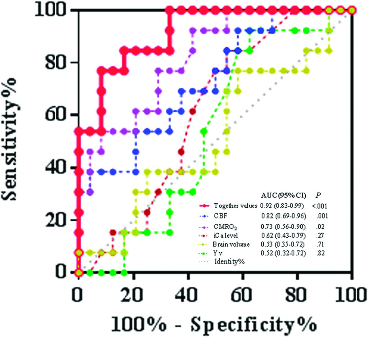
  • FIG 5.
    • Download figure
    • Open in new tab
    • Download powerpoint
    FIG 5.

    Receiver operating characteristic curve for the combined values together versus a single value (CBF, CMRO2, brain volume, or iCa levels, respectively) to analyze the association between NHC and brain injuries. The combined values together versus a single value showed superior capacity to detect NHC with brain injuries (AUC= 0.92 [95% CI, 0.83-0.99]) versus CBF (AUC = 0.82 [95% CI, 0.69-0.96]; P = .001), CMRO2 (AUC = 0.73 [95% CI, 0.56-0.90]; P = .02), brain volume (AUC = 0.53 [95% CI, 0.35-0.72]; P = .82), Yv (AUC = 0.52 [95% CI, 0.32-0.72]; P = .82), or iCa levels (AUC = 0.62 [95% CI, 0.44-0.80]; P = .23).

Tables

  • Figures
    • View popup
    Table 1:

    Demographic and clinical data of the NHC group and controls

    NHC (n = 37)NHC-a (n = 24)NHC-n (n = 13)Control (n = 19)PNHC & Control Value
    Demographics
     Males (No.) (%)24 (64.9)14 (58.3)7 (53.8)14 (73.7).11
     Birth weight (mean) (g)2024.9 (SD, 614.5)2080.8 (SD, 532.1)1676.5 (SD, 695.9)2434.7 (SD, 1047.5).13
     Scan age (mean) (wk)37.0 (SD, 2.9)37.2 (SD, 3.2)36.0 (SD, 1.8)37.0 (SD, 4.0).99
    Clinical
     Apgar score (mean)
      1 min7.6 (SD, 3.2)7.0 (SD, 3.5)9.4 (SD, 0.5)9.6 (SD, 0.8)<.05
      5 min6.2 (SD, 3.3)5.5 (SD, 3.5)8.0 (SD, 1.3)8.6 (SD, 2.1)<.05
     NRDS (No.) (%)16 (43.2)13 (54.2)3 (23.1)––
     Twins (No.) (%)6 (16.2)5 (20.8)1 (7.7)––
    iCa (mean) (mmol/L)0.9 (SD, 0.2)0.9 (SD, 0.2)0.8 (SD, 0.1)1.2 (SD, 0.1)<.05
    • Note:—The en dash indicates none.

    • View popup
    Table 2:

    MR imaging findings between the NHC group and the NHC-a subset

    Characteristics of Neonatal MR ImagingNHC (n = 37)NHC-a (n = 24)
    Periventricular WM lesions I (No.) (%)6 (16.2)6 (25)
    Periventricular WM lesions II
     +0 (No.) (%)4 (10.8)4 (16.7)
     + Intraventricular hemorrhage (No.) (%)1 (2.7)1 (4.2)
    Periventricular white matter lesions III (No.) (%)1 (2.7)1 (4.2)
    Periventricular white matter lesions IV (No.) (%)7 (18.9)7 (29.2)
    SAH without parenchymal hemorrhagic lesions (No.) (%)5 (13.5)5 (20.8)
    • View popup
    Table 3:

    Comparison of physiologic parameters between groups and their subgroups (n = 56)a

    ComparatorsYv (%)CBF (mL/100g/min)CMRO2 (mL/100g/min)Brain Volume (mL)
    NHC group (n = 37)63.4 (SD, 13.6)13.3 (SD, 5.3)31.1 (SD, 12.2)300.0 (SD, 88.6)
    Control group (n = 19)64.7 (SD, 7.1)16.5 (SD, 4.7)40.4 (SD, 14.8)290.4 (SD, 72.8)
    P value.64<.05<.05.69
    NHC-a group (n = 24)63.2 (SD, 16.1)11.2 (SD, 4.5)27.4 (SD, 10.7)296.9 (SD, 87.3)
    NHC-n group (n = 13)63.8 (SD, 7.5)17.1 (SD, 4.7)37.9 (SD, 12.1)305.6 (SD, 94.2)
    P value.88<.05<.05.78
    NHC-a group (n = 24)63.2 (SD, 16.1)11.2 (SD, 4.5)27.4 (SD, 10.7)296.9 (SD, 87.3)
    Control group (n = 19)64.7 (SD, 7.1)16.5 (SD, 4.7)40.4 (SD, 14.8)290.4 (SD, 72.8)
    P value.70<.05<.05.80
    NHC-n group (n = 13)63.8 (SD, 7.5)17.1 (SD, 4.7)37.9 (SD, 12.1)305.6 (SD, 94.2)
    Control group (n = 19)64.7 (SD, 7.1)16.5 (SD, 4.7)40.4 (SD, 14.8)290.4 (SD, 72.8)
    P value.73.70.63.61
    • ↵a Data are expressed as means. P values are based on the Wilcoxon 2-sample exact test.

PreviousNext
Back to top

In this issue

American Journal of Neuroradiology: 44 (10)
American Journal of Neuroradiology
Vol. 44, Issue 10
1 Oct 2023
  • Table of Contents
  • Index by author
  • Complete Issue (PDF)
Advertisement
Print
Download PDF
Email Article

Thank you for your interest in spreading the word on American Journal of Neuroradiology.

NOTE: We only request your email address so that the person you are recommending the page to knows that you wanted them to see it, and that it is not junk mail. We do not capture any email address.

Enter multiple addresses on separate lines or separate them with commas.
Cerebral Hemodynamic and Metabolic Abnormalities in Neonatal Hypocalcemia: Findings from Advanced MRI
(Your Name) has sent you a message from American Journal of Neuroradiology
(Your Name) thought you would like to see the American Journal of Neuroradiology web site.
CAPTCHA
This question is for testing whether or not you are a human visitor and to prevent automated spam submissions.
Cite this article
Ying Qi, Zixuan Lin, Hanzhang Lu, Jian Mao, Hongyang Zhang, Pengfei Zhao, Yang Hou
Cerebral Hemodynamic and Metabolic Abnormalities in Neonatal Hypocalcemia: Findings from Advanced MRI
American Journal of Neuroradiology Oct 2023, 44 (10) 1224-1230; DOI: 10.3174/ajnr.A7994

Citation Manager Formats

  • BibTeX
  • Bookends
  • EasyBib
  • EndNote (tagged)
  • EndNote 8 (xml)
  • Medlars
  • Mendeley
  • Papers
  • RefWorks Tagged
  • Ref Manager
  • RIS
  • Zotero
0 Responses
Respond to this article
Share
Bookmark this article
Neonatal Hypocalcemia: Cerebral MRI Abnormalities
Ying Qi, Zixuan Lin, Hanzhang Lu, Jian Mao, Hongyang Zhang, Pengfei Zhao, Yang Hou
American Journal of Neuroradiology Oct 2023, 44 (10) 1224-1230; DOI: 10.3174/ajnr.A7994
del.icio.us logo Twitter logo Facebook logo Mendeley logo
  • Tweet Widget
  • Facebook Like
  • Google Plus One
Purchase

Jump to section

  • Article
    • Abstract
    • ABBREVIATIONS:
    • MATERIALS AND METHODS
    • RESULTS
    • DISCUSSION
    • CONCLUSIONS
    • Footnotes
    • References
  • Figures & Data
  • Supplemental
  • Info & Metrics
  • Responses
  • References
  • PDF

Related Articles

  • PubMed
  • Google Scholar

Cited By...

  • No citing articles found.
  • Crossref (2)
  • Google Scholar

This article has been cited by the following articles in journals that are participating in Crossref Cited-by Linking.

  • Altered cerebral oxygen extraction and metabolism in preterm neonates and the relationship to anemia: A noncontrast MRI study
    Zixuan Lin, Dan Wu, Dengrong Jiang, Hanzhang Lu, Ying Qi
    Annals of the Child Neurology Society 2024 2 4
  • Imputation models and error analysis for phase contrast MR cerebral blood flow measurements in heterogeneous pediatric and adult populations
    Eamon K. Doyle, Isabel Torres, Joseph Liu, Abhishek Karnwal, Sudarshan Ranganathan, Bradley J. De Souza, Payal Shah, Bradley S. Peterson, John C. Wood, Matthew Thomas Borzage
    Frontiers in Physiology 2025 16

More in this TOC Section

  • Comparison of Image Quality and Radiation Dose in Pediatric Temporal Bone CT Using Photon-Counting Detector CT and Energy-Integrating Detector CT
  • SyMRI & MR Fingerprinting in Brainstem Myelination
  • Pons&Vermis Localization on Fetal MRI Using U-Net
Show more Pediatrics

Similar Articles

Advertisement

Indexed Content

  • Current Issue
  • Accepted Manuscripts
  • Article Preview
  • Past Issues
  • Editorials
  • Editors Choice
  • Fellow Journal Club
  • Letters to the Editor

Cases

  • Case Collection
  • Archive - Case of the Week
  • Archive - Case of the Month
  • Archive - Classic Case

Special Collections

  • Special Collections

Resources

  • News and Updates
  • Turn around Times
  • Submit a Manuscript
  • Author Policies
  • Manuscript Submission Guidelines
  • Evidence-Based Medicine Level Guide
  • Publishing Checklists
  • Graphical Abstract Preparation
  • Imaging Protocol Submission
  • Submit a Case
  • Become a Reviewer/Academy of Reviewers
  • Get Peer Review Credit from Publons

Multimedia

  • AJNR Podcast
  • AJNR SCANtastic
  • Video Articles

About Us

  • About AJNR
  • Editorial Board
  • Not an AJNR Subscriber? Join Now
  • Alerts
  • Feedback
  • Advertise with us
  • Librarian Resources
  • Permissions
  • Terms and Conditions

American Society of Neuroradiology

  • Not an ASNR Member? Join Now

© 2025 by the American Society of Neuroradiology All rights, including for text and data mining, AI training, and similar technologies, are reserved.
Print ISSN: 0195-6108 Online ISSN: 1936-959X

Powered by HighWire