Advertisement

Main menu

  • Home
  • Content
    • Current Issue
    • Accepted Manuscripts
    • Article Preview
    • Past Issue Archive
    • AJNR Case Collection
    • Case of the Week Archive
    • Classic Case Archive
    • Case of the Month Archive
  • Special Collections
    • Spinal CSF Leak Articles (Jan 2020-June 2024)
    • 2024 AJNR Journal Awards
    • Most Impactful AJNR Articles
  • Multimedia
    • AJNR Podcast
    • AJNR Scantastics
    • Video Articles
  • For Authors
    • Submit a Manuscript
    • Author Policies
    • Fast publishing of Accepted Manuscripts
    • Graphical Abstract Preparation
    • Manuscript Submission Guidelines
    • Imaging Protocol Submission
    • Submit a Case for the Case Collection
  • About Us
    • About AJNR
    • Editorial Board
  • More
    • Become a Reviewer/Academy of Reviewers
    • Subscribers
    • Permissions
    • Alerts
    • Feedback
    • Advertisers
    • ASNR Home
  • Other Publications
    • ajnr

User menu

  • Alerts
  • Log in

Search

  • Advanced search
American Journal of Neuroradiology

American Journal of Neuroradiology

ASHNR American Society of Functional Neuroradiology ASHNR American Society of Pediatric Neuroradiology ASSR
  • Alerts
  • Log in

Advanced Search

  • Home
  • Content
    • Current Issue
    • Accepted Manuscripts
    • Article Preview
    • Past Issue Archive
    • AJNR Case Collection
    • Case of the Week Archive
    • Classic Case Archive
    • Case of the Month Archive
  • Special Collections
    • Spinal CSF Leak Articles (Jan 2020-June 2024)
    • 2024 AJNR Journal Awards
    • Most Impactful AJNR Articles
  • Multimedia
    • AJNR Podcast
    • AJNR Scantastics
    • Video Articles
  • For Authors
    • Submit a Manuscript
    • Author Policies
    • Fast publishing of Accepted Manuscripts
    • Graphical Abstract Preparation
    • Manuscript Submission Guidelines
    • Imaging Protocol Submission
    • Submit a Case for the Case Collection
  • About Us
    • About AJNR
    • Editorial Board
  • More
    • Become a Reviewer/Academy of Reviewers
    • Subscribers
    • Permissions
    • Alerts
    • Feedback
    • Advertisers
    • ASNR Home
  • Follow AJNR on Twitter
  • Visit AJNR on Facebook
  • Follow AJNR on Instagram
  • Join AJNR on LinkedIn
  • RSS Feeds

Welcome to the new AJNR, Updated Hall of Fame, and more. Read the full announcements.


AJNR is seeking candidates for the position of Associate Section Editor, AJNR Case Collection. Read the full announcement.


Research ArticleSpine

Conebeam CT as an Adjunct to Digital Subtraction Myelography for Detection of CSF-Venous Fistulas

A.A. Madhavan, J.K. Cutsforth-Gregory, J.C. Benson, W. Brinjikji, I.T. Mark and J.T. Verdoorn
American Journal of Neuroradiology March 2023, 44 (3) 347-350; DOI: https://doi.org/10.3174/ajnr.A7794
A.A. Madhavan
aFrom the Division of Neuroradiology (A.A.M., J.C.B., W.B., I.T.M., J.T.V.)
  • Find this author on Google Scholar
  • Find this author on PubMed
  • Search for this author on this site
  • ORCID record for A.A. Madhavan
J.K. Cutsforth-Gregory
bDepartment of Radiology and Department of Neurology (J.K.C.-G.), Mayo Clinic, Rochester, Minnesota
  • Find this author on Google Scholar
  • Find this author on PubMed
  • Search for this author on this site
  • ORCID record for J.K. Cutsforth-Gregory
J.C. Benson
aFrom the Division of Neuroradiology (A.A.M., J.C.B., W.B., I.T.M., J.T.V.)
  • Find this author on Google Scholar
  • Find this author on PubMed
  • Search for this author on this site
  • ORCID record for J.C. Benson
W. Brinjikji
aFrom the Division of Neuroradiology (A.A.M., J.C.B., W.B., I.T.M., J.T.V.)
  • Find this author on Google Scholar
  • Find this author on PubMed
  • Search for this author on this site
  • ORCID record for W. Brinjikji
I.T. Mark
aFrom the Division of Neuroradiology (A.A.M., J.C.B., W.B., I.T.M., J.T.V.)
  • Find this author on Google Scholar
  • Find this author on PubMed
  • Search for this author on this site
  • ORCID record for I.T. Mark
J.T. Verdoorn
aFrom the Division of Neuroradiology (A.A.M., J.C.B., W.B., I.T.M., J.T.V.)
  • Find this author on Google Scholar
  • Find this author on PubMed
  • Search for this author on this site
  • ORCID record for J.T. Verdoorn
  • Article
  • Figures & Data
  • Supplemental
  • Info & Metrics
  • Responses
  • References
  • PDF
Loading

Article Figures & Data

Figures

  • FIG 1.
    • Download figure
    • Open in new tab
    • Download powerpoint
    FIG 1.

    A 52-year-old woman with years of orthostatic headaches and brain MR imaging demonstrating brain sag and pachymeningeal enhancement. Right lateral decubitus DSM (A) shows a large, right T10 meningeal diverticulum (A, dashed arrow), but no venous opacification was seen on dynamic imaging. Axial (B) and sagittal (C) images from CBCT obtained during contrast injection demonstrate subtle opacification of intramuscular venous branches (B and C, arrows) adjacent to the diverticulum (B and C, dashed arrows), compatible with CSF-venous fistula. Sagittal 50-keV monoenergetic reconstruction from right lateral decubitus CT obtained 15 minutes later (D) no longer shows opacification of these veins in the same location (D, arrow). The patient was treated with transvenous Onyx embolization of the right T10 fistula, with complete resolution of symptoms in 3 months.

  • FIG 2.
    • Download figure
    • Open in new tab
    • Download powerpoint
    FIG 2.

    A 56-year-old woman with several months of orthostatic headaches. Brain MR imaging (not shown) demonstrated brain sag and pachymeningeal enhancement. Unsubtracted image from right lateral decubitus DSM (A) shows a prominent right T6 meningeal diverticulum (A, arrow), with subtle flickering density along the lateral edge of the diverticulum during dynamic imaging. Axial (B) and sagittal (C) images from CBCT obtained during contrast injection demonstrate opacification of the right T6 paraspinal vein (B and C, solid arrows), distinct from the diverticulum (B and C, dashed arrows), overall compatible with CSF-venous fistula. Axial 50-keV reconstruction from delayed right lateral decubitus CT obtained 15 minutes later (D) shows a prominent right T6 meningeal diverticulum (D, dashed arrow) without convincing venous contrast. Transvenous Onyx embolization of the right T6 fistula is pending.
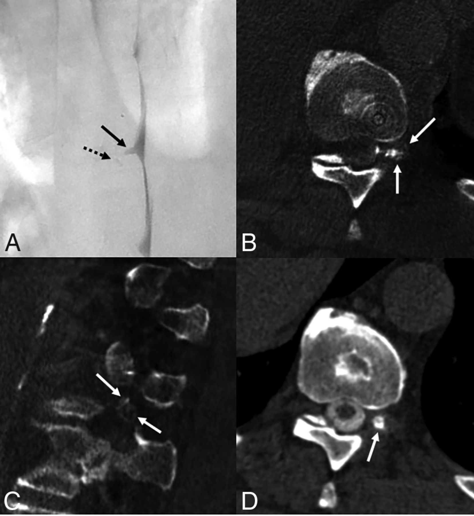
  • FIG 3.
    • Download figure
    • Open in new tab
    • Download powerpoint
    FIG 3.

    A 66-year-old man with many years of orthostatic headaches that improved after surgical treatment of a left T10 CSF-venous fistula and then recurred. Left lateral decubitus DSM (A) shows a mildly prominent left T10 meningeal diverticulum (A, arrow), with no definite venous contrast on dynamic imaging. Minimal hyperdensity seen adjacent to the diverticulum (A, dashed arrow) was initially thought to be artifactual, secondary to motion or a multilobed diverticulum. CBCT was performed to further investigate the finding. Axial (B) and sagittal (C) images from CBCT obtained during contrast injection demonstrate opacification of numerous left T10 foraminal veins (B and C, arrows), compatible with CSF-venous fistula. Axial 50-keV reconstruction from a delayed left lateral decubitus CT obtained 20 minutes later (D) shows a prominent left T10 meningeal diverticulum (D, arrow) without convincing venous contrast. The patient was treated with transvenous Onyx embolization of the left T10 fistula, with complete resolution of symptoms in 3 months.

PreviousNext
Back to top

In this issue

American Journal of Neuroradiology: 44 (3)
American Journal of Neuroradiology
Vol. 44, Issue 3
1 Mar 2023
  • Table of Contents
  • Index by author
  • Complete Issue (PDF)
Advertisement
Print
Download PDF
Email Article

Thank you for your interest in spreading the word on American Journal of Neuroradiology.

NOTE: We only request your email address so that the person you are recommending the page to knows that you wanted them to see it, and that it is not junk mail. We do not capture any email address.

Enter multiple addresses on separate lines or separate them with commas.
Conebeam CT as an Adjunct to Digital Subtraction Myelography for Detection of CSF-Venous Fistulas
(Your Name) has sent you a message from American Journal of Neuroradiology
(Your Name) thought you would like to see the American Journal of Neuroradiology web site.
CAPTCHA
This question is for testing whether or not you are a human visitor and to prevent automated spam submissions.
Cite this article
A.A. Madhavan, J.K. Cutsforth-Gregory, J.C. Benson, W. Brinjikji, I.T. Mark, J.T. Verdoorn
Conebeam CT as an Adjunct to Digital Subtraction Myelography for Detection of CSF-Venous Fistulas
American Journal of Neuroradiology Mar 2023, 44 (3) 347-350; DOI: 10.3174/ajnr.A7794

Citation Manager Formats

  • BibTeX
  • Bookends
  • EasyBib
  • EndNote (tagged)
  • EndNote 8 (xml)
  • Medlars
  • Mendeley
  • Papers
  • RefWorks Tagged
  • Ref Manager
  • RIS
  • Zotero
0 Responses
Respond to this article
Share
Bookmark this article
Conebeam CT for CSF-Venous Fistulas Detection
A.A. Madhavan, J.K. Cutsforth-Gregory, J.C. Benson, W. Brinjikji, I.T. Mark, J.T. Verdoorn
American Journal of Neuroradiology Mar 2023, 44 (3) 347-350; DOI: 10.3174/ajnr.A7794
del.icio.us logo Twitter logo Facebook logo Mendeley logo
  • Tweet Widget
  • Facebook Like
  • Google Plus One
Purchase

Jump to section

  • Article
    • Abstract
    • ABBREVIATIONS:
    • TECHNICAL REPORT
    • DISCUSSION
    • Footnotes
    • references
  • Figures & Data
  • Supplemental
  • Info & Metrics
  • Responses
  • References
  • PDF

Related Articles

  • PubMed
  • Google Scholar

Cited By...

  • Conebeam CT Myelography for the Detection of Spinal CSF Leaks
  • Assessing the Diagnostic Value of Brain White Matter Hyperintensities and Clinical Symptoms in Predicting the Detection of CSF-Venous Fistula in Patients with Suspected Spontaneous Intracranial Hypotension
  • Additional Diagnostic Value of Conebeam CT Myelography Performed after Digital Subtraction Myelography for Detecting CSF-Venous Fistulas
  • CSF-Venous Fistulas Arising Intraosseously within Bone Remodeled by Meningeal Diverticula
  • {beta}-Trace Protein as a Potential Biomarker for CSF-Venous Fistulas
  • Spontaneous Intracranial Hypotension in Children: A Multi-Institutional Review of Spinal CSF Leaks Localized on Advanced Myelography
  • Spinal CSF Leaks: The Neuroradiologist Transforming Care
  • Myelographic Techniques for the Localization of CSF-Venous Fistulas: Updates in 2024
  • Direct comparison of digital subtraction myelography versus CT myelography in lateral decubitus position: evaluation of diagnostic yield for cerebrospinal fluid-venous fistulas
  • Cone-Beam CT for the Detection of a Ventral Spinal CSF Leak in Spontaneous Intracranial Hypotension
  • Utility of Photon-Counting Detector CT Myelography for the Detection of CSF-Venous Fistulas
  • Utility of Photon-Counting Detector CT Myelography for the Detection of CSF-Venous Fistulas
  • Crossref (16)
  • Google Scholar

This article has been cited by the following articles in journals that are participating in Crossref Cited-by Linking.

  • Utility of Photon-Counting Detector CT Myelography for the Detection of CSF-Venous Fistulas
    A.A. Madhavan, L. Yu, W. Brinjikji, J.K. Cutsforth-Gregory, F.R. Schwartz, I.T. Mark, J.C. Benson, T.J. Amrhein
    American Journal of Neuroradiology 2023 44 6
  • Direct comparison of digital subtraction myelography versus CT myelography in lateral decubitus position: evaluation of diagnostic yield for cerebrospinal fluid-venous fistulas
    Niklas Lützen, Theo Demerath, Urs Würtemberger, Nebiyat Filate Belachew, Enrique Barvulsky Aleman, Katharina Wolf, Amir El Rahal, Florian Volz, Christian Fung, Jürgen Beck, Horst Urbach
    Journal of NeuroInterventional Surgery 2024 16 10
  • Myelographic Techniques for the Localization of CSF-Venous Fistulas: Updates in 2024
    Ajay A. Madhavan, Waleed Brinjikji, Jeremy K. Cutsforth-Gregory, Timothy J. Amrhein, Peter G. Kranz, John C. Benson, Felix E. Diehn, Ben A. Johnson-Tesch, Greta B. Liebo, Vance T. Lehman, Ian T. Mark, Pearse P. Morris, Michael P. Oien, Darya P. Shlapak, Jared T. Verdoorn
    American Journal of Neuroradiology 2024 45 10
  • Fluctuating hearing loss secondary to spontaneous intracranial hypotension: A case report and review of the literature
    Michal Cahal, Jonathan Roth, Omer J Ungar, Waleed Brinjikji
    Interventional Neuroradiology 2023
  • Cerebrospinal Fluid-Venous Fistulas
    Nitesh P. Patel, Waleed Brinjikji
    Neurosurgery Clinics of North America 2024 35 3
  • Spinal CSF Leaks: The Neuroradiologist Transforming Care
    Mark D. Mamlouk, Andrew L. Callen, Ajay A. Madhavan, Niklas Lützen, Lalani Carlton Jones, Ian T. Mark, Waleed Brinjikji, John C. Benson, Jared T. Verdoorn, D.K. Kim, Timothy J. Amrhein, Linda Gray, William P. Dillon, Marcel M. Maya, Thien J. Huynh, Vinil N. Shah, Tomas Dobrocky, Eike I. Piechowiak, Joseph Levi Chazen, Michael D. Malinzak, Jessica L. Houk, Peter G. Kranz
    American Journal of Neuroradiology 2024 45 11
  • Spontaneous Intracranial Hypotension in Children: A Multi-Institutional Review of Spinal CSF Leaks Localized on Advanced Myelography
    Ajay A. Madhavan, Peter G. Kranz, Lalani Carlton Jones, Edward S. Ahn, Timothy J. Amrhein, Waleed Brinjikji, Andrew Callen, Jeremy K. Cutsforth-Gregory, Mark D. Mamlouk, V. Michelle Silvera, Julie B. Guerin
    American Journal of Neuroradiology 2024 45 12
  • Additional Diagnostic Value of Conebeam CT Myelography Performed after Digital Subtraction Myelography for Detecting CSF-Venous Fistulas
    Ajay A. Madhavan, Niklas Lutzen, Jeremy K. Cutsforth-Gregory, Wouter I. Schievink, Michelle L. Kodet, Ian T. Mark, Pearse P. Morris, Steven A. Messina, John T. Wald, Waleed Brinjikji
    American Journal of Neuroradiology 2025 46 5
  • Cone-Beam CT for the Detection of a Ventral Spinal CSF Leak in Spontaneous Intracranial Hypotension
    Niklas Lützen, Theo Demerath, Florian Volz, Jürgen Beck, Horst Urbach
    Neurology 2023 101 15
  • Myelographic Techniques for the Localization of Cerebrospinal Fluid Leaks
    Ajay A. Madhavan, Vinil Shah, Waleed Brinjikji, Jeremy K. Cutsforth-Gregory
    Neuroimaging Clinics of North America 2025 35 1

More in this TOC Section

  • Bern Score Validity for SIH
  • MP2RAGE 7T in MS Lesions of the Cervical Spine
  • Deep Learning for STIR Spine MRI Quality
Show more Spine

Similar Articles

Advertisement

Indexed Content

  • Current Issue
  • Accepted Manuscripts
  • Article Preview
  • Past Issues
  • Editorials
  • Editors Choice
  • Fellow Journal Club
  • Letters to the Editor

Cases

  • Case Collection
  • Archive - Case of the Week
  • Archive - Case of the Month
  • Archive - Classic Case

Special Collections

  • Special Collections

Resources

  • News and Updates
  • Turn around Times
  • Submit a Manuscript
  • Author Policies
  • Manuscript Submission Guidelines
  • Evidence-Based Medicine Level Guide
  • Publishing Checklists
  • Graphical Abstract Preparation
  • Imaging Protocol Submission
  • Submit a Case
  • Become a Reviewer/Academy of Reviewers
  • Get Peer Review Credit from Publons

Multimedia

  • AJNR Podcast
  • AJNR SCANtastic
  • Video Articles

About Us

  • About AJNR
  • Editorial Board
  • Not an AJNR Subscriber? Join Now
  • Alerts
  • Feedback
  • Advertise with us
  • Librarian Resources
  • Permissions
  • Terms and Conditions

American Society of Neuroradiology

  • Not an ASNR Member? Join Now

© 2025 by the American Society of Neuroradiology All rights, including for text and data mining, AI training, and similar technologies, are reserved.
Print ISSN: 0195-6108 Online ISSN: 1936-959X

Powered by HighWire