
Article Figures & Data
Figures
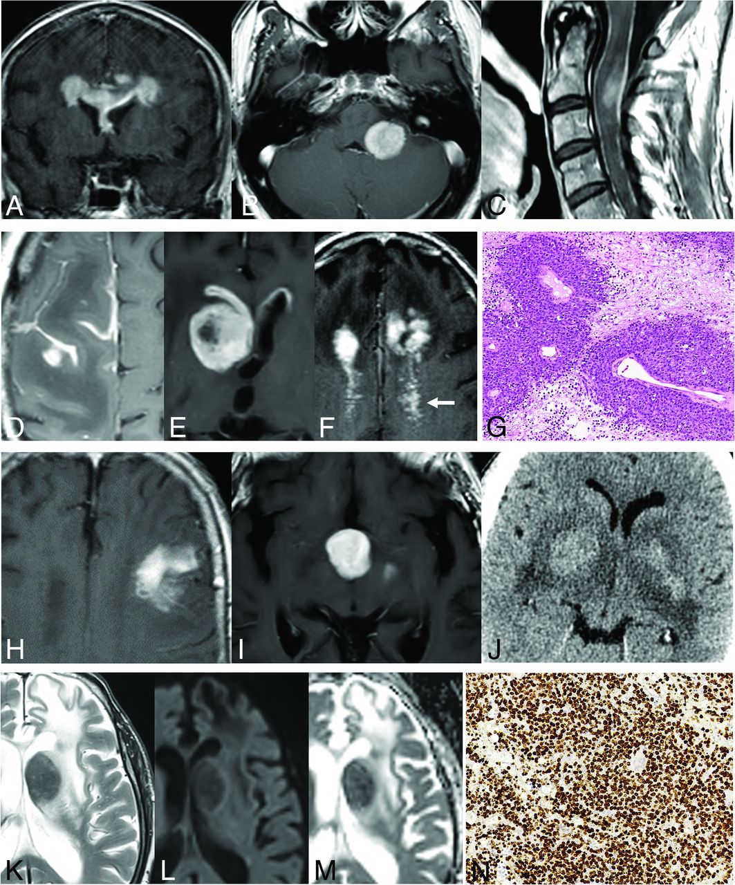
- FIG 1.
Primary DLBCLs of the CNS, EBV-negative. A–C: Usual deep, periventricular, corpus callosum, and midline location (A); a rare location in the posterior fossa (B); and exceptional in the spinal cord (C). D–G: Parenchymal lesions with associated characteristic leptomeningeal (D), subependymal (E), and perivascular (arrow, F) enhancement patterns. Histologic hematoxylin-eosin stain (original magnification ×20) shows highly cellular, perivascular accumulation of lymphoma cells (G). H–J: Mass lesion with ill-defined infiltrative (H) or well-defined expansive (I) margins. Hyperattenuated lesions on NCCT (J). K–N: Deep T2 hypointensity of a lesion (K) with a T2-blackout effect at b = 1000 image (L) but a low signal of actual diffusion restriction on the ADC map (M). Ki-67 proliferation index by immunohistochemistry (original magnification ×20) exceeding 90% (N).
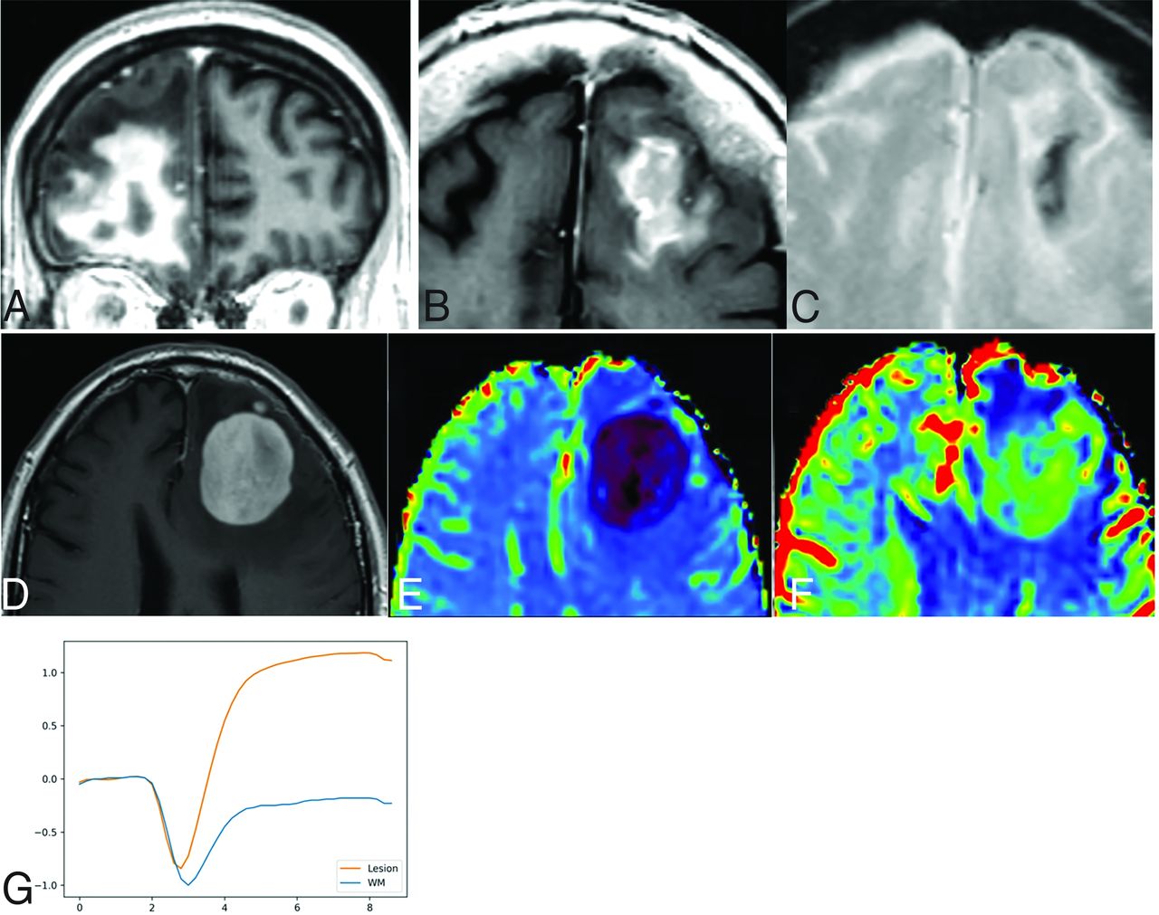
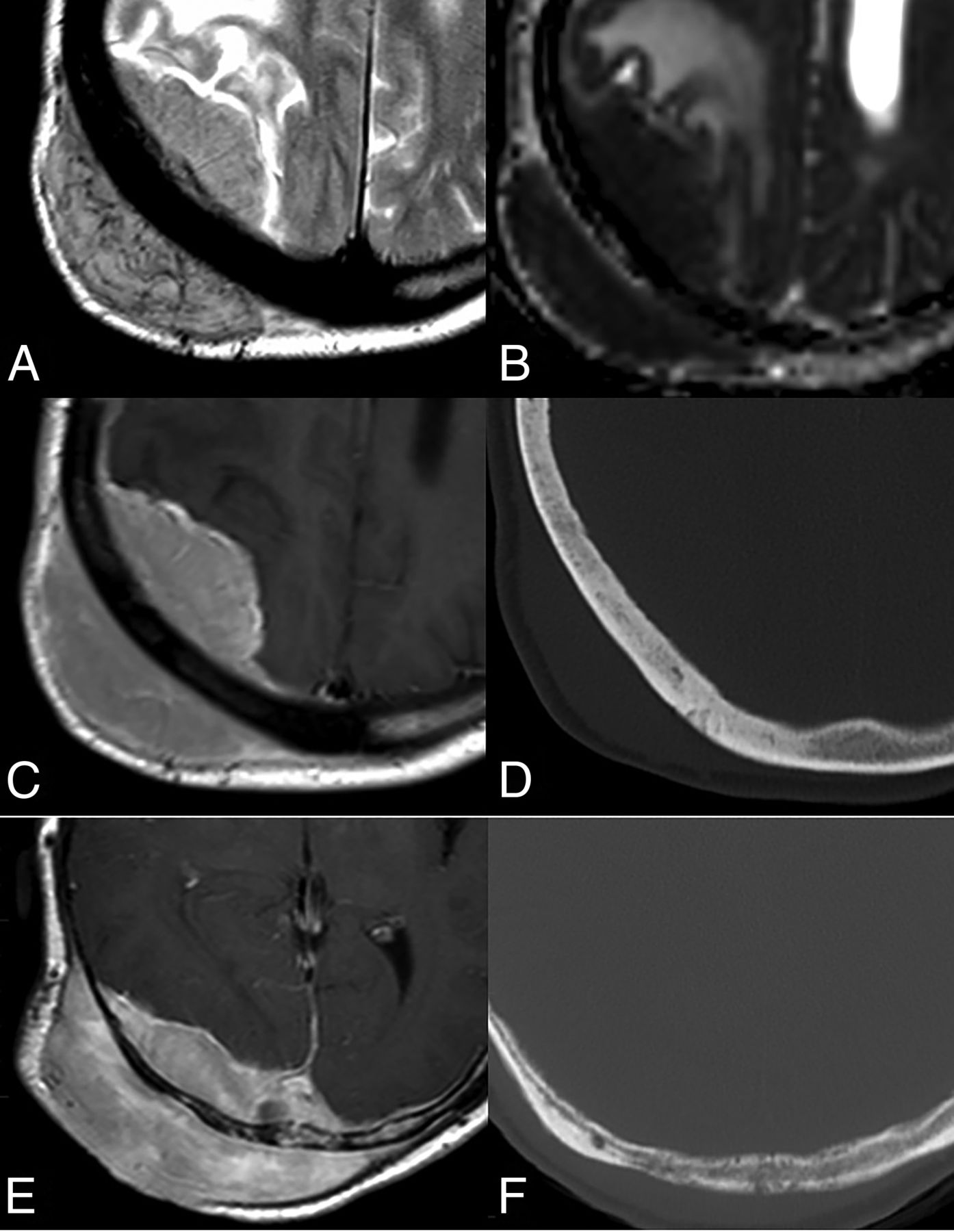
- FIG 2.
A–C: Primary DLBCLs of the CNS, EBV-negative, with imaging signs of central necrosis (A) and tumoral hemorrhage (B and C). D–G: DSC-PWI features of a left frontal primary DLBCL of the CNS, EBV-negative (D). Low-to-intermediate CBV on noncorrected (E) and corrected (F) color maps. Characteristic lymphoma DSC-PWI time-intensity curve morphology with ascending-part of the curve recovering signal intensity far above the baseline (high PSR) (G).
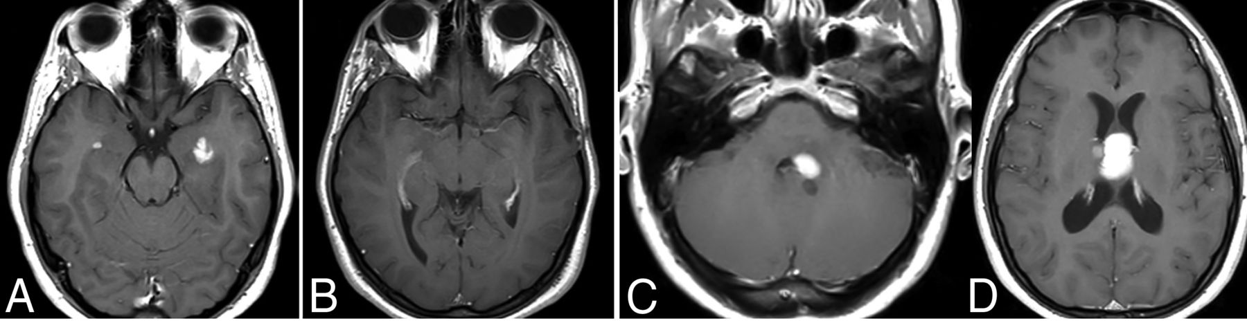
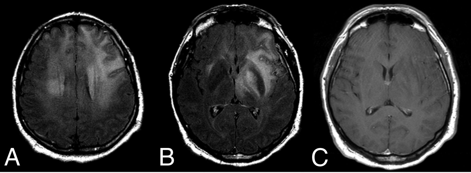
- FIG 3.
Sentinel inflammatory lesions preceding primary DLBCL of the CNS. Two enhancing periventricular temporal lesions were detected in a patient (A). A biopsy was obtained, and histopathology showed an inflammatory infiltrate without evidence of neoplasia. The lesions disappeared on further imaging controls during the following 2 years (B). In a subsequent MR imaging control, new masslike lesions reappeared (C and D). A biopsy of the new lesions yielded the final histopathologic diagnosis of primary DLBCL of the CNS.
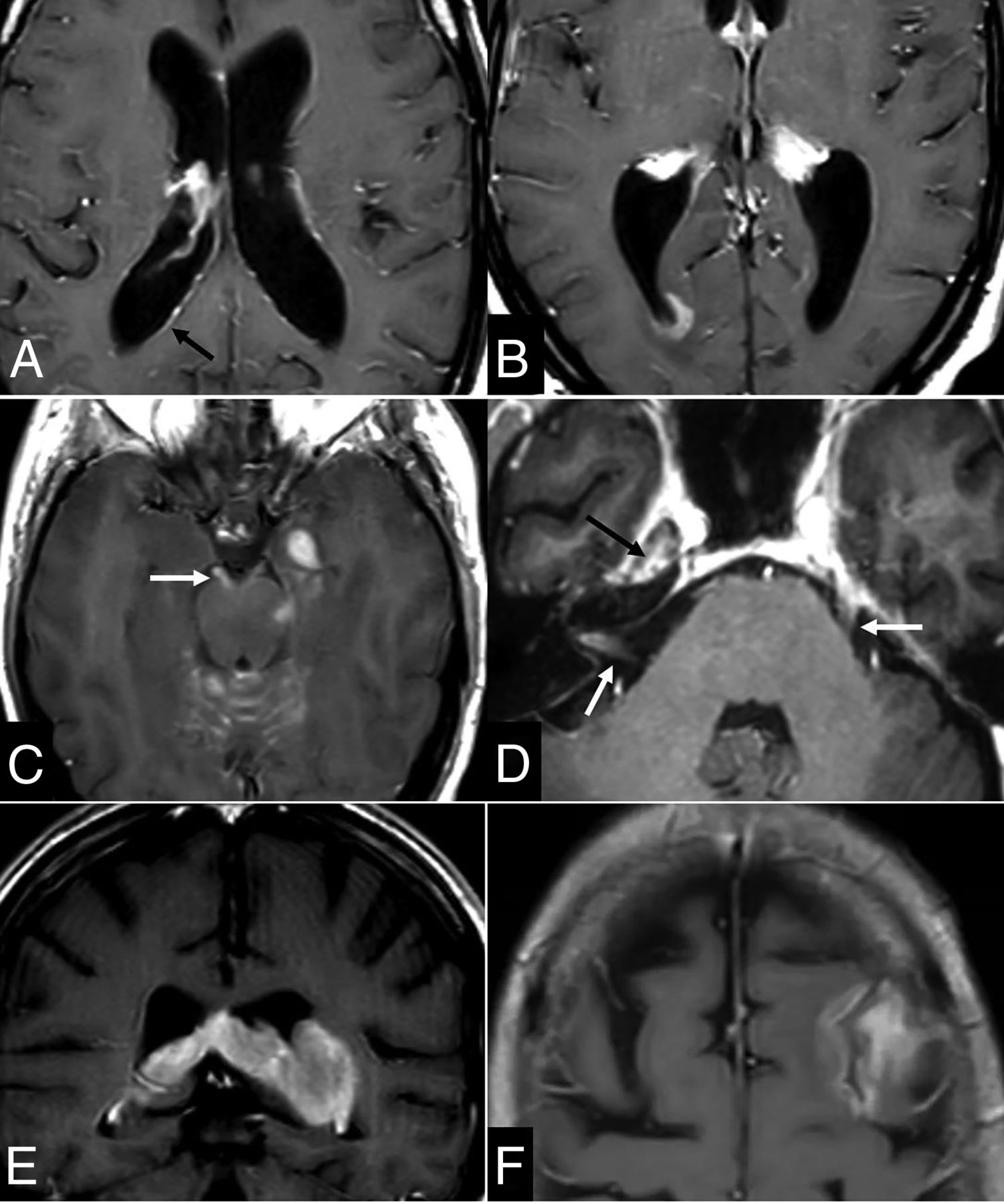
- FIG 4.
Primary DLBCLs of the CNS, EBV-positive (immunodeficiency/dysregulation-associated). Single (A) and multiple (B) lesions with prominent necrosis (C and E) and tumoral hemorrhage (D and F). Heterogeneous deep T2 hypointensity (H) of the nonenhancing central content (G) of lesions, so-called necrosis. Low-intermediate CBV on the corrected color map (I) and DSC-PWI time-intensity curve with high PSR (J), also very characteristic of this lymphoma subtype.
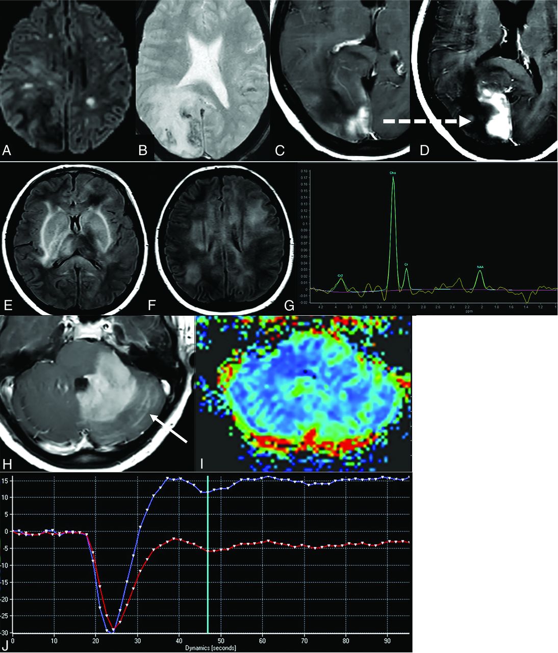
- FIG 5.
Intravascular lymphoma (A–D). Acute patched ischemia-like lesions on DWI (A), hemorrhages (B), and an area of enhancement (C), which grows on the subsequent few days of imaging control (D). Dashed arrow in C–D indicates the growth of the same enhancing-lesion in few days. DLBCL following a lymphomatosis cerebri pattern (E–J): extensive, patched, bilateral, and diffuse FLAIR hyperintensity on the basal ganglia (E) and white matter (F), with an area of enhancement in the left cerebellum (H) and associated leptomeningeal disease (arrow in H). Intermediate CBV in DSC-PWI color maps (I) and characteristic high PSR and time-intensity curve morphology (J). Tumoral pattern on 1H-MR spectroscopy at long TE with a high Cho-to-NAA ratio (H) and absent mIns at the short TE (not shown), helpful in the differential diagnosis with nontumoral entities and gliomatosis cerebri, respectively.
- FIG 6.
Dural lymphomas. MALT dural lymphoma (A–D) with extra-axial lesion features such as a CSF cleft (A) and a wide-implantation dural base with soft marginal angles (C), as well as T2-hypointensity (A) and diffusion restriction (B). Almost normal calvarial bone; only subtle sclerosis seen (D), despite the great soft-tissue component on both sides of the diploe (A–C). Similar imaging features with minimal bone destruction and a subtle permeative pattern (F) in comparison with the prominent soft-tissue component (E) in another diffuse large B-cell dural lymphoma (E and F).
- FIG 7.
NK/T-cell lymphoma presenting with a lymphomatosis cerebri radiologic pattern (A–C). Patched and diffuse, bilateral and asymmetric, deep and subcortical, hyperintense lesions on FLAIR (A and B) without contrast enhancement (C).
- FIG 8.
Imaging findings in secondary lymphomas of the CNS. A–B: Thin subtle linear (arrow in A) and nodular (B) subependymal enhancements. C–D: Prominent leptomeningeal disease along the superior vermian and cerebellar folia and third cranial nerve (arrow in C) as well as inside the right internal auditory canal—cranial nerves VII and VIII—and along the trigeminal nerve in the right Meckel cave and the left cisternal segment (arrows in D). Associated parenchymal mass in the left temporal lobe (C). E–F: Secondary lymphomas presenting as predominant intraparenchymal lesions with associated adjacent subependymal (E) and leptomeningeal (F) disease.


Jump to section
Related Articles
Cited By...
- Light-Chain Deposition Diseases of the CNS: Review of Pathogenesis, Imaging Features, and Radiographic Mimics
- Clinical manifestations and outcomes of patients with intravascular large B-cell lymphoma with neurological involvement: highlighting longitudinally extensive myelopathy as a distinct feature
- Glial Fibrillary Acidic Protein Astrocytopathy: Review of Pathogenesis, Imaging Features, and Radiographic Mimics
- Unpacking the CNS Manifestations of Epstein-Barr Virus: An Imaging Perspective