
Article Figures & Data
Figures
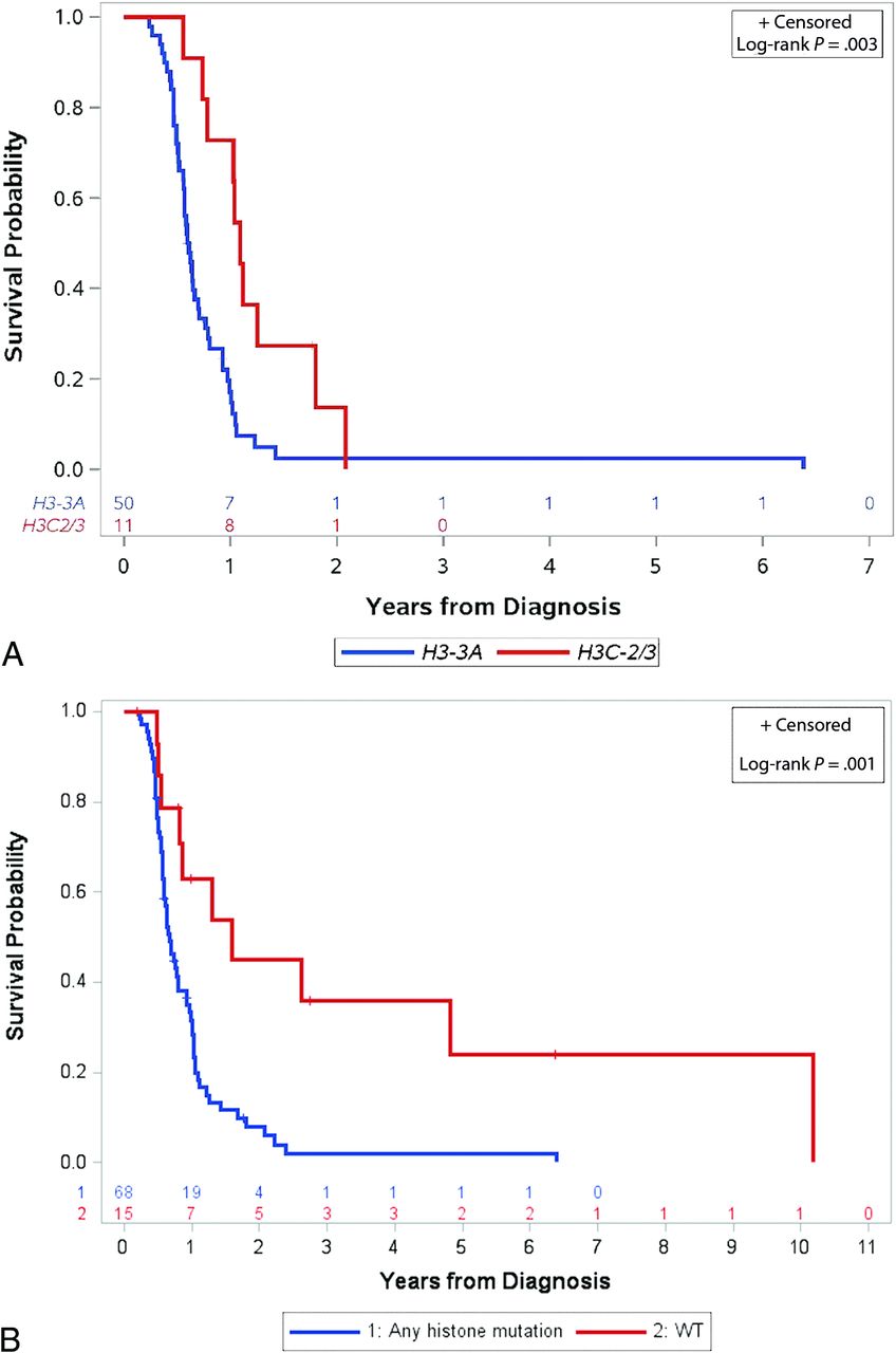
- FIG 1.
Differences in OS between variant histone profiles. A, Kaplan-Meier curves of a patient’s OS by histone H3 mutation: H3-3A (n = 50) versus H3C2/3 (n = 11). B, Kaplan-Meier curves of OS for patients with tumors with histone H3 mutation (H3-3A, H3C2/3, and H3-mutant, specific gene unknown) (n = 68) versus H3 wild-type (n = 15) tumors.
- FIG 2.
Differences in ADC histogram parameters among histone H3 K27–altered tumors. H3-3A-mutant tumor (A) shows lower ADC_total mode (1252.531 × 10–6 mm2/s), higher ADC_total skewness (0.673), and higher ADC_total kurtosis (2.721) relative to H3C2/3-altered tumor (B) (ADC_total mode: 1703.875 × 10–6 mm2/s; ADC_total skewness: −0.231; and ADC_total kurtosis: 0.399). H3-3A-mutated tumor (A) shows lower mean (PG) ADC_enhancement (1216.092 × 10 mm2/s), median (PG) ADC_enhancement (1206 × 10–6 mm2/s), and mode (PG) ADC_enhancement (1103.723 × 10–6 mm2/s), relative to H3C2/3-mutated tumor (B): mean (PG) ADC_enhancement (1461.677 × 10–6 mm2/s), median (PG) ADC_enhancement (1500 × 10–6 mm2/s), and mode (PG) ADC_enhancement (1665.322 × 10–6 mm2/s). A, A 3-year-old girl with H3-3A-mutant diffuse midline glioma and OS = 217 days. B, An 8-year-old girl with H3C2/3-mutant diffuse midline glioma and OS = 761 days.


Tables
Patient Characteristic No. (%) or Median (Range) Sex Male 43 (52) Female 40 (48) Race White 60 (72) Black 19 (23) Other 4 (5) Age at registration (yr) 6 (0.7–17) Histone mutation status H3-3A 50 (60) H3C2/3 11 (13) Wild-type 15 (18) H3 K27–altered, specific gene unknown 7 (8) Presence of enhancement Yes 54 No 25 Unknown 4 Median follow-up time of surviving patients (mo) 10.3 (2.3–76.2) - Table 2:
Association of measures of continuous imaging features with variant histone H3–altered tumors
Imaging Metric H3-3A H3C2/3 P Valuea No. Median (Range) No. Median (Range) ADC total (×10–6 mm2/s) Mean 46 1227.51 (538.06–1710.88) 9 1586.56 (1224.37–1797.58) .001 Median 46 1185 (532–1755) 9 1654 (1215–1842) .001 Mode 46 1168.14 (450.92–1902.53) 9 1703.88 (1263.52–1961.24) .001 Skewness 38 0.91 (−0.49–2.16) 9 –0.20 (−0.89–0.88) .003 Kurtosis 38 1.57 (−0.60–9.71) 9 0.40 (−0.78–1.79) .002 ADC_enhancement (×10–6 mm2/s) Mean 33 1138.81 (689.44–1509.96) 7 1390.54 (1033.58–1898.26) .004 Median 33 1085 (667–1532) 7 1364 (995–1890) .003 Mode 33 1012.56 (577.86–1617.56) 7 1393.38 (966.31–1746.86) .002 Skewness 31 1.27 (−0.56–2.82) 7 0.52 (−0.71–2.61) .1 Kurtosis 31 2.08 (−0.94–13.03) 7 0.85 (−0.84–9.09) .3 FLAIR/T2/T1 PG volume (×103 mm3) FLAIR/T2 44 35.75 (7.97–77.53) 10 41.10 (22.84–77.57) .2 Enhancing 32 1.90 (0.07–14.10) 9 5.04 (0.04–8.25) .6 ↵a Wilcoxon rank-sum test.
- Table 3:
Association of measures of continuous imaging features with variant histone H3–altered versus H3 wild-type tumors
Imaging Metric Any Histone Mutation Wild-Type P Valuea No. Median (Range) No. Median (Range) ADC total (×10–6 mm2/s) Mean 61 1302.42 (538.06–1797.58) 12 1350.77 (922.96–1729.62) .98 Median 61 1245 (532–1842) 12 1345 (917–1781) .97 Mode 61 1250.51 (450.92–1961.24) 12 1305.29 (863.39–1805.47) .9 Skewness 52 0.68 (−0.89–3.11) 11 0.98 (−0.30–2.57) .3 Kurtosis 52 1.4 (−0.78–15.62) 11 2.82 (−0.78–10.77) .1 ADC_enhancement (×10–6 mm2/s) Mean 45 1152.88 (689.44–1898.26) 6 1199.86 (956.62–1723.18) .7 Median 45 1142.00 (667–1890) 6 1187.00 (807–1762) .7 Mode 45 1062.49 (577.86–1746.86) 6 1175.10 (719.70–1805.78) .6 Skewness 43 0.86 (−0.85–4.73) 6 0.67 (−0.14–1.86) .7 Kurtosis 43 1.69 (−0.94–29.06) 6 1.35 (0.098–12.45) .9 FLAIR/T2/T1 PG volume (×103 mm3) FLAIR/T2 60 35.13 (7.97–77.57) 13 32.77 (10.13–79.62) .8 Enhancing 46 1.96 (0.04–14.10) 8 1.95 (0.01–4.71) .4 ↵a Wilcoxon rank-sum test.


Jump to section
Related Articles
Cited By...
- No citing articles found.