Advertisement

Main menu

  • Home
  • Content
    • Current Issue
    • Accepted Manuscripts
    • Article Preview
    • Past Issue Archive
    • Video Articles
    • AJNR Case Collection
    • Case of the Week Archive
    • Case of the Month Archive
    • Classic Case Archive
  • Special Collections
    • AJNR Awards
    • Low-Field MRI
    • Alzheimer Disease
    • ASNR Foundation Special Collection
    • Photon-Counting CT
    • View All
  • Multimedia
    • AJNR Podcasts
    • AJNR SCANtastic
    • Trainee Corner
    • MRI Safety Corner
    • Imaging Protocols
  • For Authors
    • Submit a Manuscript
    • Submit a Video Article
    • Submit an eLetter to the Editor/Response
    • Manuscript Submission Guidelines
    • Statistical Tips
    • Fast Publishing of Accepted Manuscripts
    • Graphical Abstract Preparation
    • Imaging Protocol Submission
    • Author Policies
  • About Us
    • About AJNR
    • Editorial Board
    • Editorial Board Alumni
  • More
    • Become a Reviewer/Academy of Reviewers
    • Subscribers
    • Permissions
    • Alerts
    • Feedback
    • Advertisers
    • ASNR Home

User menu

  • Alerts
  • Log in

Search

  • Advanced search
American Journal of Neuroradiology

American Journal of Neuroradiology

ASHNR American Society of Functional Neuroradiology ASHNR American Society of Pediatric Neuroradiology ASSR
  • Alerts
  • Log in

Advanced Search

  • Home
  • Content
    • Current Issue
    • Accepted Manuscripts
    • Article Preview
    • Past Issue Archive
    • Video Articles
    • AJNR Case Collection
    • Case of the Week Archive
    • Case of the Month Archive
    • Classic Case Archive
  • Special Collections
    • AJNR Awards
    • Low-Field MRI
    • Alzheimer Disease
    • ASNR Foundation Special Collection
    • Photon-Counting CT
    • View All
  • Multimedia
    • AJNR Podcasts
    • AJNR SCANtastic
    • Trainee Corner
    • MRI Safety Corner
    • Imaging Protocols
  • For Authors
    • Submit a Manuscript
    • Submit a Video Article
    • Submit an eLetter to the Editor/Response
    • Manuscript Submission Guidelines
    • Statistical Tips
    • Fast Publishing of Accepted Manuscripts
    • Graphical Abstract Preparation
    • Imaging Protocol Submission
    • Author Policies
  • About Us
    • About AJNR
    • Editorial Board
    • Editorial Board Alumni
  • More
    • Become a Reviewer/Academy of Reviewers
    • Subscribers
    • Permissions
    • Alerts
    • Feedback
    • Advertisers
    • ASNR Home
  • Follow AJNR on Twitter
  • Visit AJNR on Facebook
  • Follow AJNR on Instagram
  • Join AJNR on LinkedIn
  • RSS Feeds

AJNR Awards, New Junior Editors, and more. Read the latest AJNR updates

Research ArticleUltra-High-Field MRI/Imaging of Epilepsy/Demyelinating Diseases/Inflammation/Infection

Macro- and Microstructural White Matter Differences in Neurologic Postacute Sequelae of SARS-CoV-2 Infection

Erin E. O’Connor, Rosangela Salerno-Goncalves, Nikita Rednam, Rory O’Brien, Peter Rock, Andrea R. Levine and Thomas A. Zeffiro
American Journal of Neuroradiology December 2024, 45 (12) 1910-1918; DOI: https://doi.org/10.3174/ajnr.A8481
Erin E. O’Connor
aFrom the Department of Diagnostic Radiology & Nuclear Medicine (E.E.O., N.R., T.A.Z.), University of Maryland School of Medicine, Baltimore, Maryland
  • Find this author on Google Scholar
  • Find this author on PubMed
  • Search for this author on this site
  • ORCID record for Erin E. O’Connor
Rosangela Salerno-Goncalves
bDepartment of Pediatrics (R.S.-G.), University of Maryland School of Medicine, Baltimore, Maryland
  • Find this author on Google Scholar
  • Find this author on PubMed
  • Search for this author on this site
Nikita Rednam
aFrom the Department of Diagnostic Radiology & Nuclear Medicine (E.E.O., N.R., T.A.Z.), University of Maryland School of Medicine, Baltimore, Maryland
  • Find this author on Google Scholar
  • Find this author on PubMed
  • Search for this author on this site
Rory O’Brien
cLantern Lab (R.O.), Fulton, Maryland
  • Find this author on Google Scholar
  • Find this author on PubMed
  • Search for this author on this site
Peter Rock
dDepartment of Anesthesiology (P.R.), University of Maryland School of Medicine, Baltimore, Maryland
  • Find this author on Google Scholar
  • Find this author on PubMed
  • Search for this author on this site
  • ORCID record for Peter Rock
Andrea R. Levine
eDepartment of Medicine (A.R.L.), Division of Pulmonary and Critical Care Medicine, University of Maryland School of Medicine, Baltimore, Maryland
  • Find this author on Google Scholar
  • Find this author on PubMed
  • Search for this author on this site
Thomas A. Zeffiro
aFrom the Department of Diagnostic Radiology & Nuclear Medicine (E.E.O., N.R., T.A.Z.), University of Maryland School of Medicine, Baltimore, Maryland
  • Find this author on Google Scholar
  • Find this author on PubMed
  • Search for this author on this site
  • Article
  • Figures & Data
  • Supplemental
  • Info & Metrics
  • Responses
  • References
  • PDF
Loading

Article Figures & Data

Figures

  • Tables
  • FIG 1.
    • Download figure
    • Open in new tab
    • Download powerpoint
    FIG 1.

    In NeuroPASC compared with NoCOVID participants, NeuroQoL symptoms were higher in all domains, reflecting greater disability. Group differences are shown as a forest plot by using θ scores, with values of 1 representing 1 standard deviation (SD) of difference.

  • FIG 2.
    • Download figure
    • Open in new tab
    • Download powerpoint
    FIG 2.

    On the d2 Test of Attention between-group comparisons, NeuroPASC participants showed significantly: (A) lower processing speed (β = −0.30; 95% CI: −0.51–−0.09; t = −2.80), (B) concentration (β = −0.22; 95% CI: −0.43–−0.01; t = −2.09), and (C) accuracy (β = −0.27; 95% CI: −0.48–−0.06; t = −2.60).

  • FIG 3.
    • Download figure
    • Open in new tab
    • Download powerpoint
    FIG 3.

    The total intracranial tissue compartments show interrelated changes consistent with maintenance of constant intracranial volume for each participant. In NeuroPASC participants, cerebral global WM volume is significantly higher, GM is higher, and CSF is lower (left). A voxelwise map of WM volume revealed widespread, predominantly frontal and subcortical, higher regional WM volume in the NeuroPASC group (red-yellow; P = .005, by using TFCE FWE-correction (right).

  • FIG 4.
    • Download figure
    • Open in new tab
    • Download powerpoint
    FIG 4.

    In NeuroPASC participants, regional GM volume was higher in the prefrontal cortex. Voxelwise analysis of group differences in GM volume (red-yellow) are superimposed on the group mean T1-weighted structural images. Suprathreshold voxels that represent areas of higher GM volume in NeuroPASC compared with NoCOVID (P = .005, TFCE FWE-corrected) are shown in coronal slices with MNI coordinates and participant left on viewer left. The color bar shows T statistic values.

  • FIG 5.
    • Download figure
    • Open in new tab
    • Download powerpoint
    FIG 5.

    In NeuroPASC participants, WM mean kurtosis is significantly higher. A voxelwise analysis demonstrates group differences. Areas of higher mean kurtosis (red-yellow) superimposed on the DTI fractional anisotropy skeleton (blue) represent areas of higher WM mean kurtosis in NeuroPASC compared with NoCOVID (left) axial view, and (right) coronal view. The critical threshold was (P = .05, TFCE FWE-corrected).
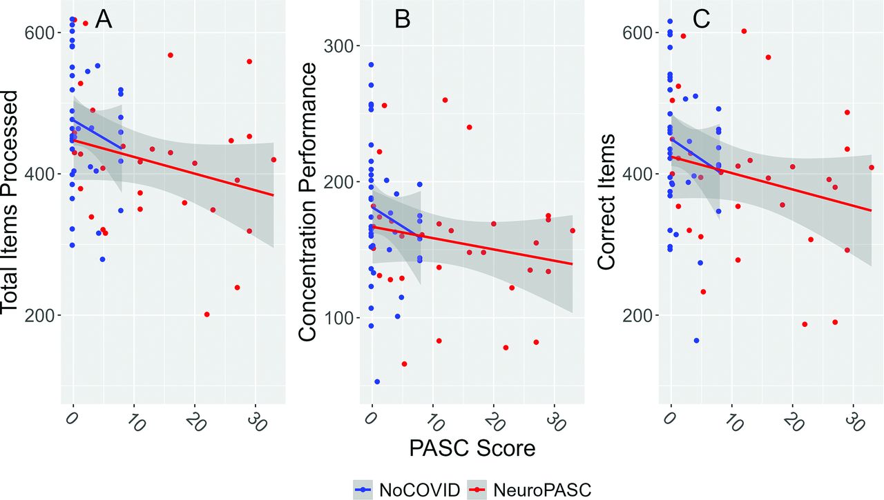
  • FIG 6.
    • Download figure
    • Open in new tab
    • Download powerpoint
    FIG 6.

    Higher clinical PASC scores were associated with significantly lower processing speed (A: β = −0.30; 95% CI: −0.51–−0.09; t = −2.80), lower concentration (B: β = −0.22; 95% CI: −0.43–−0.01; t = −2.09), and fewer correct responses (C: β = −0.27; 95% CI: −0.48–−0.06; t = −2.60).

  • FIG 7.
    • Download figure
    • Open in new tab
    • Download powerpoint
    FIG 7.

    Normalized cytokine profiles differ between the NeuroPASC and NoCOVID groups (P <.05). IFN-λ1, IFN-λ2/3, and IL-1β concentrations were higher in the NeuroPASC group.

Tables

  • Figures
  • Participant characteristics

    NoCOVIDNeuroPASCSMD
    n4131
    PASC score21.6 (3.1)13.0 (10.8)1.43
    Age in years (mean [SD])46.8 (15.6)45.0 (13.5)0.12
    Sex = F (%)19 (46.3)14 (45.2)0.024
    Education years (SD)16.7 (3.1)16.7 (3.9)0.009
    CES-D score (mean [SD])6.4 (5.4)14.3 (11.0)0.913
    Headache6 (14.6)17 (54.8)0.931
    Vaccination = Yes (%)40 (100.0)29 (93.5)–
    Hospitalization = Yes (%)–9 (29.0)–
    Supplemental oxygen = Yes (%)–7 (23.3)–
    Mechanical ventilation = Yes (%)–3 (10.0)–
    Days from infection (mean [SD])–322.13 (179.52)–
    • Note:—SD indicates standard deviation; SMD, standardized mean difference.

PreviousNext
Back to top

In this issue

American Journal of Neuroradiology: 45 (12)
American Journal of Neuroradiology
Vol. 45, Issue 12
1 Dec 2024
  • Table of Contents
  • Index by author
  • Complete Issue (PDF)
Advertisement
Print
Download PDF
Email Article

Thank you for your interest in spreading the word on American Journal of Neuroradiology.

NOTE: We only request your email address so that the person you are recommending the page to knows that you wanted them to see it, and that it is not junk mail. We do not capture any email address.

Enter multiple addresses on separate lines or separate them with commas.
Macro- and Microstructural White Matter Differences in Neurologic Postacute Sequelae of SARS-CoV-2 Infection
(Your Name) has sent you a message from American Journal of Neuroradiology
(Your Name) thought you would like to see the American Journal of Neuroradiology web site.
CAPTCHA
This question is for testing whether or not you are a human visitor and to prevent automated spam submissions.
Cite this article
Erin E. O’Connor, Rosangela Salerno-Goncalves, Nikita Rednam, Rory O’Brien, Peter Rock, Andrea R. Levine, Thomas A. Zeffiro
Macro- and Microstructural White Matter Differences in Neurologic Postacute Sequelae of SARS-CoV-2 Infection
American Journal of Neuroradiology Dec 2024, 45 (12) 1910-1918; DOI: 10.3174/ajnr.A8481

Citation Manager Formats

  • BibTeX
  • Bookends
  • EasyBib
  • EndNote (tagged)
  • EndNote 8 (xml)
  • Medlars
  • Mendeley
  • Papers
  • RefWorks Tagged
  • Ref Manager
  • RIS
  • Zotero
0 Responses
Respond to this article
Share
Bookmark this article
White Matter Changes Post-SARS CoV-2 Infection
Erin E. O’Connor, Rosangela Salerno-Goncalves, Nikita Rednam, Rory O’Brien, Peter Rock, Andrea R. Levine, Thomas A. Zeffiro
American Journal of Neuroradiology Dec 2024, 45 (12) 1910-1918; DOI: 10.3174/ajnr.A8481
del.icio.us logo Twitter logo Facebook logo Mendeley logo
  • Tweet Widget
  • Facebook Like
  • Google Plus One
Purchase

Jump to section

  • Article
    • Abstract
    • ABBREVIATIONS:
    • MATERIALS AND METHODS
    • RESULTS
    • DISCUSSION
    • CONCLUSIONS
    • Acknowledgments
    • Footnotes
    • References
  • Figures & Data
  • Supplemental
  • Info & Metrics
  • Responses
  • References
  • PDF

Related Articles

  • PubMed
  • Google Scholar

Cited By...

  • No citing articles found.
  • Crossref
  • Google Scholar

This article has not yet been cited by articles in journals that are participating in Crossref Cited-by Linking.

More in this TOC Section

  • DL Image Reconstruction in T2-Weighted TSE at 7T
  • Automated Lesion Segmentation Software in MS
  • Interpretation Errors in Adults with MRE
Show more Ultra-High-Field MRI/Imaging of Epilepsy/Demyelinating Diseases/Inflammation/Infection

Similar Articles

Advertisement

Indexed Content

  • Current Issue
  • Accepted Manuscripts
  • Article Preview
  • Past Issues
  • Editorials
  • Editor's Choice
  • Fellows' Journal Club
  • Letters to the Editor
  • Video Articles

Cases

  • Case Collection
  • Archive - Case of the Week
  • Archive - Case of the Month
  • Archive - Classic Case

More from AJNR

  • Trainee Corner
  • Imaging Protocols
  • MRI Safety Corner
  • Book Reviews

Multimedia

  • AJNR Podcasts
  • AJNR Scantastics

Resources

  • Turnaround Time
  • Submit a Manuscript
  • Submit a Video Article
  • Submit an eLetter to the Editor/Response
  • Manuscript Submission Guidelines
  • Statistical Tips
  • Fast Publishing of Accepted Manuscripts
  • Graphical Abstract Preparation
  • Imaging Protocol Submission
  • Evidence-Based Medicine Level Guide
  • Publishing Checklists
  • Author Policies
  • Become a Reviewer/Academy of Reviewers
  • News and Updates

About Us

  • About AJNR
  • Editorial Board
  • Editorial Board Alumni
  • Alerts
  • Permissions
  • Not an AJNR Subscriber? Join Now
  • Advertise with Us
  • Librarian Resources
  • Feedback
  • Terms and Conditions
  • AJNR Editorial Board Alumni

American Society of Neuroradiology

  • Not an ASNR Member? Join Now

© 2025 by the American Society of Neuroradiology All rights, including for text and data mining, AI training, and similar technologies, are reserved.
Print ISSN: 0195-6108 Online ISSN: 1936-959X

Powered by HighWire