Advertisement

Main menu

  • Home
  • Content
    • Current Issue
    • Accepted Manuscripts
    • Article Preview
    • Past Issue Archive
    • AJNR Case Collection
    • Case of the Week Archive
    • Classic Case Archive
    • Case of the Month Archive
  • Special Collections
    • Spinal CSF Leak Articles (Jan 2020-June 2024)
    • 2024 AJNR Journal Awards
    • Most Impactful AJNR Articles
  • Multimedia
    • AJNR Podcast
    • AJNR Scantastics
    • Video Articles
  • For Authors
    • Submit a Manuscript
    • Author Policies
    • Fast publishing of Accepted Manuscripts
    • Graphical Abstract Preparation
    • Manuscript Submission Guidelines
    • Imaging Protocol Submission
    • Submit a Case for the Case Collection
  • About Us
    • About AJNR
    • Editorial Board
  • More
    • Become a Reviewer/Academy of Reviewers
    • Subscribers
    • Permissions
    • Alerts
    • Feedback
    • Advertisers
    • ASNR Home
  • Other Publications
    • ajnr

User menu

  • Alerts
  • Log in

Search

  • Advanced search
American Journal of Neuroradiology

American Journal of Neuroradiology

ASHNR American Society of Functional Neuroradiology ASHNR American Society of Pediatric Neuroradiology ASSR
  • Alerts
  • Log in

Advanced Search

  • Home
  • Content
    • Current Issue
    • Accepted Manuscripts
    • Article Preview
    • Past Issue Archive
    • AJNR Case Collection
    • Case of the Week Archive
    • Classic Case Archive
    • Case of the Month Archive
  • Special Collections
    • Spinal CSF Leak Articles (Jan 2020-June 2024)
    • 2024 AJNR Journal Awards
    • Most Impactful AJNR Articles
  • Multimedia
    • AJNR Podcast
    • AJNR Scantastics
    • Video Articles
  • For Authors
    • Submit a Manuscript
    • Author Policies
    • Fast publishing of Accepted Manuscripts
    • Graphical Abstract Preparation
    • Manuscript Submission Guidelines
    • Imaging Protocol Submission
    • Submit a Case for the Case Collection
  • About Us
    • About AJNR
    • Editorial Board
  • More
    • Become a Reviewer/Academy of Reviewers
    • Subscribers
    • Permissions
    • Alerts
    • Feedback
    • Advertisers
    • ASNR Home
  • Follow AJNR on Twitter
  • Visit AJNR on Facebook
  • Follow AJNR on Instagram
  • Join AJNR on LinkedIn
  • RSS Feeds

Welcome to the new AJNR, Updated Hall of Fame, and more. Read the full announcements.


AJNR is seeking candidates for the position of Associate Section Editor, AJNR Case Collection. Read the full announcement.


  • Getting new auth cookie, if you see this message a lot, tell someone!
  • Getting new auth cookie, if you see this message a lot, tell someone!
Research ArticleNeurointervention
Open Access

Lateral Compression Manipulation: A Simple Approach for Sizing Taller-Than-Wide Intracranial Aneurysms with the Woven EndoBridge Device

Han Seng Chew, Ming Yao Chong, Waleed Butt, Samer Al-Ali, Benjamin Butler, Mohammad Al-Tibi, Swarupsinh Chavda and Saleh Lamin
American Journal of Neuroradiology June 2024, 45 (6) 731-736; DOI: https://doi.org/10.3174/ajnr.A8172
Han Seng Chew
aFrom the Department of Neuroradiology Queen Elizabeth Hospital Birmingham, Birmingham, UK
  • Find this author on Google Scholar
  • Find this author on PubMed
  • Search for this author on this site
  • ORCID record for Han Seng Chew
Ming Yao Chong
aFrom the Department of Neuroradiology Queen Elizabeth Hospital Birmingham, Birmingham, UK
  • Find this author on Google Scholar
  • Find this author on PubMed
  • Search for this author on this site
  • ORCID record for Ming Yao Chong
Waleed Butt
aFrom the Department of Neuroradiology Queen Elizabeth Hospital Birmingham, Birmingham, UK
  • Find this author on Google Scholar
  • Find this author on PubMed
  • Search for this author on this site
Samer Al-Ali
aFrom the Department of Neuroradiology Queen Elizabeth Hospital Birmingham, Birmingham, UK
  • Find this author on Google Scholar
  • Find this author on PubMed
  • Search for this author on this site
  • ORCID record for Samer Al-Ali
Benjamin Butler
aFrom the Department of Neuroradiology Queen Elizabeth Hospital Birmingham, Birmingham, UK
  • Find this author on Google Scholar
  • Find this author on PubMed
  • Search for this author on this site
  • ORCID record for Benjamin Butler
Mohammad Al-Tibi
aFrom the Department of Neuroradiology Queen Elizabeth Hospital Birmingham, Birmingham, UK
  • Find this author on Google Scholar
  • Find this author on PubMed
  • Search for this author on this site
Swarupsinh Chavda
aFrom the Department of Neuroradiology Queen Elizabeth Hospital Birmingham, Birmingham, UK
  • Find this author on Google Scholar
  • Find this author on PubMed
  • Search for this author on this site
  • ORCID record for Swarupsinh Chavda
Saleh Lamin
aFrom the Department of Neuroradiology Queen Elizabeth Hospital Birmingham, Birmingham, UK
  • Find this author on Google Scholar
  • Find this author on PubMed
  • Search for this author on this site
  • Article
  • Figures & Data
  • Supplemental
  • Info & Metrics
  • Responses
  • References
  • PDF
Loading

Article Figures & Data

Figures

  • Tables
  • FIG 1.
    • Download figure
    • Open in new tab
    • Download powerpoint
    FIG 1.

    The manufacturer’s sizing chart for the WEB device does not cover aneurysms with a height-to-dome ratio of >1. Our proposed approach expands the original range of treatable aneurysms to include those shown in the red zone, effectively doubling the spectrum of aneurysms without the need for additional fixed-sized WEB devices.

  • FIG 2.
    • Download figure
    • Open in new tab
    • Download powerpoint
    FIG 2.

    Diagram depicting the proposed sizing strategy for aneurysms with a high height-to-dome ratio. The appropriate WEB size is determined by strategically swapping the height and width dimensions of the aneurysm. For an aneurysm measuring w × h, it is advisable to use a h × w WEB. When fully deployed, the device undergoes significant lateral compression, leading to a substantial increase in its vertical extension, which helps to achieve an optimal fit for the aneurysm.
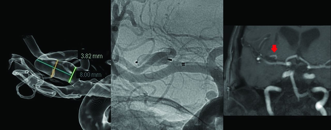
  • FIG 3.
    • Download figure
    • Open in new tab
    • Download powerpoint
    FIG 3.

    An elective embolization was performed on a right cylindrical MCA aneurysm, with measurements indicating a maximum width of 3.82 mm and a height of 8 mm. An 8 × 4 mm WEB SL device was successfully deployed without impeding the adjacent temporal branch. As a result of intense lateral compression, the base of the SL device assumed a cup-shaped configuration, leaving a small “dog ear” at the neck of the aneurysm. At the 6-month follow-up, the aneurysm continued to show satisfactory results (red arrow).
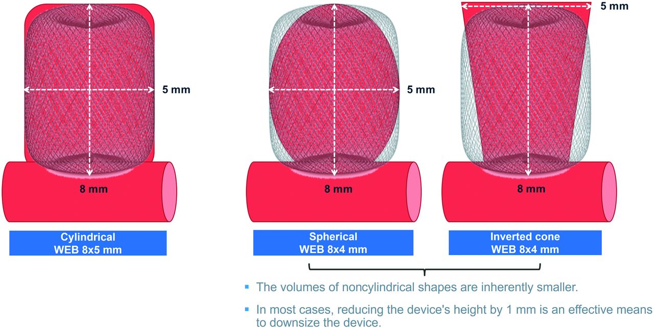
  • FIG 4.
    • Download figure
    • Open in new tab
    • Download powerpoint
    FIG 4.

    When one deals with aneurysms of noncylindrical shapes, interobserver discrepancies in image interpretation and aneurysm measurement can result in inconsistent results. Aneurysms with noncylindrical shapes generally have smaller actual volumes than their cylindrical counterparts with the same maximum width and height. To ensure an adequate fit within the aneurysms, one must downsize the WEB device by reducing the device height. In this visual representation, all 3 aneurysms share the same maximum width and height dimensions of 5 × 8 mm, suggesting that they should be treated with an 8 × 5 mm WEB device according to the recommended sizing method. However, the actual volumes of the 2 noncylindrical aneurysms are smaller and would not provide sufficient room for an 8 × 5 mm device. The WEB device can be downsized by reducing the height by 1 mm, making an 8 × 4 mm device a more appropriate choice for these aneurysms to address this issue.

  • FIG 5.
    • Download figure
    • Open in new tab
    • Download powerpoint
    FIG 5.

    WEB embolization of an acute anterior communicating artery aneurysm, characterized by a maximum width of 5.4 mm and a height of 8.5 mm. A strategic downsizing was performed due to the noncylindrical shape of the aneurysm. Instead of using an 8 × 5 mm device, an 8 × 4 mm WEB SL was selected for the procedure. A follow-up at 6 months showed that the aneurysm remained fully occluded (red arrow).

  • FIG 6.
    • Download figure
    • Open in new tab
    • Download powerpoint
    FIG 6.

    When subjected to extreme, lateral compression, the WEB SL device tends to assume a characteristic hourglass shape, while the WEB SLS is more likely to take on a cylindrical configuration. As a result, the base of the WEB SL device frequently adopts a concave, cup-shaped appearance (open arrow), whereas the WEB SLS device tends to create a flat base (solid arrow). This inversion of configurations can potentially affect the neck coverage during the treatment of the aneurysm.

Tables

  • Figures
  • Comparison of patient demographics and details of operative procedures between group A, which adhered to the proposed sizing method, and group B, which did not

    Group AGroup BP Value
    Patient demographics
     No. (total =25)205
     Age (mean) (yr)63.8 (SD, 11.2)58.6 (SD, 14.5)
     Sex (female/male)13:7 (65.0%)4:1 (80.0%).642
    Rupture status.289
     Acute SAH14 (70%)5 (100%)
     Elective6 (30%)
    Location of aneurysm
     MCA7 (35%)3 (60%)
     AcomA11 (55%)
     Ophthalmic ICA1 (5%)1 (20%)
     Pericallosal1 (20%)
     Anterior choroidal1 (5%)
    Morphology of aneurysm
     Cylindrical6 (30%)
     Teardrop1 (5%)1 (20%)
     Spindle2 (10%)
     Irregular11 (55%)4 (80%)
    Aneurysm dimensions (mean, range)
     Width 4.95 mm (3.32–8.40 mm)5.40 mm (3.55–7.73 mm)
     Height7.11 mm (5.24–10.60 mm)8.44 mm (6.60–11.39 mm)
     Neck3.70 mm (2.36–6.00 mm)3.81 mm (2.36–4.70 mm)
    Degree of lateral compression2.05 mm (1.4–4.0 mm)2.00 mm (0.03–3.27 mm)
    2-Year imaging follow-up10 (50%)5 (100%).061
    Occlusion rate.001 (<.05)
     WOS grade A14 (70%)1 (20%)a
     WOS grade B6 (30%)
     WOS grade C4 (80%)
    Operative procedure
     WEB sizes (width).822
      3–7 mm14 (70%)3 (60%)
      8–9 mm4 (20%)1 (20%)
      10–11 mm2 (10%)1 (20%)
    WEB models.012 (<.05)
     SL/SLS18 (90%)2 (40%)
     SL/SLS 172 (10%)3 (60%)
    • Note:—AcomA indicates anterior communicating artery.

    • ↵a Successful replacement of an oversized WEB device conducted during the same procedure resulted in complete angiographic occlusion on subsequent follow-up.

PreviousNext
Back to top

In this issue

American Journal of Neuroradiology: 45 (6)
American Journal of Neuroradiology
Vol. 45, Issue 6
1 Jun 2024
  • Table of Contents
  • Index by author
  • Complete Issue (PDF)
Advertisement
Print
Download PDF
Email Article

Thank you for your interest in spreading the word on American Journal of Neuroradiology.

NOTE: We only request your email address so that the person you are recommending the page to knows that you wanted them to see it, and that it is not junk mail. We do not capture any email address.

Enter multiple addresses on separate lines or separate them with commas.
Lateral Compression Manipulation: A Simple Approach for Sizing Taller-Than-Wide Intracranial Aneurysms with the Woven EndoBridge Device
(Your Name) has sent you a message from American Journal of Neuroradiology
(Your Name) thought you would like to see the American Journal of Neuroradiology web site.
CAPTCHA
This question is for testing whether or not you are a human visitor and to prevent automated spam submissions.
Cite this article
Han Seng Chew, Ming Yao Chong, Waleed Butt, Samer Al-Ali, Benjamin Butler, Mohammad Al-Tibi, Swarupsinh Chavda, Saleh Lamin
Lateral Compression Manipulation: A Simple Approach for Sizing Taller-Than-Wide Intracranial Aneurysms with the Woven EndoBridge Device
American Journal of Neuroradiology Jun 2024, 45 (6) 731-736; DOI: 10.3174/ajnr.A8172

Citation Manager Formats

  • BibTeX
  • Bookends
  • EasyBib
  • EndNote (tagged)
  • EndNote 8 (xml)
  • Medlars
  • Mendeley
  • Papers
  • RefWorks Tagged
  • Ref Manager
  • RIS
  • Zotero
0 Responses
Respond to this article
Share
Bookmark this article
Lateral Compression for Intracranial Aneurysms
Han Seng Chew, Ming Yao Chong, Waleed Butt, Samer Al-Ali, Benjamin Butler, Mohammad Al-Tibi, Swarupsinh Chavda, Saleh Lamin
American Journal of Neuroradiology Jun 2024, 45 (6) 731-736; DOI: 10.3174/ajnr.A8172
del.icio.us logo Twitter logo Facebook logo Mendeley logo
  • Tweet Widget
  • Facebook Like
  • Google Plus One
Purchase

Jump to section

  • Article
    • Abstract
    • ABBREVIATIONS:
    • MATERIALS AND METHODS
    • RESULTS
    • DISCUSSION
    • CONCLUSIONS
    • Footnotes
    • References
  • Figures & Data
  • Supplemental
  • Info & Metrics
  • Responses
  • References
  • PDF

Related Articles

  • PubMed
  • Google Scholar

Cited By...

  • No citing articles found.
  • Crossref
  • Google Scholar

This article has not yet been cited by articles in journals that are participating in Crossref Cited-by Linking.

More in this TOC Section

  • Vascular Injury After Stent Retriever Retraction
  • Contrast-Induced Sialadenitis After NeuroIR
  • Mechanic Thrombectomy with ANA Device: ANAIS Study
Show more Neurointervention

Similar Articles

Advertisement

Indexed Content

  • Current Issue
  • Accepted Manuscripts
  • Article Preview
  • Past Issues
  • Editorials
  • Editors Choice
  • Fellow Journal Club
  • Letters to the Editor

Cases

  • Case Collection
  • Archive - Case of the Week
  • Archive - Case of the Month
  • Archive - Classic Case

Special Collections

  • Special Collections

Resources

  • News and Updates
  • Turn around Times
  • Submit a Manuscript
  • Author Policies
  • Manuscript Submission Guidelines
  • Evidence-Based Medicine Level Guide
  • Publishing Checklists
  • Graphical Abstract Preparation
  • Imaging Protocol Submission
  • Submit a Case
  • Become a Reviewer/Academy of Reviewers
  • Get Peer Review Credit from Publons

Multimedia

  • AJNR Podcast
  • AJNR SCANtastic
  • Video Articles

About Us

  • About AJNR
  • Editorial Board
  • Not an AJNR Subscriber? Join Now
  • Alerts
  • Feedback
  • Advertise with us
  • Librarian Resources
  • Permissions
  • Terms and Conditions

American Society of Neuroradiology

  • Not an ASNR Member? Join Now

© 2025 by the American Society of Neuroradiology All rights, including for text and data mining, AI training, and similar technologies, are reserved.
Print ISSN: 0195-6108 Online ISSN: 1936-959X

Powered by HighWire