Advertisement

Main menu

  • Home
  • Content
    • Current Issue
    • Accepted Manuscripts
    • Article Preview
    • Past Issue Archive
    • Video Articles
    • AJNR Case Collection
    • Case of the Week Archive
    • Case of the Month Archive
    • Classic Case Archive
  • Special Collections
    • AJNR Awards
    • Low-Field MRI
    • Alzheimer Disease
    • ASNR Foundation Special Collection
    • Photon-Counting CT
    • View All
  • Multimedia
    • AJNR Podcasts
    • AJNR SCANtastic
    • Trainee Corner
    • MRI Safety Corner
    • Imaging Protocols
  • For Authors
    • Submit a Manuscript
    • Submit a Video Article
    • Submit an eLetter to the Editor/Response
    • Manuscript Submission Guidelines
    • Statistical Tips
    • Fast Publishing of Accepted Manuscripts
    • Graphical Abstract Preparation
    • Imaging Protocol Submission
    • Author Policies
  • About Us
    • About AJNR
    • Editorial Board
    • Editorial Board Alumni
  • More
    • Become a Reviewer/Academy of Reviewers
    • Subscribers
    • Permissions
    • Alerts
    • Feedback
    • Advertisers
    • ASNR Home

User menu

  • Alerts
  • Log in

Search

  • Advanced search
American Journal of Neuroradiology

American Journal of Neuroradiology

ASHNR American Society of Functional Neuroradiology ASHNR American Society of Pediatric Neuroradiology ASSR
  • Alerts
  • Log in

Advanced Search

  • Home
  • Content
    • Current Issue
    • Accepted Manuscripts
    • Article Preview
    • Past Issue Archive
    • Video Articles
    • AJNR Case Collection
    • Case of the Week Archive
    • Case of the Month Archive
    • Classic Case Archive
  • Special Collections
    • AJNR Awards
    • Low-Field MRI
    • Alzheimer Disease
    • ASNR Foundation Special Collection
    • Photon-Counting CT
    • View All
  • Multimedia
    • AJNR Podcasts
    • AJNR SCANtastic
    • Trainee Corner
    • MRI Safety Corner
    • Imaging Protocols
  • For Authors
    • Submit a Manuscript
    • Submit a Video Article
    • Submit an eLetter to the Editor/Response
    • Manuscript Submission Guidelines
    • Statistical Tips
    • Fast Publishing of Accepted Manuscripts
    • Graphical Abstract Preparation
    • Imaging Protocol Submission
    • Author Policies
  • About Us
    • About AJNR
    • Editorial Board
    • Editorial Board Alumni
  • More
    • Become a Reviewer/Academy of Reviewers
    • Subscribers
    • Permissions
    • Alerts
    • Feedback
    • Advertisers
    • ASNR Home
  • Follow AJNR on Twitter
  • Visit AJNR on Facebook
  • Follow AJNR on Instagram
  • Join AJNR on LinkedIn
  • RSS Feeds

AJNR Awards, New Junior Editors, and more. Read the latest AJNR updates

  • Getting new auth cookie, if you see this message a lot, tell someone!
  • Getting new auth cookie, if you see this message a lot, tell someone!
Review ArticleReview Article

Imaging Findings in Giant Cell Arteritis: Don’t Turn a Blind Eye to the Obvious!

Girish Bathla, Amit K. Agarwal, Steven A. Messina, David F. Black, Neetu Soni, Felix E. Diehn, Norbert G. Campeau, Vance T. Lehman, Kenneth J. Warrington, Rennie L. Rhee and Thorsten A. Bley
American Journal of Neuroradiology March 2025, 46 (3) 457-464; DOI: https://doi.org/10.3174/ajnr.A8388
Girish Bathla
aFrom the Department of Radiology (G.B., S.A.M., D.F.B., F.E.D., N.G.C., V.T.L.), Mayo Clinic, Rochester, Minnesota
  • Find this author on Google Scholar
  • Find this author on PubMed
  • Search for this author on this site
  • ORCID record for Girish Bathla
Amit K. Agarwal
bDepartment of Radiology (A.K.A., N.S.), Mayo Clinic, Jacksonville, Florida
  • Find this author on Google Scholar
  • Find this author on PubMed
  • Search for this author on this site
  • ORCID record for Amit K. Agarwal
Steven A. Messina
aFrom the Department of Radiology (G.B., S.A.M., D.F.B., F.E.D., N.G.C., V.T.L.), Mayo Clinic, Rochester, Minnesota
  • Find this author on Google Scholar
  • Find this author on PubMed
  • Search for this author on this site
  • ORCID record for Steven A. Messina
David F. Black
aFrom the Department of Radiology (G.B., S.A.M., D.F.B., F.E.D., N.G.C., V.T.L.), Mayo Clinic, Rochester, Minnesota
  • Find this author on Google Scholar
  • Find this author on PubMed
  • Search for this author on this site
  • ORCID record for David F. Black
Neetu Soni
bDepartment of Radiology (A.K.A., N.S.), Mayo Clinic, Jacksonville, Florida
  • Find this author on Google Scholar
  • Find this author on PubMed
  • Search for this author on this site
  • ORCID record for Neetu Soni
Felix E. Diehn
aFrom the Department of Radiology (G.B., S.A.M., D.F.B., F.E.D., N.G.C., V.T.L.), Mayo Clinic, Rochester, Minnesota
  • Find this author on Google Scholar
  • Find this author on PubMed
  • Search for this author on this site
  • ORCID record for Felix E. Diehn
Norbert G. Campeau
aFrom the Department of Radiology (G.B., S.A.M., D.F.B., F.E.D., N.G.C., V.T.L.), Mayo Clinic, Rochester, Minnesota
  • Find this author on Google Scholar
  • Find this author on PubMed
  • Search for this author on this site
  • ORCID record for Norbert G. Campeau
Vance T. Lehman
aFrom the Department of Radiology (G.B., S.A.M., D.F.B., F.E.D., N.G.C., V.T.L.), Mayo Clinic, Rochester, Minnesota
  • Find this author on Google Scholar
  • Find this author on PubMed
  • Search for this author on this site
  • ORCID record for Vance T. Lehman
Kenneth J. Warrington
cDepartment of Internal Medicine (Rheumatology) (K.J.W.), Mayo Clinic, Rochester, Minnesota
  • Find this author on Google Scholar
  • Find this author on PubMed
  • Search for this author on this site
Rennie L. Rhee
dDepartment of Medicine/Rheumatology (R.L.R.), Hospital of the University of Pennsylvania, Philadelphia, Pennsylvania
  • Find this author on Google Scholar
  • Find this author on PubMed
  • Search for this author on this site
Thorsten A. Bley
eDepartment of Diagnostic and Interventional Radiology (T.A.B.), University Medical Center Würzburg, Würzburg, Germany
  • Find this author on Google Scholar
  • Find this author on PubMed
  • Search for this author on this site
  • Article
  • Figures & Data
  • Supplemental
  • Info & Metrics
  • Responses
  • References
  • PDF
Loading

Article Figures & Data

Figures

  • Tables
  • Figure
    • Download figure
    • Open in new tab
    • Download powerpoint
  • FIG 1.
    • Download figure
    • Open in new tab
    • Download powerpoint
    FIG 1.

    H&E (A) and Verhoeff-van Giesson (VVG) (B) stain photomicrographs from a temporal artery biopsy in a positive GCA case reveal severe arteritis with inflammatory lymphocytic cells throughout the vessel wall (A and B). There is loss of the internal elastic membrane (B, arrow) with marked intimal fibroplasia (asterisk), resulting in complete obliteration of lumen. Inserts with magnified views show multinucleated giant cells (arrowheads) interspersed between lymphocytes.

  • FIG 2.
    • Download figure
    • Open in new tab
    • Download powerpoint
    FIG 2.

    Halo sign and compressibility test in a patient with biopsy-proved GCA. Ultrasound images (A and B) show a dark hypoechoic halo around the STA lumen (arrows), representing the vessel wall inflammation with partial loss of normal compressibility and decrease in flow.
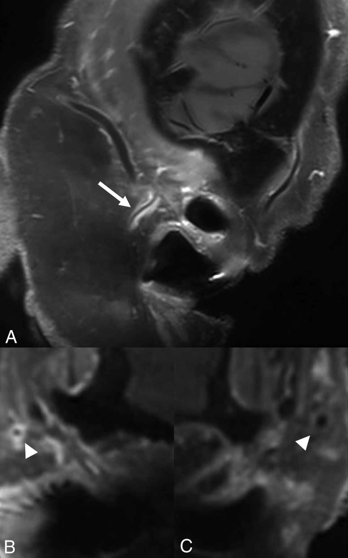
  • FIG 3.
    • Download figure
    • Open in new tab
    • Download powerpoint
    FIG 3.

    Sagittal postcontrast T1-SPACE image shows circumferential wall enhancement along the right superficial temporal artery (STA, arrow, A). Oblique MPR images perpendicular to the involved vessel (in A) show corresponding circumferential enhancement (arrowhead, B). The left STA (arrowhead, C) is normal.

  • FIG 4.
    • Download figure
    • Open in new tab
    • Download powerpoint
    FIG 4.

    Axial DWI in a treatment-naïve patient with GCA showing scattered foci of increased diffusion signal along bilateral scalp vessels (arrowheads). Inset in bottom left shows magnified diffusion-weighted signal changes along the right frontal STA branch.

  • FIG 5.
    • Download figure
    • Open in new tab
    • Download powerpoint
    FIG 5.

    Long-standing GCA in a 66-year-old woman with pseudoaneurysm of the superficial temporal artery. Frontal (A) and lateral (B) projections on catheter angiogram (External carotid artery injection) reveal multifocal areas of narrowing involving the frontal and parietal branches of STA (A and B, arrows). A small pseudoaneurysm is noted along the frontal branch of STA (A and B, arrowheads). Temporal artery biopsy with H&E (C) and Verhoeff-van Giesson (VVG) (D) stains reveals marked atrophy of the arterial wall (C and D, arrows), suggesting healing with pseudoaneurysm (C and D, arrowheads).

  • FIG 6.
    • Download figure
    • Open in new tab
    • Download powerpoint
    FIG 6.

    Axial postcontrast images reveal flow artifacts in the right occipital vein (arrows), which should not be misinterpreted as mural inflammatory changes. There is circumferential enhancement along the right superficial occipital artery (arrowheads).

  • FIG 7.
    • Download figure
    • Open in new tab
    • Download powerpoint
    FIG 7.

    Sagittal CTA-MPR image (A) reveals blurring of vessel margins and subtle fat stranding along the frontal branch of the right STA. Volume-rendered image (B) shows scattered areas of vessel irregularity and stenosis along the STA branches (arrows).

  • FIG 8.
    • Download figure
    • Open in new tab
    • Download powerpoint
    FIG 8.

    Axial CTA image (A) in a patient with newly diagnosed GCA shows reduced contrast opacification along the left STA (arrow) and a normal-appearing right STA (arrowhead). Fused PET-CT image (B) from the same patient at a slightly cranial level shows prominent radiotracer uptake on the left (circled).

  • FIG 9.
    • Download figure
    • Open in new tab
    • Download powerpoint
    FIG 9.

    Axial T1-SPACE postcontrast image through the orbits in a patient with newly diagnosed GCA shows bilateral retrobulbar intraconal enhancement near the apex with involvement of bilateral ophthalmic arteries (arrows).

  • FIG 10.
    • Download figure
    • Open in new tab
    • Download powerpoint
    FIG 10.

    Axial T1-SPACE postcontrast image (same patient as Fig 9) shows asymmetrically increased enhancement along the right temporalis muscle (arrows) and along the deep temporal artery (arrowhead).

Tables

  • Figures
  • Updated classification criteria for GCA diagnosis

    Classification Criteria for Giant Cell Arteritisa
    2022 American College of Rheumatology & European Alliance of Associations for Rheumatology
    Absolute requirement
     • Age ≥50 years at time of diagnosis
    Additional clinical criteria
     • Morning stiffness in shoulder/neck area +2
     • Sudden visual loss +3
     • Jaw or tongue claudication +2
     • New temporal headache +2
     • Scalp tenderness +2
     • Abnormal examination of temporal artery +2
    Laboratory, imaging & biopsy criteria
     • Maximum ESR ≥50 mm/h or maximum CRP ≥10 mg/L +3
     • Positive temporal artery biopsy or halo sign on ultrasound +5
     • Bilateral axillary involvement +2
     • FDG-PET activity throughout aorta +2
    Sum the scores for 10 items, if present. A score of ≥6 points is needed for diagnosis of giant cell arteritis
    aConsideration while applying the criteria
     • Classification criteria should be applied when a diagnosis of medium-vessel or large-vessel vasculitis has been made
     • Alternate diagnosis mimicking vasculitis should be excluded before applying the criteria
    • ↵a Adapted from Ponte C, Grayson PC, Robson JC; DCVAS Study Group, et al. 2022 American College of Rheumatology/EULAR classification criteria for giant cell arteritis. Arthritis Rheumatol 2022;74:1881–89.

PreviousNext
Back to top

In this issue

American Journal of Neuroradiology: 46 (3)
American Journal of Neuroradiology
Vol. 46, Issue 3
1 Mar 2025
  • Table of Contents
  • Index by author
  • Complete Issue (PDF)
Advertisement
Print
Download PDF
Email Article

Thank you for your interest in spreading the word on American Journal of Neuroradiology.

NOTE: We only request your email address so that the person you are recommending the page to knows that you wanted them to see it, and that it is not junk mail. We do not capture any email address.

Enter multiple addresses on separate lines or separate them with commas.
Imaging Findings in Giant Cell Arteritis: Don’t Turn a Blind Eye to the Obvious!
(Your Name) has sent you a message from American Journal of Neuroradiology
(Your Name) thought you would like to see the American Journal of Neuroradiology web site.
CAPTCHA
This question is for testing whether or not you are a human visitor and to prevent automated spam submissions.
Cite this article
Girish Bathla, Amit K. Agarwal, Steven A. Messina, David F. Black, Neetu Soni, Felix E. Diehn, Norbert G. Campeau, Vance T. Lehman, Kenneth J. Warrington, Rennie L. Rhee, Thorsten A. Bley
Imaging Findings in Giant Cell Arteritis: Don’t Turn a Blind Eye to the Obvious!
American Journal of Neuroradiology Mar 2025, 46 (3) 457-464; DOI: 10.3174/ajnr.A8388

Citation Manager Formats

  • BibTeX
  • Bookends
  • EasyBib
  • EndNote (tagged)
  • EndNote 8 (xml)
  • Medlars
  • Mendeley
  • Papers
  • RefWorks Tagged
  • Ref Manager
  • RIS
  • Zotero
0 Responses
Respond to this article
Share
Bookmark this article
Giant Cell Arteritis: Important Imaging Findings
Girish Bathla, Amit K. Agarwal, Steven A. Messina, David F. Black, Neetu Soni, Felix E. Diehn, Norbert G. Campeau, Vance T. Lehman, Kenneth J. Warrington, Rennie L. Rhee, Thorsten A. Bley
American Journal of Neuroradiology Mar 2025, 46 (3) 457-464; DOI: 10.3174/ajnr.A8388
del.icio.us logo Twitter logo Facebook logo Mendeley logo
  • Tweet Widget
  • Facebook Like
  • Google Plus One
Purchase

Jump to section

  • Article
    • Graphical Abstract
    • SUMMARY:
    • ABBREVIATIONS:
    • VASCULAR FINDINGS IN CRANIAL GCA
    • CONCLUSIONS
    • Footnotes
    • References
  • Figures & Data
  • Supplemental
  • Info & Metrics
  • Responses
  • References
  • PDF

Related Articles

  • PubMed
  • Google Scholar

Cited By...

  • No citing articles found.
  • Crossref
  • Google Scholar

This article has not yet been cited by articles in journals that are participating in Crossref Cited-by Linking.

More in this TOC Section

  • DCE MRI in Spinal Disease Assessment
  • Cerebrovascular Anomalies in Fetal Imaging
Show more Review Article

Similar Articles

Advertisement

Indexed Content

  • Current Issue
  • Accepted Manuscripts
  • Article Preview
  • Past Issues
  • Editorials
  • Editor's Choice
  • Fellows' Journal Club
  • Letters to the Editor
  • Video Articles

Cases

  • Case Collection
  • Archive - Case of the Week
  • Archive - Case of the Month
  • Archive - Classic Case

More from AJNR

  • Trainee Corner
  • Imaging Protocols
  • MRI Safety Corner
  • Book Reviews

Multimedia

  • AJNR Podcasts
  • AJNR Scantastics

Resources

  • Turnaround Time
  • Submit a Manuscript
  • Submit a Video Article
  • Submit an eLetter to the Editor/Response
  • Manuscript Submission Guidelines
  • Statistical Tips
  • Fast Publishing of Accepted Manuscripts
  • Graphical Abstract Preparation
  • Imaging Protocol Submission
  • Evidence-Based Medicine Level Guide
  • Publishing Checklists
  • Author Policies
  • Become a Reviewer/Academy of Reviewers
  • News and Updates

About Us

  • About AJNR
  • Editorial Board
  • Editorial Board Alumni
  • Alerts
  • Permissions
  • Not an AJNR Subscriber? Join Now
  • Advertise with Us
  • Librarian Resources
  • Feedback
  • Terms and Conditions
  • AJNR Editorial Board Alumni

American Society of Neuroradiology

  • Not an ASNR Member? Join Now

© 2025 by the American Society of Neuroradiology All rights, including for text and data mining, AI training, and similar technologies, are reserved.
Print ISSN: 0195-6108 Online ISSN: 1936-959X

Powered by HighWire