
Article Figures & Data
Figures
- Fig 1.
Box-and-whisker plot depicting the reference point air kerma of biplane DSA and 3DRA protocols across all patients. Boxes denote median, lower, and upper quartiles, with whiskers capturing values outside the interquartile range.
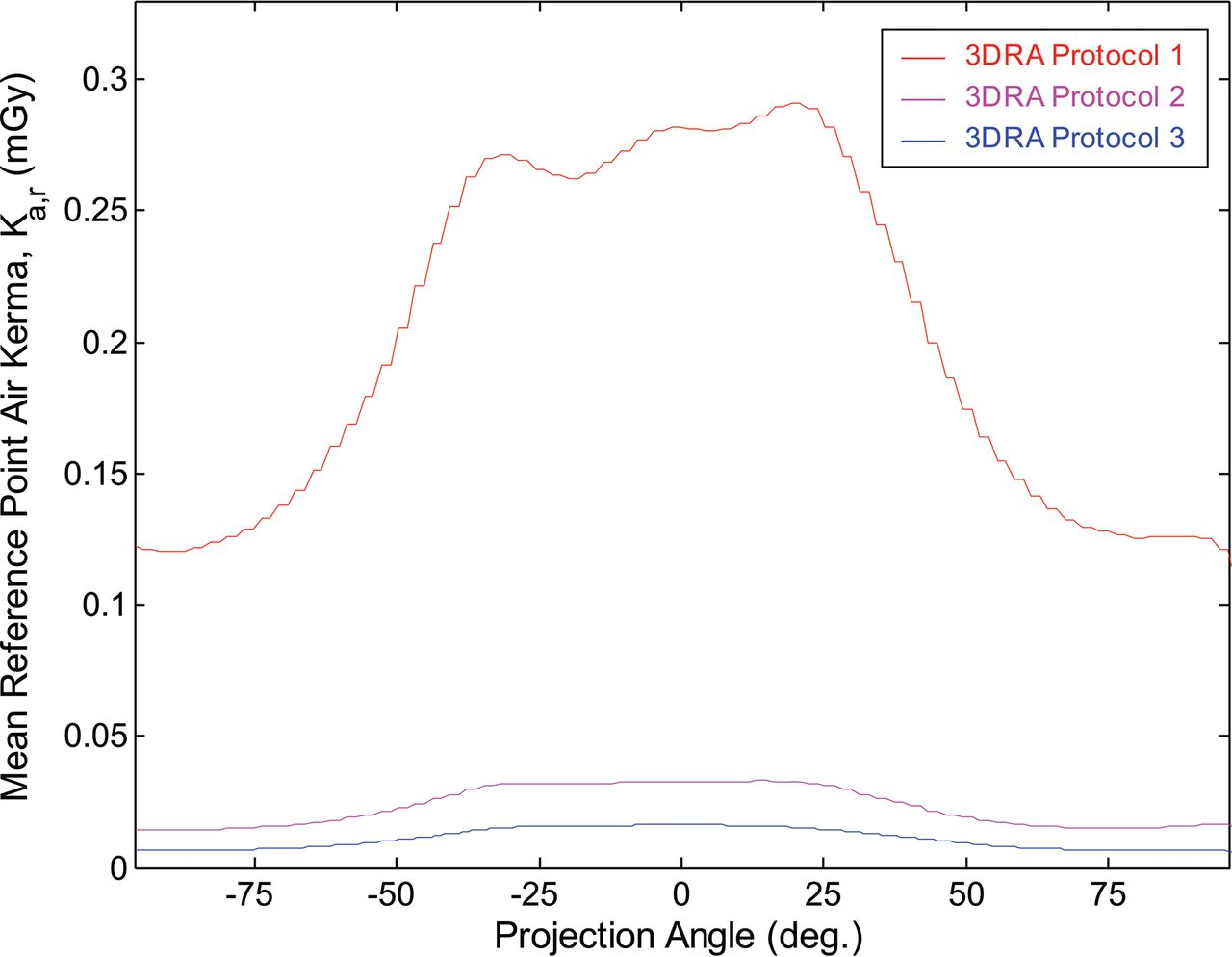
- Fig 2.
Mean reference point air kerma as a function of projection angle for 3DRA protocols 1, 2, and 3.
- Fig 3.
Effective dose as a function of projection angle for 3DRA protocols 1, 2, and 3. Each line indicates the effective dose for an individual rotation.
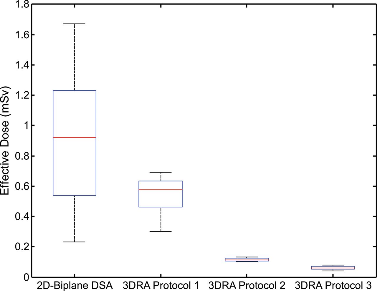
- Fig 4.
Box-and-whisker plot depicting the effective dose of biplane DSA and 3DRA protocols across all patients. Boxes denote median, lower, and upper quartiles, with whiskers capturing values outside the interquartile range.
- Fig 5.
Box-and-whisker plot depicting the relative effective dose comparing 3DRA and biplane DSA acquisitions in the same patient. Relative comparison with the original 2D-DSA effective dose (left) and normalized DSA effective dose (right) for each 3DRA protocol. Boxes denote median, lower, and upper quartiles, with whiskers capturing values outside the interquartile range.
- Fig 6.
An example of the use of a 3D rotational angiographic sequence (protocol 3). A 12-year-old girl was referred from an outside institution 4 months after hemorrhage from a right frontal arteriovenous malformation. A, Postgadolinium axial 3D T1-weighted MR image shows the AVM nidus within a collapsed hemorrhagic cavity in the right superior frontal gyrus. B, Diagnostic angiography in the right internal cerebral artery in an oblique frontal projection shows the nidus fed from posterior internal frontal branches of the anterior cerebral artery, with early filling of an inferiorly draining cortical vein (arrow). The AVM was surgically resected, following which DSA (not shown) confirmed no residual nidus. C, Postgadolinium axial 3D T1-weighted MR image obtained at 6-month follow-up shows linear enhancement within the surgical cavity. Diagnostic angiography subsequently performed showed no arteriovenous shunting. D, 3D rotational angiography performed in the right internal cerebral artery using protocol 3 with 30% contrast concentration, subsequently reconstructed into CTA. E and F, 3D rotational angiography fused with the pregadolinium 3D T1-weighted MR imaging sequence, which confirmed no vascular structures within the surgical cavity in multiple planes.


Tables
Parameter Value Age (mean) (yr) 10.4 ± 5.1 Male/female 18:6 Procedure indication (No.) AVM 28 Intra-arterial chemotherapy 2 Aneurysm 1 Aberrant neurovasculature 1 Presurgical embolization 1 Projections per Rotation Rotation Rotations Required per Acquisition kVp Target Detector Dose (nGy/projection) Filtration (mm Cu) Half-Value Layer (mm Al) Protocol 1 (subtracted) 133 200° 2 (mask, contrast) 70 360 0.0 2.9 Protocol 2 (subtracted) 133 200° 2 (mask, contrast) 90 200 0.3 7.1 Protocol 3 (unsubtracted) 248 200° 1 (contrast) 90 100 0.6 8.2 - Table 3:
Mean, median, and range of effective dose as a function of acquisition type and protocol
Effective Dose (mSv) Mean Median Range 2D Biplane DSA 0.89 0.92 0.23–1.7 3D Protocol 1 0.54 0.57 0.3–0.69 Protocol 2 0.12 0.11 0.1–0.13 Protocol 3 0.06 0.06 0.04–0.08 - Table 4:
Relative effective dose comparison indicating mean, median, and range for direct and normalized comparisons
Relative Effective Dose E3DRA/E2D-DSA (%) (E3DRA/E2D-DSA)Normalized (%) Mean Median Range Mean Median Range Protocol 1 68 65 27–127 61 67 25–103 Protocol 2 15 13 10–24 14 13 7.4–23 Protocol 3 8.9 6.1 3.3–29 7.5 6.8 3.4–12


