Advertisement

Main menu

  • Home
  • Content
    • Current Issue
    • Accepted Manuscripts
    • Article Preview
    • Past Issue Archive
    • Video Articles
    • AJNR Case Collection
    • Case of the Week Archive
    • Case of the Month Archive
    • Classic Case Archive
  • Special Collections
    • AJNR Awards
    • Low-Field MRI
    • Alzheimer Disease
    • ASNR Foundation Special Collection
    • Photon-Counting CT
    • View All
  • Multimedia
    • AJNR Podcasts
    • AJNR SCANtastic
    • Trainee Corner
    • MRI Safety Corner
    • Imaging Protocols
  • For Authors
    • Submit a Manuscript
    • Submit a Video Article
    • Submit an eLetter to the Editor/Response
    • Manuscript Submission Guidelines
    • Statistical Tips
    • Fast Publishing of Accepted Manuscripts
    • Graphical Abstract Preparation
    • Imaging Protocol Submission
    • Author Policies
  • About Us
    • About AJNR
    • Editorial Board
    • Editorial Board Alumni
  • More
    • Become a Reviewer/Academy of Reviewers
    • Subscribers
    • Permissions
    • Alerts
    • Feedback
    • Advertisers
    • ASNR Home

User menu

  • Alerts
  • Log in

Search

  • Advanced search
American Journal of Neuroradiology

American Journal of Neuroradiology

ASHNR American Society of Functional Neuroradiology ASHNR American Society of Pediatric Neuroradiology ASSR
  • Alerts
  • Log in

Advanced Search

  • Home
  • Content
    • Current Issue
    • Accepted Manuscripts
    • Article Preview
    • Past Issue Archive
    • Video Articles
    • AJNR Case Collection
    • Case of the Week Archive
    • Case of the Month Archive
    • Classic Case Archive
  • Special Collections
    • AJNR Awards
    • Low-Field MRI
    • Alzheimer Disease
    • ASNR Foundation Special Collection
    • Photon-Counting CT
    • View All
  • Multimedia
    • AJNR Podcasts
    • AJNR SCANtastic
    • Trainee Corner
    • MRI Safety Corner
    • Imaging Protocols
  • For Authors
    • Submit a Manuscript
    • Submit a Video Article
    • Submit an eLetter to the Editor/Response
    • Manuscript Submission Guidelines
    • Statistical Tips
    • Fast Publishing of Accepted Manuscripts
    • Graphical Abstract Preparation
    • Imaging Protocol Submission
    • Author Policies
  • About Us
    • About AJNR
    • Editorial Board
    • Editorial Board Alumni
  • More
    • Become a Reviewer/Academy of Reviewers
    • Subscribers
    • Permissions
    • Alerts
    • Feedback
    • Advertisers
    • ASNR Home
  • Follow AJNR on Twitter
  • Visit AJNR on Facebook
  • Follow AJNR on Instagram
  • Join AJNR on LinkedIn
  • RSS Feeds

AJNR Awards, New Junior Editors, and more. Read the latest AJNR updates

Research ArticleHead and Neck Imaging

Submandibular Gland Transfer: A Potential Imaging Pitfall

X. Wu, S.S. Yom, P.K. Ha, C.M. Heaton and C.M. Glastonbury
American Journal of Neuroradiology March 2018, DOI: https://doi.org/10.3174/ajnr.A5609
X. Wu
aFrom the Department of Radiology and Imaging Science (X.W.), Emory University, Atlanta, Georgia
  • Find this author on Google Scholar
  • Find this author on PubMed
  • Search for this author on this site
  • ORCID record for X. Wu
S.S. Yom
bDepartments of Radiation Oncology (S.S.Y., C.M.G.)
cOtolaryngology-Head and Neck Surgery (S.S.Y., P.K.H., C.M.H., C.M.G.)
  • Find this author on Google Scholar
  • Find this author on PubMed
  • Search for this author on this site
  • ORCID record for S.S. Yom
P.K. Ha
cOtolaryngology-Head and Neck Surgery (S.S.Y., P.K.H., C.M.H., C.M.G.)
  • Find this author on Google Scholar
  • Find this author on PubMed
  • Search for this author on this site
  • ORCID record for P.K. Ha
C.M. Heaton
cOtolaryngology-Head and Neck Surgery (S.S.Y., P.K.H., C.M.H., C.M.G.)
  • Find this author on Google Scholar
  • Find this author on PubMed
  • Search for this author on this site
  • ORCID record for C.M. Heaton
C.M. Glastonbury
bDepartments of Radiation Oncology (S.S.Y., C.M.G.)
cOtolaryngology-Head and Neck Surgery (S.S.Y., P.K.H., C.M.H., C.M.G.)
dClinical Radiology (C.M.G.), University of California, San Francisco, San Francisco, California.
  • Find this author on Google Scholar
  • Find this author on PubMed
  • Search for this author on this site
  • ORCID record for C.M. Glastonbury
  • Article
  • Figures & Data
  • Info & Metrics
  • Responses
  • References
  • PDF
Loading

Article Figures & Data

Figures

  • Tables
  • Fig 1.
    • Download figure
    • Open in new tab
    • Download powerpoint
    Fig 1.

    Illustrative schematic demonstrating the key steps in the SMG transfer operation, including mobilization or ligation of the facial artery and vein proximal to the SMG, anterior and inferior translation of the gland into the submental space, and bisection of the mylohyoid muscle to allow repositioning of the submandibular duct and ganglion.3,7
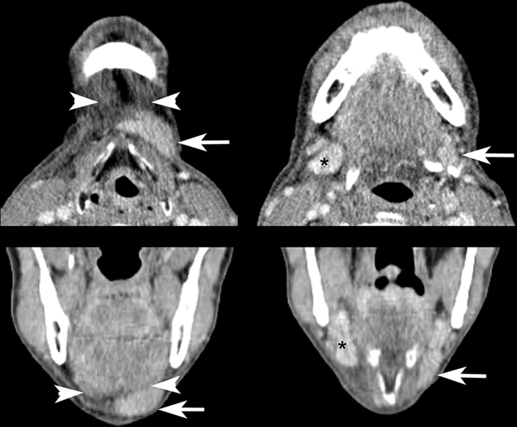
  • Fig 2.
    • Download figure
    • Open in new tab
    • Download powerpoint
    Fig 2.

    Contrast-enhanced CT images demonstrating the typical asymmetric appearance of the submental and submandibular spaces after SMG transfer. The left transferred SMG (arrows) is elongated and displaced inferiorly and anteriorly into the submental space superficial to the anterior belly of the digastric muscle (arrowheads), resulting in an asymmetric soft-tissue density in the submental space and diminished soft-tissue volume in the submandibular space relative to the contralateral gland (asterisks). Note also edema of surrounding tissues in this patient who was 3 months postchemoradiation with cisplatin and NRG-HN002 (NCT02254278; ClinicalTrials.gov) de-escalation protocol at time of imaging.

  • Fig 3.
    • Download figure
    • Open in new tab
    • Download powerpoint
    Fig 3.

    Appearance of SMG (arrows) transferred deep to the anterior belly of the digastric muscle (arrowheads) on axial T2-weighted, fat-suppressed imaging and coronal T1-weighted imaging. The patient was 2 months postchemoradiation with cisplatin and intensity-modulated radiation therapy at imaging.

  • Fig 4.
    • Download figure
    • Open in new tab
    • Download powerpoint
    Fig 4.

    Axial and coronal fat-suppressed postcontrast T1-weighted imaging performed 28 days postoperatively for staging purposes demonstrated platysma enhancement (arrows) adjacent to the transferred SMG. The patient had not yet undergone chemoradiation at imaging.

  • Fig 5.
    • Download figure
    • Open in new tab
    • Download powerpoint
    Fig 5.

    Postoperative asymmetry within the submandibular space results in misinterpretation of the superior aspect of the normal contralateral SMG (arrows) as a parapharyngeal mass (axial T2 fat-suppressed and postcontrast imaging). The patient was 2 months postchemoradiation with cisplatin and intensity-modulated radiation therapy at imaging.

  • Fig 6.
    • Download figure
    • Open in new tab
    • Download powerpoint
    Fig 6.

    PET/CT images demonstrating mildly increased FDG uptake in the left transferred SMG (arrows) compared with the contralateral gland (asterisk) 5 months after SMG transfer surgery and 3 months following conclusion of chemoradiation. These findings are congruent with previously published PET findings in a SMG transfer operation and may reflect relatively preserved function in the transferred gland.8

Tables

  • Figures
    • View popup
    Table 1:

    SMG morphologic measurement definitions

    Morphologic MeasurementDefinitionDirection of Measurement
    AP lengthAs measured between anteriormost border of gland and posteriormost border of SMGOn axial images, perpendicular to axis connecting the mandibular condyles
    AP length differenceDifference between the AP lengths of the SMGs by subtraction of the AP length of the contralateral gland from the transferred glandOn axial images, perpendicular to axis connecting the mandibular condyles
    Posterior margin differenceDistance between the posteriormost border of the gland and that of the contralateral glandOn axial images, perpendicular to axis formed by connecting the mandibular condyles
    Superior margin differenceDistance between the superiormost border of the gland and that of the contralateral glandOn coronal images, perpendicular to axis formed by connecting the mandibular condyles
    Anteroinferior margin differenceDistance between the anteroinferior-most border of the gland and that of the contralateral glandOn axial images, perpendicular to axis formed by connecting the mandibular condyles
    • Note:—AP indicates anteroposterior.

    • View popup
    Table 2:

    Radiation therapy detailsa

    Patient No.Primary Tumor SitePrescribed Dose to Primary Tumor (Gy)Prescribed Dose to Involved Neck (Gy)Mean Dose to SMG on Involved Side (Gy)Prescribed Dose to Uninvolved Neck (Gy)Mean Dose to Transferred SMG (Gy)
    1R BOT605458.644820.45
    2R tonsil69.9659.461.3354.1228.99
    3R tonsil69.9659.468.854.1243.97
    4R BOT69.9659.466.0159.441.44
    5R BOT605460.014844.95
    6L tonsil6659.466.754.1216.59
    7R tonsil6659.459.454.1238.8
    8R tonsil69.9659.472.754.1227.17
    9Nasopharynx69.9659.461.0259.460.77
    Average dose (Gy)66.8758.2063.8553.9335.90
    SD4.242.384.894.0413.88
    • Note:—R indicates right; BOT, base of tongue; L, left.

    • ↵a Postsurgical radiation dosages for the 9 out of 11 patients who received radiation therapy at our institution following the SMG transfer procedure. Per the Student t test, the transferred SMG received a significantly lower radiation dose than the contralateral SMG (P < .001).

    • View popup
    Table 3:

    SMG locationa

    Preoperative (mm)P ValuePostoperative (mm)P Value
    Anteroposterior length difference2.5 (−4–5).2810.5 (−1–17)<.001
    Anteroinferior margin difference0 (0–0)13.5 (10–16)<.001
    Posterior margin difference−1.8 (−9–3).107.2 (0–16)<.001
    Superior margin difference0.2 (−3–6).79−7.5 (−15–0)<.001
    • ↵a Preoperative and postoperative morphologic features of the transferred SMGs, presented as averaged length and location differences between the SMGs in each patient (transferred gland–contralateral gland) followed by ranges of the differences. Positive values indicate anterior and superior directions, respectively. P values for significance of length differences are derived from paired pre- and post-t tests with a reference value of 0 mm (no difference).

Next
Back to top
Advertisement
Print
Download PDF
Email Article

Thank you for your interest in spreading the word on American Journal of Neuroradiology.

NOTE: We only request your email address so that the person you are recommending the page to knows that you wanted them to see it, and that it is not junk mail. We do not capture any email address.

Enter multiple addresses on separate lines or separate them with commas.
Submandibular Gland Transfer: A Potential Imaging Pitfall
(Your Name) has sent you a message from American Journal of Neuroradiology
(Your Name) thought you would like to see the American Journal of Neuroradiology web site.
CAPTCHA
This question is for testing whether or not you are a human visitor and to prevent automated spam submissions.
Cite this article
X. Wu, S.S. Yom, P.K. Ha, C.M. Heaton, C.M. Glastonbury
Submandibular Gland Transfer: A Potential Imaging Pitfall
American Journal of Neuroradiology Mar 2018, DOI: 10.3174/ajnr.A5609

Citation Manager Formats

  • BibTeX
  • Bookends
  • EasyBib
  • EndNote (tagged)
  • EndNote 8 (xml)
  • Medlars
  • Mendeley
  • Papers
  • RefWorks Tagged
  • Ref Manager
  • RIS
  • Zotero
0 Responses
Respond to this article
Share
Bookmark this article
Submandibular Gland Transfer: A Potential Imaging Pitfall
X. Wu, S.S. Yom, P.K. Ha, C.M. Heaton, C.M. Glastonbury
American Journal of Neuroradiology Mar 2018, DOI: 10.3174/ajnr.A5609
del.icio.us logo Twitter logo Facebook logo Mendeley logo
  • Tweet Widget
  • Facebook Like
  • Google Plus One
Purchase

Jump to section

  • Article
    • Abstract
    • ABBREVIATION:
    • Materials and Methods
    • Results
    • Discussion
    • Conclusions
    • Footnotes
    • References
  • Figures & Data
  • Info & Metrics
  • Responses
  • References
  • PDF

Related Articles

  • No related articles found.
  • PubMed
  • Google Scholar

Cited By...

  • CT and MR Imaging Appearance of the Pedicled Submandibular Gland Flap: A Potential Imaging Pitfall in the Posttreatment Head and Neck
  • Crossref
  • Google Scholar

This article has not yet been cited by articles in journals that are participating in Crossref Cited-by Linking.

More in this TOC Section

  • Hydrops Herniation into the Semicircular Canals
  • ASL Sensitivity for Head and Neck Paraganglioma
  • Post SRS Peritumoral Hyperintense Signal of VSs
Show more HEAD AND NECK IMAGING

Similar Articles

Advertisement

Indexed Content

  • Current Issue
  • Accepted Manuscripts
  • Article Preview
  • Past Issues
  • Editorials
  • Editor's Choice
  • Fellows' Journal Club
  • Letters to the Editor
  • Video Articles

Cases

  • Case Collection
  • Archive - Case of the Week
  • Archive - Case of the Month
  • Archive - Classic Case

More from AJNR

  • Trainee Corner
  • Imaging Protocols
  • MRI Safety Corner
  • Book Reviews

Multimedia

  • AJNR Podcasts
  • AJNR Scantastics

Resources

  • Turnaround Time
  • Submit a Manuscript
  • Submit a Video Article
  • Submit an eLetter to the Editor/Response
  • Manuscript Submission Guidelines
  • Statistical Tips
  • Fast Publishing of Accepted Manuscripts
  • Graphical Abstract Preparation
  • Imaging Protocol Submission
  • Evidence-Based Medicine Level Guide
  • Publishing Checklists
  • Author Policies
  • Become a Reviewer/Academy of Reviewers
  • News and Updates

About Us

  • About AJNR
  • Editorial Board
  • Editorial Board Alumni
  • Alerts
  • Permissions
  • Not an AJNR Subscriber? Join Now
  • Advertise with Us
  • Librarian Resources
  • Feedback
  • Terms and Conditions
  • AJNR Editorial Board Alumni

American Society of Neuroradiology

  • Not an ASNR Member? Join Now

© 2025 by the American Society of Neuroradiology All rights, including for text and data mining, AI training, and similar technologies, are reserved.
Print ISSN: 0195-6108 Online ISSN: 1936-959X

Powered by HighWire