Advertisement

Main menu

  • Home
  • Content
    • Current Issue
    • Accepted Manuscripts
    • Article Preview
    • Past Issue Archive
    • Video Articles
    • AJNR Case Collection
    • Case of the Week Archive
    • Case of the Month Archive
    • Classic Case Archive
  • Special Collections
    • AJNR Awards
    • Low-Field MRI
    • Alzheimer Disease
    • ASNR Foundation Special Collection
    • Photon-Counting CT
    • View All
  • Multimedia
    • AJNR Podcasts
    • AJNR SCANtastic
    • Trainee Corner
    • MRI Safety Corner
    • Imaging Protocols
  • For Authors
    • Submit a Manuscript
    • Submit a Video Article
    • Submit an eLetter to the Editor/Response
    • Manuscript Submission Guidelines
    • Statistical Tips
    • Fast Publishing of Accepted Manuscripts
    • Graphical Abstract Preparation
    • Imaging Protocol Submission
    • Author Policies
  • About Us
    • About AJNR
    • Editorial Board
    • Editorial Board Alumni
  • More
    • Become a Reviewer/Academy of Reviewers
    • Subscribers
    • Permissions
    • Alerts
    • Feedback
    • Advertisers
    • ASNR Home

User menu

  • Alerts
  • Log in

Search

  • Advanced search
American Journal of Neuroradiology

American Journal of Neuroradiology

ASHNR American Society of Functional Neuroradiology ASHNR American Society of Pediatric Neuroradiology ASSR
  • Alerts
  • Log in

Advanced Search

  • Home
  • Content
    • Current Issue
    • Accepted Manuscripts
    • Article Preview
    • Past Issue Archive
    • Video Articles
    • AJNR Case Collection
    • Case of the Week Archive
    • Case of the Month Archive
    • Classic Case Archive
  • Special Collections
    • AJNR Awards
    • Low-Field MRI
    • Alzheimer Disease
    • ASNR Foundation Special Collection
    • Photon-Counting CT
    • View All
  • Multimedia
    • AJNR Podcasts
    • AJNR SCANtastic
    • Trainee Corner
    • MRI Safety Corner
    • Imaging Protocols
  • For Authors
    • Submit a Manuscript
    • Submit a Video Article
    • Submit an eLetter to the Editor/Response
    • Manuscript Submission Guidelines
    • Statistical Tips
    • Fast Publishing of Accepted Manuscripts
    • Graphical Abstract Preparation
    • Imaging Protocol Submission
    • Author Policies
  • About Us
    • About AJNR
    • Editorial Board
    • Editorial Board Alumni
  • More
    • Become a Reviewer/Academy of Reviewers
    • Subscribers
    • Permissions
    • Alerts
    • Feedback
    • Advertisers
    • ASNR Home
  • Follow AJNR on Twitter
  • Visit AJNR on Facebook
  • Follow AJNR on Instagram
  • Join AJNR on LinkedIn
  • RSS Feeds

AJNR Awards, New Junior Editors, and more. Read the latest AJNR updates

Research ArticlePediatric Neuroimaging
Open Access

MR Imaging Scoring System for White Matter Injury after Deep Medullary Vein Thrombosis and Infarction in Neonates

K.L. Benninger, N.L. Maitre, L. Ruess and J.A. Rusin
American Journal of Neuroradiology January 2019, DOI: https://doi.org/10.3174/ajnr.A5940
K.L. Benninger
aFrom the Departments of Pediatrics and Center for Perinatal Research (K.L.B., N.L.M.)
cWexner Medical Center (K.L.B., N.L.M., L.R., J.A.R.), Ohio State University, Columbus, Ohio.
  • Find this author on Google Scholar
  • Find this author on PubMed
  • Search for this author on this site
  • ORCID record for K.L. Benninger
N.L. Maitre
aFrom the Departments of Pediatrics and Center for Perinatal Research (K.L.B., N.L.M.)
cWexner Medical Center (K.L.B., N.L.M., L.R., J.A.R.), Ohio State University, Columbus, Ohio.
  • Find this author on Google Scholar
  • Find this author on PubMed
  • Search for this author on this site
  • ORCID record for N.L. Maitre
L. Ruess
bRadiology (L.R., J.A.R.), Nationwide Children's Hospital, Columbus, Ohio
cWexner Medical Center (K.L.B., N.L.M., L.R., J.A.R.), Ohio State University, Columbus, Ohio.
  • Find this author on Google Scholar
  • Find this author on PubMed
  • Search for this author on this site
  • ORCID record for L. Ruess
J.A. Rusin
bRadiology (L.R., J.A.R.), Nationwide Children's Hospital, Columbus, Ohio
cWexner Medical Center (K.L.B., N.L.M., L.R., J.A.R.), Ohio State University, Columbus, Ohio.
  • Find this author on Google Scholar
  • Find this author on PubMed
  • Search for this author on this site
  • ORCID record for J.A. Rusin
  • Article
  • Figures & Data
  • Info & Metrics
  • Responses
  • References
  • PDF
Loading

Article Figures & Data

Figures

  • Tables
  • Fig 1.
    • Download figure
    • Open in new tab
    • Download powerpoint
    Fig 1.

    Coronal T2 (A) and axial T1 FLAIR (B), T2 (C), and SWI (D) MR images of a 6-day-old boy. Global severity score = 3 (first quartile) (E). There is diffuse bilateral WM signal abnormality of cerebral edema without infarction (3 points) (A and B). The DMVs are prominent bilaterally, seen on T2 (C) and SWI (D) (ovals). No WM lesions are visible. R indicates right; L, left.

  • Fig 2.
    • Download figure
    • Open in new tab
    • Download powerpoint
    Fig 2.

    Coronal T2 (A), axial T1 (B), ADC (C and D), and SWI (E and F) MR images of a 7-day-old girl. Global severity score = 10 (second quartile) (G). There is diffuse bilateral WM cerebral edema (3 points) (A and B) and multiple acute bilateral, asymmetric (left > right) hemorrhagic WM lesions (T1 bright and T2 dark) without restricted diffusion (C–F). On the right, there are mild punctate lesions in the frontal (1 point), parietal (1 point), and occipital WM (1 point, not shown) (E and F, boxes), and on the left, there are moderate punctate lesions in the frontal (2 points) and parietal WM (2 points) (E and F, circles). No corpus callosum or temporal region WM lesions are visible. R indicates right; L, left.

  • Fig 3.
    • Download figure
    • Open in new tab
    • Download powerpoint
    Fig 3.

    Coronal T2 (A) and axial TI FLAIR (B), ADC (C), and T2 (D) MR images of a 21-day-old boy. Global severity score = 22 (third quartile) (E). There is diffuse bilateral WM cerebral edema (3 points) (A–D) and multiple acute bilateral symmetric hemorrhagic WM lesions (T1 bright and T2 dark): frontal (moderate punctate, 2 points, and severe linear, 3 points) (A, boxes), parietal (moderate punctate, 2 points) (D, circles), and temporal regions (moderate punctate, 2 points, not shown). There are no visible occipital WM lesions. R indicates right; L, left.
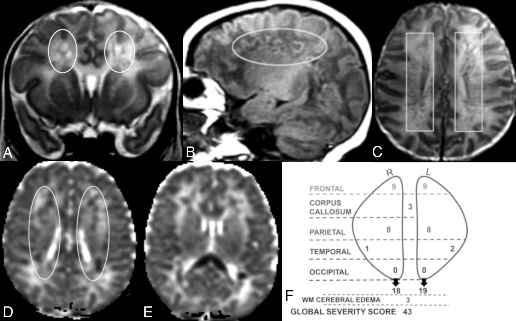
  • Fig 4.
    • Download figure
    • Open in new tab
    • Download powerpoint
    Fig 4.

    Coronal T2 (A) and sagittal T1 (B), axial T2 (C), and axial ADC (D and E) MR images of a 12-day-old boy. Global severity score = 43 (fourth quartile) (F). There is diffuse bilateral WM cerebral edema (3 points) and multiple bilateral hemorrhagic WM lesions with restricted diffusion (A–F). There are symmetric bilateral frontal and parietal lesions: severe linear lesions (3 points; C, boxes) and severe cavitary lesions with restricted diffusion (6 points; A, B, and D, ovals), There are bilateral temporal lesions: right (mild punctate, 1 point) and left (moderate punctate, 2 points, not shown). There is restricted diffusion throughout the corpus callosum (3 points, E). No occipital WM lesions are visible. R indicates right; L, left.

Tables

  • Figures
    • View popup
    Table 1:

    DMV white matter injury severity scoring system

    PointsScoring
    Cerebral edema0–3None = 0, right or left = 1, bilateral asymmetric = 2, bilateral symmetric = 3
    Corpus callosum0–31 point each for genu, body, and/or splenium lesion
    Right cerebral WMScore each cerebral hemisphere region for punctate, linear, and/or cavitary lesions
        Frontal0–12Punctate: mild (1–3 lesions) = 1, moderate (4–6 lesions) = 2, severe (>6 lesions) = 3
        Parietal0–12Linear: mild (1–3 lesions) = 1, moderate (4–6 lesions) = 2, severe (>6 lesions) = 3
        Occipital0–12Cavitary: mild (1–3 lesions, <15 mm) = 2, moderate (4–6 lesions, <15 mm) = 4
        Temporal0–12severe (>6 lesions, <15 mm, or 1+ lesion >15 mm) = 6
    Left cerebral WM
        Frontal0–12
        Parietal0–12
        Occipital0–12
        Temporal0–12
    0–102Global severity score
    • View popup
    Table 2:

    Injury severity scores: WM cerebral edema and corpus callosuma

    RegionScore 0Score 1Score 2Score 3P Value
    WM cerebral edema4 (8)2 (4)9 (18)36 (71).52
    Corpus callosum26 (51)10 (20)8 (16)7 (14)<.001
    • ↵a Data are number of patients (%).

    • View popup
    Table 3:

    Injury severity scores by regiona

    RegionScore 0–2Score 3–5Score 6–8Score ≥9P Value
    Right frontal24 (47)14 (28)8 (16)5 (10)<.001
    Right parietal33 (65)8 (16)9 (18)1 (2)<.001
    Right occipital50 (98)1 (2)0 (0)0 (0).68
    Right temporal43 (84)6 (12)2 (4)0 (0).006
    Left frontal31 (61)10 (20)7 (14)3 (6)<.001
    Left parietal37 (73)5 (10)7 (14)2 (4)<.001
    Left occipital49 (96)0 (0)2 (4)0 (0).67
    Left temporal43 (84)7 (14)1 (2)0 (0)<.001
    • ↵a Data are number of patients (%).

    • View popup
    Table 4:

    Global severity score and perinatal variablesa

    1st Quartile (Score = 1–5) (n = 14)2nd Quartile (Score = 6–11) (n = 13)3rd Quartile (Score = 12–25) (n = 12)4th Quartile (Score ≥ 26) (n = 12)P Value
    Gestation at birth (wk)38.2 ± 1.336.3 ± 2.436.7 ± 2.538 ± 1.9.05d
    Birth weight (g)3445.8 ± 6183066 ± 9892947 ± 6193247 ± 570.34d
    Male sex8 (57)8 (62)9 (75)7 (58).87e
    5-Minute Apgar7.8 ± 1.67.9 ± 1.48 ± 1.57.4 ± 2.4.92d
    Compressions/epinephrineb1 (7)0 (0)0 (0)1 (8).97e
    Chorioamnionitis0 (0)0 (0)0 (0)1 (8).98e
    Small for gestational age0 (0)2 (15)3 (25)2 (17).75e
    Neonatal infectionc1 (7)2 (15)3 (25)3 (25).84e
    Ventilator requirement2 (14)7 (54)6 (50)7 (58).18e
    Inotrope use0 (0)3 (23)2 (17)4 (33).52e
    Seizures7 (50)5 (38)6 (50)10 (83).25e
    • ↵a Data are mean or number (%).

    • ↵b Compressions or epinephrine or both were given in the delivery room during resuscitation.

    • ↵c Neonatal infection is defined as systemic/serious infection in the first week of life.

    • ↵d The ANOVA was used to measure variance in means among 4 quartiles in continuous variables.

    • ↵e The Kruskal-Wallis test was used for frequency distribution of categoric variables.

PreviousNext
Back to top
Advertisement
Print
Download PDF
Email Article

Thank you for your interest in spreading the word on American Journal of Neuroradiology.

NOTE: We only request your email address so that the person you are recommending the page to knows that you wanted them to see it, and that it is not junk mail. We do not capture any email address.

Enter multiple addresses on separate lines or separate them with commas.
MR Imaging Scoring System for White Matter Injury after Deep Medullary Vein Thrombosis and Infarction in Neonates
(Your Name) has sent you a message from American Journal of Neuroradiology
(Your Name) thought you would like to see the American Journal of Neuroradiology web site.
CAPTCHA
This question is for testing whether or not you are a human visitor and to prevent automated spam submissions.
Cite this article
K.L. Benninger, N.L. Maitre, L. Ruess, J.A. Rusin
MR Imaging Scoring System for White Matter Injury after Deep Medullary Vein Thrombosis and Infarction in Neonates
American Journal of Neuroradiology Jan 2019, DOI: 10.3174/ajnr.A5940

Citation Manager Formats

  • BibTeX
  • Bookends
  • EasyBib
  • EndNote (tagged)
  • EndNote 8 (xml)
  • Medlars
  • Mendeley
  • Papers
  • RefWorks Tagged
  • Ref Manager
  • RIS
  • Zotero
0 Responses
Respond to this article
Share
Bookmark this article
MR Imaging Scoring System for White Matter Injury after Deep Medullary Vein Thrombosis and Infarction in Neonates
K.L. Benninger, N.L. Maitre, L. Ruess, J.A. Rusin
American Journal of Neuroradiology Jan 2019, DOI: 10.3174/ajnr.A5940
del.icio.us logo Twitter logo Facebook logo Mendeley logo
  • Tweet Widget
  • Facebook Like
  • Google Plus One
Purchase

Jump to section

  • Article
    • Abstract
    • ABBREVIATION:
    • Materials and Methods
    • Results
    • Discussion
    • Conclusions
    • Footnotes
    • REFERENCES
  • Figures & Data
  • Info & Metrics
  • Responses
  • References
  • PDF

Related Articles

  • No related articles found.
  • PubMed
  • Google Scholar

Cited By...

  • No citing articles found.
  • Crossref
  • Google Scholar

This article has not yet been cited by articles in journals that are participating in Crossref Cited-by Linking.

More in this TOC Section

  • FRACTURE MR in Congenital Vertebral Anomalies
  • Comparing MRI Perfusion in Pediatric Brain Tumors
  • Sodium MRI in Pediatric Brain Tumors
Show more Pediatric Neuroimaging

Similar Articles

Advertisement

Indexed Content

  • Current Issue
  • Accepted Manuscripts
  • Article Preview
  • Past Issues
  • Editorials
  • Editor's Choice
  • Fellows' Journal Club
  • Letters to the Editor
  • Video Articles

Cases

  • Case Collection
  • Archive - Case of the Week
  • Archive - Case of the Month
  • Archive - Classic Case

More from AJNR

  • Trainee Corner
  • Imaging Protocols
  • MRI Safety Corner
  • Book Reviews

Multimedia

  • AJNR Podcasts
  • AJNR Scantastics

Resources

  • Turnaround Time
  • Submit a Manuscript
  • Submit a Video Article
  • Submit an eLetter to the Editor/Response
  • Manuscript Submission Guidelines
  • Statistical Tips
  • Fast Publishing of Accepted Manuscripts
  • Graphical Abstract Preparation
  • Imaging Protocol Submission
  • Evidence-Based Medicine Level Guide
  • Publishing Checklists
  • Author Policies
  • Become a Reviewer/Academy of Reviewers
  • News and Updates

About Us

  • About AJNR
  • Editorial Board
  • Editorial Board Alumni
  • Alerts
  • Permissions
  • Not an AJNR Subscriber? Join Now
  • Advertise with Us
  • Librarian Resources
  • Feedback
  • Terms and Conditions
  • AJNR Editorial Board Alumni

American Society of Neuroradiology

  • Not an ASNR Member? Join Now

© 2025 by the American Society of Neuroradiology All rights, including for text and data mining, AI training, and similar technologies, are reserved.
Print ISSN: 0195-6108 Online ISSN: 1936-959X

Powered by HighWire