
Article Figures & Data
Figures
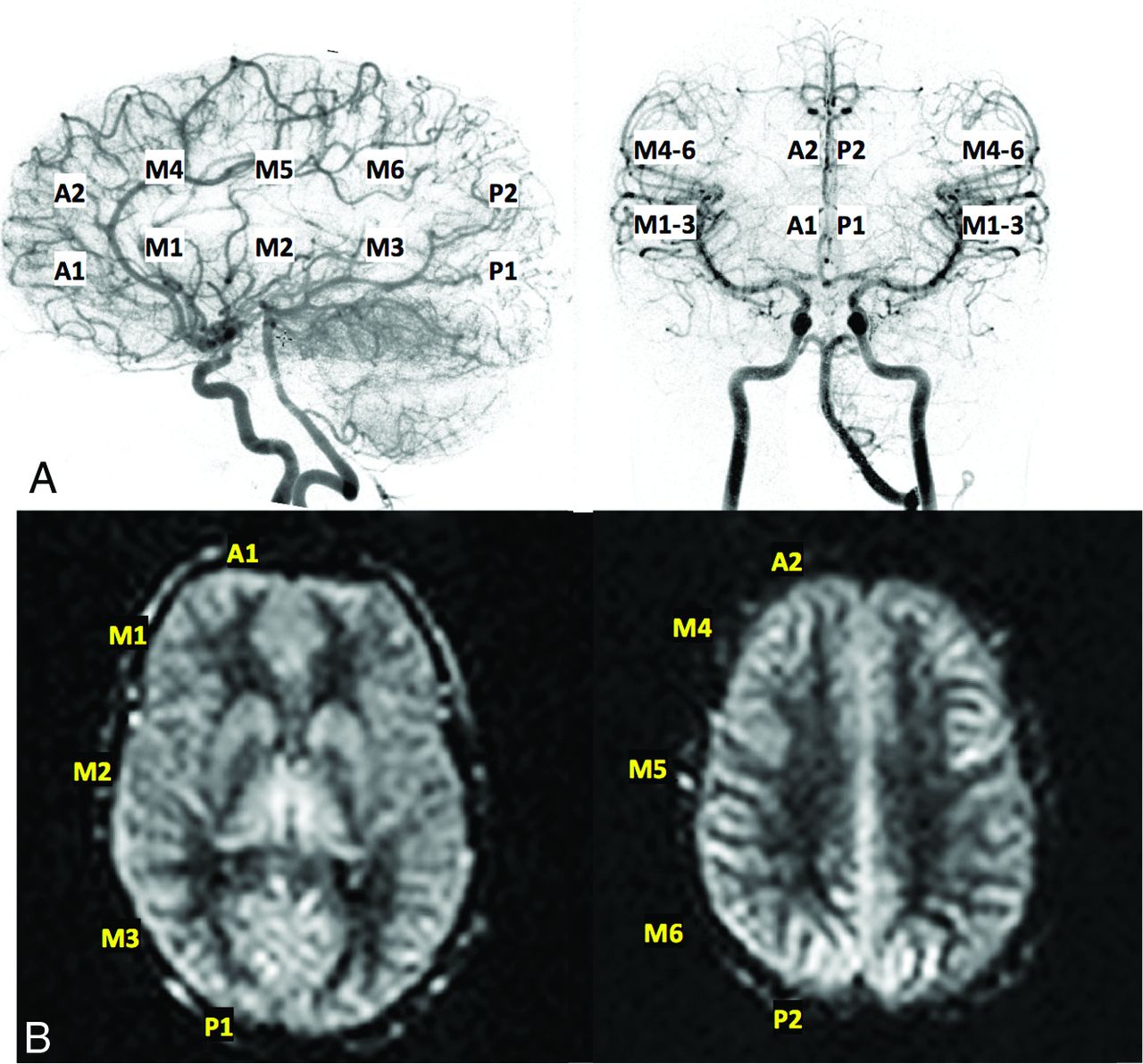
- Fig 1.
Representative ASPECTS regions used for scoring of both DSA (A) and ASL data (B); specifically, 2 ACA regions, 6 MCA regions, and 2 posterior cerebral artery regions per side were evaluated.
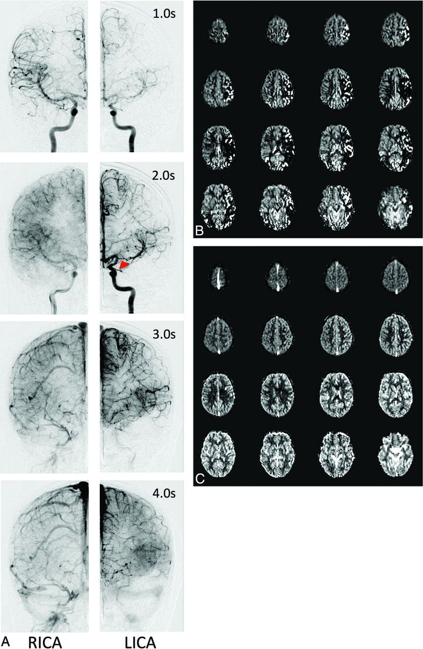
- Fig 2.
Representative DSA data (A), PASL CBF maps (B), and VSASL CBF maps (C) for a preoperative patient with left-sided moyamoya. A, Frontal DSA data from bilateral ICA injections at various postinjection times. The right side appears relatively normal, with early arterial filling and ACA/MCA parenchymal blush at 2.0 seconds. In contrast, the left side demonstrates delayed anterograde filling through a proximal M1 MCA stenosis (red arrowhead), retrograde filling via ACA-MCA collaterals, and delayed parenchymal perfusion of the MCA territory. Parenchymal perfusion of the left MCA territory is finally reached by 4.0 seconds. PASL maps (B) reflect the DSA appearance at 2.0 seconds bilaterally, including areas of curvilinear hyperintensity corresponding to macrovascular flow and perfusion deficit, while VSASL maps (C) reflect parenchymal DSA phases, despite these occurring at different times (2.0 seconds on the right, 4.0 seconds on the left). RICA indicates right ICA injection; LICA, left ICA injection.
- Fig 3.
Average PASL and VSASL-CBF CV (a marker of perfusion variability) calculated for patient vascular territories demonstrating delayed-but-complete parenchymal perfusion by DSA, with normal sibling values provided for comparison. Patient VSASL values are similar to those of healthy siblings, consistent with the DSA appearance, but PASL values are markedly different. Error bars denote 95% CI.
- Fig 4.
Standard ASL label propagation with patent proximal vessels (A) and steno-occlusive disease with secondary collateralization (B). A, The ASL label travels from the labeling band to the distant microvasculature during the standard PLD, resulting in symmetric, homogeneous gray matter perfusion (C). B, The label is delayed due to slow flow through the stenosis and circuitous collateral pathways. Consequently, the label does not fully reach the distal microvasculture during PLD and remains caught in the macrovasculature, resulting in areas of apparent perfusion deficit and hyperintense arterial transit artifacts (D).


Tables
Pt. Moyamoya Etiology Age (yr) Sex Laterality Operative Status Suzuki37 (R/L), Preoperative Treatment Matsushima38 (R/L) 1 Idiopathic 11 F Left Pre IV 12 Post Left synangiosis C 2 NF1 6 M Right Post IV–V Right synangiosis A 3 SCD 13 F Bilateral Post NA Bil synangiosis A 4 Idiopathic 14 F Bilateral Post III–IV/III–IV Bil synangiosis A/A 5 Idiopathic 19 F Bilateral Post V–VI/III–IV Bil synangiosis A/C 6 NF1 17 F Bilateral Pre I/III–IV 7 SCD 11 M Bilateral Post II/II Bil synangiosis A/B 8 Idiopathic 14 M Bilateral Post IV/IV Bil synangiosis A/A 9 Idiopathic 13 F Bilateral Post II/III Bil synangiosis B/C 10 Idiopathic 16 F Bilateral Pre I/I 11 Idiopathic 7 F Bilateral Pre II/II 12 NF1a 5 F Right Pre I 13 SCD 11 F Right Pre II 14 NF1 8 M Left Post III Left synangiosis A 15 Idiopathic 7 F Right Post III Right synangiosis A 16 Idiopathic 13 F Left Post NA Left synangiosis B 17 Idiopathic 19 F Bilateral Pre III/I–II 18 Idiopathic 3 M Bilateral Post II–III/II–III Bil synangiosis B/A–B 19 NF1 15 M Left Post V Left synangiosis B 20 Idiopathic 12 M Right Post III–IV Right synangiosis B–C 21 Idiopathic 15 M Bilateral Post III/III Bil synangiosis B/B 22 Idiopathic 2 M Bilateral Post IV/II Bil synangiosis A/A–B Note:—NF1 indicates neurofibromatosis type 1; SCD, sickle cell disease; Pre, before; Post, after; R, right; L, left; NA, not applicable; Bil, bilateral.
↵a Patient with NF1 with radiated right masticator space tumor; arteriopathy possibly related to radiation.
DSA Score ASL Score Totals 0 1 2 3 0 0 1 0 0 1 (<1%) 1 0 6 17 1 24 (5%) 2 0 7 192 21 220 (46%) 3 0 0 13 222 235 (49%) Totals 0 (0%) 14 (3%) 222 (46%) 244 (51%) 480 ↵a Agreement = 88%, Cohen κ = 0.77, P < .0001.


Jump to section
Related Articles
Cited By...
- Macrovascular blood flow and microvascular cerebrovascular reactivity are regionally coupled in adolescence
- Velocity-selective arterial spin labelling bolus duration measurements: Implications for consensus recommendations
- Blood oxygenation-level dependent cerebrovascular reactivity imaging as strategy to monitor CSF-hemoglobin toxicity