
Article Figures & Data
Figures
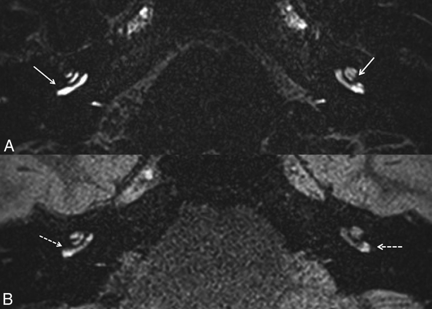
- FIG 1.
A 42-year-old woman with right possible MD. Axial 3D-FLAIR CFA at the level of the lateral semicircular canal (A) shows a normal utricle (white arrow) and lateral ampulla (right dotted arrow). C, Axial 3D-FLAIR CFA through the inferior part of the vestibule shows a normal saccule (white arrow) and posterior ampulla (white dotted arrow). Axial 3D-FLAIR heavily-T2 VFA at the level of the lateral semicircular canal (B) shows a normal utricle (white arrow), while the lateral ampulla (right dotted arrow) is barely visible. D, Axial 3D-FLAIR heavily-T2 VFA through the inferior part of the vestibule shows a normal saccule (white arrow) and posterior ampulla (white dotted arrow).
- FIG 2.
A 63-year-old woman with right definite MD. A, Axial CFA at the level of the basal turn of the cochlea shows a marked right cochlear BLB impairment compared with the left side (white arrow). B, The right cochlear BLB impairment (white dotted arrow) is less obvious with hVFA.
- FIG 3.
A 42-year-old woman with right possible MD. Right axial CFA (A) and hVFA (B) sequences through the inferior part of the vestibule show a normal saccule (white arrow) and posterior ampulla (white dotted arrow) in both sequences. A 43-year-old man with a right definite MD. Right axial CFA (C) and hVFA (D) sequences through the inferior part of the vestibule show saccular hydrops (white arrow) in both sequences. A 35-year-old woman with a right possible MD. Right axial CFA (E) and hVFA (F) sequences through the inferior part of the vestibule and the posterior ampulla (white dotted arrow). With the CFA sequence, the right saccule appears normal (white arrow). With the hVFA sequence, reader A described a right saccular hydrops, while reader B defined the right saccule as normal. The saccule (white arrow) and the posterior and lower parts of the utricle (white arrowhead) were confluent without expansion of these 2 structures.


