
Article Figures & Data
Figures
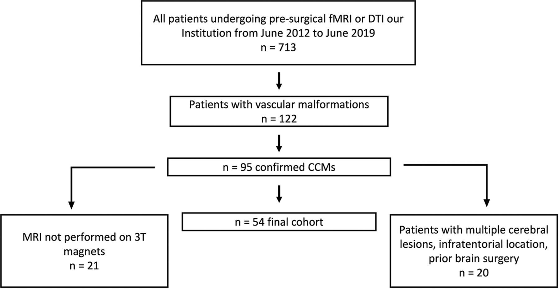
- FIG 1.
Flow chart of study selection.
- FIG 2.
Conventional T2 TSE showing a left frontal subcortical cavernous hemangioma. White and gray matter segmentations of ROIs of the first and second expansion shells (4 mm each) are seen in light blue. On the contralateral hemisphere, computation of control ROIs is shown in light red.
- FIG 3.
Images exemplifying qualitative data analysis in 2 different patients (A and C) with T2 TSE and T2 gradient sequences (B and C). A 43-year-old woman with a superficial, previously ruptured left parietal CCM with a collection of qualitative characteristics. CCM maximum diameters (red and green, A and B), hematoma maximum diameters (yellow and orange, A and B), and maximum thickness of hemosiderin rim thickness (light blue, A and B). A 52-year-old man with a left subcortical CCM with measurement of maximum diameters (green and red, C and D) and maximum hemosiderin rim thickness (light blue, C and D).
- FIG 4.
Error, bias, and variance of ReHo and FA measurements near CCMs. A, Absolute errors of each ROI compared with their contralateral controls (median with interquartile range and adjusted P values from the Wilcoxon matched-pairs signed-rank test). B, Biases of each ROI compared with their contralateral controls. Mean with standard error of mean is shown. P values from 1-sample t tests are shown below the bars, and adjusted P values from Welch t tests are shown above the bars (C). Variances of each ROI compared with their contralateral controls and SDs are shown. Adjusted P values from F-tests for equality of variance are shown. P values or adjusted P values > .05 were deemed nonsignificant and marked as ns.


Tables
Sequences Parameters 3D turbo field-echo T1-weighted Sagittal acquisition; TR/TE = 8.1, 3.7 ms; section thickness = 1-mm isovoxel; matrix = 256 × 256 Turbo spin-echo T2-weighted Axial acquisition; TR/TE = 3000/80 ms; section thickness = 3 mm; matrix = 420 × 272 FLAIR Axial acquisition; TR/TE = 11,000/125 ms; matrix = 320 × 200 ms; TI = 2800 ms T2*-weighted fast-field echo Axial acquisition; TR/TE = 1061/16 ms; matrix = 232 × 141 SWI Axial acquisition; TR/TE = 31/7.2 ms; Δ TE = 6.2 ms; matrix = 288 × 235 DTI Single-shot spin-echo echo-planar imaging; TR/TE = 6502/70 ms; matrix = 112 × 110; b-values = 0–800 mm2/s; 32 diffusion-sensitive directions BOLD functional imaging T2*-weighted echo-planar; TR/TE = 2000/35 ms; matrix = 96 × 96; flip angle = 90°; section thickness = 3 mm fMRI (ReHo) Variable Shell 1: 0–4 mm Shell 2: 4–8 mm Error, median APE (IQR) 25.1% (13.5–43.6) 15.0% (6.9–26.4) Bias, mean PE (95% CI) −27.2% (−33.8 to −19.8)b −8.3% (−14.9 to −1.1)c Variance, % deviation from mean 28.3%−39.5% 22.6%−29.2% DTI (FA) Variable Shell 1: 0–4 mm Shell 2: 4–8 mm Error, median APE (IQR) 9.2% (4.4–17.6) 6.5% (2.1–11.7) Bias, mean PE (95% CI) −0.3% (−4.1–4.8)d −0.1% (–3.1–2.9)e Variance, % deviation from mean 14.6%−17.1% 10.4%−11.6% ReHo vs FA: P value Variable Shell 1: 0– 4mm Shell 2: 4–8 mm Error <.001 <.001 Bias <.001 .16 Variance <.001 <.001


Jump to section
Related Articles
Cited By...
- No citing articles found.